AbbVie seeks USFDA nod for combination regimen of Venclexta, Acalabrutinib for previously untreated patients with Chronic Lymphocytic Leukemia

North Chicago: AbbVie has announced the submission of a supplemental New Drug Application (sNDA) to the U.S. Food and Drug Administration (FDA) for the fixed-duration, all-oral combination regimen of VENCLEXTA (venetoclax) and acalabrutinib in previously untreated patients with Chronic Lymphocytic Leukemia (CLL), offering CLL patients another VENCLEXTA combination regimen with the potential for time-limited treatment.

The submission is based on the positive results from the Phase 3 AMPLIFY trial. The combination regimen of VENCLEXTA and acalabrutinib improved progression-free survival (PFS) compared to standard chemoimmunotherapy in previously untreated patients with CLL.

“This FDA submission marks a milestone for CLL treatment with the potential approval for the first oral combination regimen of VENCLEXTA and acalabrutinib for previously untreated patients with chronic blood cancer. This new fixed-treatment duration approach could allow patients the opportunity for time off treatment, if approved, and be potentially practice-changing in frontline CLL care,” said Svetlana Kobina, vice president, global medical affairs, oncology, AbbVie.

AMPLIFY is an AstraZeneca-sponsored, global, multi-center Phase 3 trial evaluating VENCLEXTA plus acalabrutinib alone or combined with obinutuzumab versus chemoimmunotherapy in patients with previously untreated CLL without del(17p) or TP53 mutation.

Data presented at the 2024 American Society of Hematology Annual Meeting showed that the fixed-duration combination regimen of VENCLEXTA and acalabrutinib reduced the risk of disease progression or death by 35% vs chemoimmunotherapy (HR 0.65; 95% CI: 0.49-0.87; p=0.004). The safety profile of the VENCLEXTA and acalabrutinib combination regimen is consistent with the known safety profile of each individual therapy alone.

VENCLEXTA (venetoclax) is a first-in-class medicine that selectively binds and inhibits the B-cell lymphoma-2 (BCL-2) protein. In some blood cancers, BCL-2 prevents cancer cells from undergoing their natural death or self-destruction process, called apoptosis. VENCLEXTA targets the BCL-2 protein and works to help restore the process of apoptosis.

VENCLEXTA is being developed by AbbVie and Roche. It is jointly commercialized by AbbVie and Genentech, a member of the Roche Group, in the U.S. and by AbbVie outside of the U.S. Venetoclax is approved in more than 80 countries, including the U.S.

Powered by WPeMatico

Telanagana Medical Council mandates QR codes to combat Quackery

Hyderabad: To ensure that patients get treatment only from qualified medical practitioners, the Telangana Medical Council (TSMC) has directed all registered doctors to display their QR codes at their clinics and even on prescriptions. 

These QR codes, issued by the council to qualified doctors at the time of registration and are printed on the registration certificate. When scanned, the code provides essential background information about the registered doctor, ensuring transparency.

This will help the patients verify a doctor’s credentials before seeking medical treatment to avoid falling prey to fake doctors, which are rising at an increasing rate in Telangana. 

Also read- Illegal clinics run by Quacks busted in Telangana Medical Council Inspections

Since many patients visit their nearest clinics in the area for emergency treatment, the council has asked the doctors to voluntarily start displaying the QR codes so that they don’t visit a fake doctor’s clinic and risk their health. 

In case the general public is interested in finding the registration details of a doctor, they too can visit the website of the TGMC and search for the details by using the doctor’s registration number. The council’s website has a search option for doctors.  

It has been alleged that the Council had adopted this practice of providing QR codes for the past several months. Speaking in this regard, vice-chairman, Dr G Srinivas told Telangana Today, “QR codes are already there in every registration certificate issued by TGMC. All doctors can voluntarily display their QR codes on their clinic board or even prescriptions. This goes a long way in ensuring there is transparency.”

“We have a doctor search option on our TSMC online website. There is a facility available where the details of the doctor can be searched with a registration number,” Dr Srinivas pointed out.

Medical Dialogues had previously reported that the much-awaited ‘Know Your Doctor’, an exclusive and one-of-a-kind platform created to combat the growing menace of quacks in the state and ensure the highest standards of patient safety, was ultimately launched by the Maharashtra Medical Council in February this year. This innovative mobile application will allow patients to verify a doctor’s credentials before seeking medical treatment.

Also read- Know Your Doctor: Maharashtra Medical Council to combat quackery with QR Codes

Powered by WPeMatico

Primary Health Services to be available within 3 km radius in 4 Years: CM Fadnavis

Nagpur: Chief Minister Devendra Fadnavis on Sunday announced a comprehensive plan to develop the country’s best health services in Maharashtra in the next four years. The initiative aims to provide primary facilities to every person within a 3-kilometre radius.

Addressing a gathering after inaugurating the Late Bhanutai Gadkari Memorial Diagnostic Centre here, he said the cost of treatment is going up along with advancements in technology, which highlighted the need to give more people affordable and subsidised care under the government health system.

The Maharashtra government and the Centre are working on this aspect, Fadnavis added.

Also Read:Rs 1000 crore sanctioned to upgrade GMCH, IGGMCH Nagpur: CM Devendra Fadnavis

“A study of health services has shown that the government has developed very good tertiary treatment facilities in Maharashtra, but we are lacking to some extent when it comes to primary health services. If we go by the ratio, then 60 per cent of funds should go to primary health services and 40 per cent to tertiary services,” he said, reports PTI.

If the primary health services are strengthened, then it lessens the load on tertiary services, because 60 to 70 per cent patients get treated for their diseases in the primary health segment, he pointed out.

“The state government has prepared a plan to provide primary health services to every person within a 3-kilometre radius in urban and rural areas. We are developing a network of primary, secondary and tertiary health services. With the help of external aided agencies and the Union government, we will try to establish the best health services in the country in Maharashtra in the next four years,” Fadnavis asserted.

Union Minister Nitin Gadkari also addressed the event at the Late Bhanutai Gadkari Memorial Diagnostic Centre, which is named after his mother.

Gadkari emphasised that whatever he has achieved and has been able to do in life is because of the blessings and teachings of his mother.

His mother always taught him to serve the poor, the Union Minister of Road Transport and Highways and local Lok Sabha MP told the gathering.

This diagnostic centre will greatly help the needy and poor since services will be available at very affordable prices, Gadkari informed.

“Through such social initiatives, I learnt that the meaning of politics is not just power politics. I always tell my colleagues that politics is an instrument for social and economic reforms in society,” he said.

Also Read:Mumbai’s Tata Memorial Hospital receives bomb threat email

Powered by WPeMatico

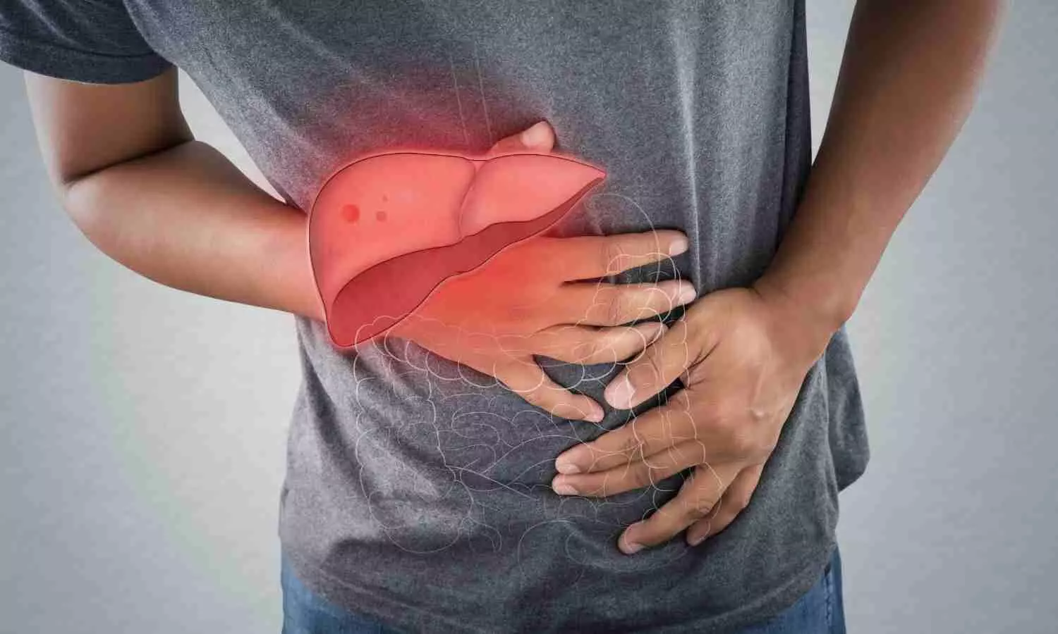
Fatty Liver Index Predicts Hepatocellular Carcinoma Risk in T2DM Without Cirrhosis: Study

A new study has established that the follow-up of the fatty liver index (FLI), both baseline and longitudinally, is an effective predictor of the risk of hepatocellular carcinoma (HCC) in type 2 diabetes mellitus (T2DM) patients even without cirrhosis. The research points out that T2DM is a leading risk factor for HCC, especially among patients with nonalcoholic fatty liver disease (NAFLD), but existing surveillance guidelines tend to disregard non-cirrhotic patients. These results emphasize the value of adding FLI as an inexpensive yet useful instrument for early cancer risk detection and intervention. The study was published in the journal Endocrinology and Metabolism by Eun-Hee Cho and colleagues.

HCC is still among the top cancer killers globally, and T2DM plays a major role in its development, especially when it occurs with fatty liver. Although there is known association, patients with T2DM without cirrhosis are generally not considered for routine screening for liver cancer, leaving a gap for early detection. This new evidence puts the FLI, a scoring system from waist circumference, body mass index (BMI), triglyceride, and gamma-glutamyl transferase (GGT) levels as a promising solution.

In this population-based study, health screening data of 92,761 people with T2DM aged 40-79 years who had two standardized health examinations between 2012 and 2015 were investigated. FLI was computed for each participant according to validated clinical parameters. Participants were stratified by both baseline FLI and FLI change according to these scores.

To detect new cases of HCC, investigators employed International Classification of Diseases (ICD) codes and billing records gathered from 2016 through 2020. The research sought to assess how alterations in FLI values either a rise or decline between screenings influenced the eventual risk of liver cancer development.

Key Findings

The research presented robust evidence that high baseline FLI values and rising FLI over time are associated with greater risks of developing HCC among T2DM patients:

• Those with a baseline FLI of 30 to 59.9 had a 1.90-fold higher risk of developing HCC than those with FLI <30 (P < 0.01).

• Those with baseline FLI ≥60 had a yet greater risk—2.94-fold—of developing HCC (P < 0.01).

• Individuals whose FLI rose from <30 to ≥30 during the interval between the two screenings had a 2.10-fold greater risk of HCC (P < 0.01).

• On the other hand, patients whose FLI decreased from ≥30 to <30 had 36% reduced risk for developing HCC (HR = 0.64, P = 0.03).

• Significantly, the protective effect of reduced FLI became apparent only after approximately 3 years, indicating that long-term follow-up is essential.

Overall, the research shows that baseline FLI values, as well as their time-dependent variations, are strong risk predictors of hepatocellular carcinoma in type 2 diabetic patients even if without liver cirrhosis. The rising FLI intensifies HCC risk, whereas its decline is linked to a lower risk, although with several years’ delay. These observations imply that FLI monitoring at regular intervals may be particularly useful and effective as a screening tool for early detection of high-risk individuals, enabling timely intervention and a possible decrease in liver cancer mortality in T2DM patients.

Reference:

Cho EH, Kang MG, Lee CH, Oh S, Shen C, Oh HR, Park YR, Lee H, Kim JS, Park JH. Fatty Liver Index Dynamics as a Predictor of Hepatocellular Carcinoma in Patients with Type 2 Diabetes Mellitus and Non-Cirrhotic Livers. Endocrinol Metab (Seoul). 2025 May 29. doi: 10.3803/EnM.2024.2286. Epub ahead of print. PMID: 40437794.

Powered by WPeMatico

GLP-1 Therapies Most Effective for Preoperative Weight Loss in High-BMI Patients: Study

A new study published in the journal of Surgical Endoscopy that GLP-1 therapies lead to greatest preoperative weight and BMI reduction when compared to other strategies, making them effective for high-risk, high-BMI patients before surgery. However further research is needed to determine the optimal use and cost-effectiveness of anti-obesity medications before and after metabolic procedures.

From April 2018 to February 2023, a total of 206 patients underwent bariatric procedures at the Tulsa center. Of these, 79 individuals had an initial body mass index equal to or over 49.5 kg/m², making them part of a high-risk surgical group. The average age of the participants was 44 years, with females representing roughly 76% of the sample. Also, 43% of the patients were found to have obesity-associated genetic markers.

The patients in the program were offered a comprehensive weight loss approach, including lifestyle interventions, anti-obesity medications (AOMs), or a combination thereof. The medications included oral AOMs, GLP-1 receptor agonists, GLP-1/GIP co-agonists, and combination medical therapies. The individuals who followed lifestyle modification alone lost a median of 3.1 kg before surgery. Oral AOMs improved that figure to 8.5 kg. However, the most substantial weight loss occurred among the patients treated with GLP-1 or GLP-1/GIP agents, who lost a median of 10.3 kg.

The patients using combination therapy, which included GLP-1s alongside other treatments, showed a similar median loss of 10.4 kg. The statistical difference between these groups was significant (P = 0.01), demonstrating the added value of pharmacologic support. The benefits of these medications extended beyond the operating table. At the one-year postoperative mark, the average total weight loss among all patients was approximately 62 kg, amounting to 65.6% excess weight loss.

This study emphasized the importance of GLP-1 therapies as part of a multimodal prehabilitation plan for high-BMI individuals. These results confirm that GLP-1 receptor agonists not only improve surgical readiness but may also enhance long-term outcomes in metabolic surgery. Overall, these results suggest that introducing GLP-1-based medications into preoperative protocols can help optimize weight loss and potentially reduce surgical risks in patients with severe obesity. 

Source:

Obermark, A. D., Seo, D. Y., Bernazard, A., Richards, J. R., Phillippe, S., Khorgami, Z., & Chow, G. S. (2025). Bariatric outcomes of high BMI patients with preoperative anti-obesity medications. Surgical Endoscopy. https://doi.org/10.1007/s00464-025-11851-w

Powered by WPeMatico

Incidence of wound complications is very low in TJA when using the Silk Fibroin adhesive for wound closure: study

Allergic contact dermatitis (ACD) from adhesive wound closure systems has garnered particular attention for its potential role in increasing wound complications in total joint arthroplasty (TJA).

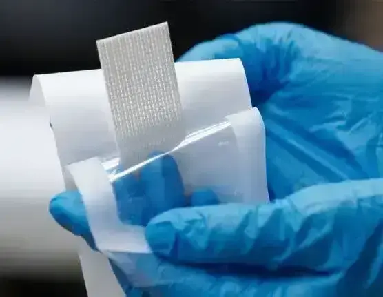
The study by Justin P. Moo Young et al performed at a high-volume orthopaedic specialty hospital, investigates the incidence of wound complications among 2 adhesive systems: a cyanoacrylate mesh (CM) adhesive and a silk fibroin (SF) adhesive. The study has been published in ‘Arthroplasty Today’

All TJAs with at least 6 weeks postoperative follow-up were retrospectively reviewed. Demographics and surgical outcomes were collected and analyzed. Statistical analyses were performed using Fisher’s exact tests and t-tests.

Key findings of the study were:

• A sample size of 170 CM and 85 SF subjects was calculated to achieve a power of 80%.

• Of the 257 patients identified (172 CM and 85 SF), 46.7% were females and 53.3% were males, with a mean age of 65.3 ± 9.0 years and a mean body mass index of 28.0 ± 4.6. Bivariate analyses revealed no significant differences in demographics or comorbidities between the CM and SF cohorts, except for frequency of American Society of Anesthesiologists 1 classification.

• The CM cohort exhibited a significantly higher incidence of ACD (6.4% vs 0%; P = .018), while differences in all other clinical outcomes were nonsignificant.

The authors concluded – “There is a very low incidence of wound complications in TJA when using the SF adhesive for wound closure. There was a statistically significant increase in ACD when using the CM adhesive. SF adhesives appear to be a superior wound closure option to consider in patients undergoing TJA.”

Further reading:

Silk Fibroin Closure Eliminates the Incidence of Allergic Contact Dermatitis Compared to Cyanoacrylate Mesh in Total Joint Arthroplasty

Justin P. Moo Young et al

Arthroplasty Today 33 (2025) 101668

https://doi.org/10.1016/j.artd.2025.101668

Powered by WPeMatico

Point-of-care ultrasonography found to be effective in identifying pneumothorax: Study

A new study published in The Journal of Emergency Medicine showed that in emergency trauma patients, point-of-care ultrasonography (POCUS) can reliably detect clinically severe pneumothorax (PTX).

Although CT is the gold standard for identifying PTX, researches pointed out that moving patients to imaging, particularly if they are unwell, can take a lot of time. Furthermore, although chest x-rays are another method for PTX identification, prior research indicates that they may overlook as many as 50% of PTX patients.

POCUS is now a practical emergency imaging technique that may be applied to trauma evaluation at the patient’s bedside. Prior research demonstrated the excellent sensitivity and specificity of the modality for PTX. Recent studies, however, have called into doubt how sensitive POCUS is for diagnosing PTX in trauma patients.

Therefore, using the red criteria of the National Expert Panel on Field Triage, 2021, Daniel Singer and team carried out this study to ascertain the accuracy of emergency physician (EP) POCUS in recognizing clinically important PTX in patients with high-severity trauma.

This research looked back at a level 1 trauma center’s high-severity trauma cases over a 3-year period. CT scan results or the clinician’s statement of a “rush of air” after tube thoracostomy implantation were used to identify whether PTX was present or not. If a patient needed a tube thoracostomy within 2.5 hours of triage, they considered PTX to be clinically severe. Emergency providers’ diagnostic test characteristics for POCUS and CXR were computed.

PTX was clinically significant in 40 out of 924 patients. For a sensitivity of 68% (95%CI, 52-80), specificity of 100% (95%CI, 19.8-100), positive predictive value (PPV) of 100% (95%CI, 84-100), and negative predictive value (NPV) of 14.3% (95%CI 2.5-43.9), POCUS found 26/38 who had survived before CT.

The sensitivity to 32/38, 84% (95%CI, 70-93), specificity, PPV 100% (95% CI 87-100), and NPV 25% (95%CI, 4.5-64) were all increased by fellowship-trained EPs reviewing the POCUS. The overall sensitivity and specificity of plain CXR were 48.1% (CI 34.2-62.2) and 99% (CI 97.5-99.6), respectively.

Overall, these findings suggest that POCUS can accurately identify clinically severe PTX, particularly when it is overseen by doctors with fellowship training. Less experienced EPs may need more supervision from more experienced EPs and should use appropriate technique when acquiring and interpreting images. 

Reference:

Daniel D Singer, Hayley Scott, Ali Khan, Alexandra Donnelly, Adam J Singer, Isadora Botwinick, Randeep Jawa, Ambika Mukhi, Henry C Thode, Michael Secko, ED accuracy of POCUS in identifying clinically significant PTX in high severity trauma patients., The Journal of Emergency Medicine, 2025, https://doi.org/10.1016/j.jemermed.2025.07.009.

Powered by WPeMatico

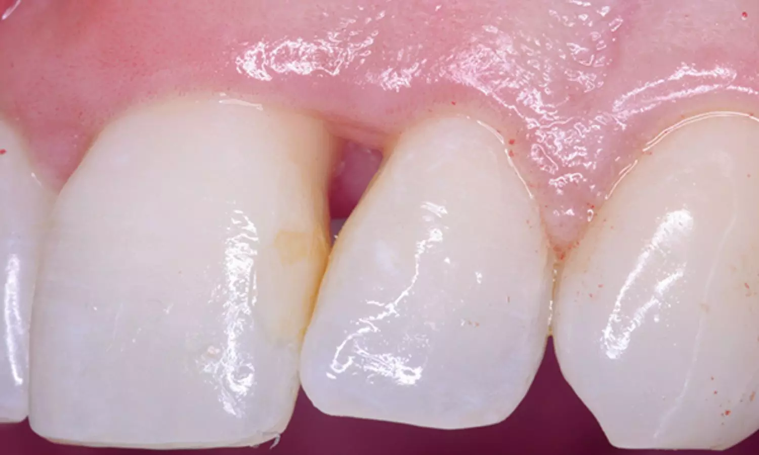
Hemolaser Therapy Effectively Reduces Interdental Black Spaces and Improves Patient Comfort: Study

Brazil: Researchers have found in a new study that hemolaser therapy significantly reduced interdental black spaces within a short follow-up period and led to improvements in patient discomfort and quality of life over time.

The study, published in Lasers in Medical Science, was conducted by Alessandra Areas e Souza and colleagues from Fluminense Federal University, Nova Friburgo, Brazil. The pilot study aimed to assess the clinical effectiveness, aesthetic impact, and quality of life outcomes of patients undergoing hemolaser therapy (HLT) for the treatment of papillary loss, a condition that results in unsightly black spaces between teeth due to the shrinking of the gum papilla.

Twelve patients were enrolled and divided into two groups. The HLT group (n=6) received gentle stimulation of gingival bleeding using a periodontal curette, followed by a red laser application (660 nm). The control group (n=6) received only the red laser without prior gingival stimulation. The procedure was carried out in two sessions spaced one week apart, and patients were followed up for 180 days.

Based on the study, the researchers reported the following findings:

  • Findings from the study demonstrated that hemolaser therapy resulted in a notable decrease in both papillary height and papillary area in the treatment group at both 90 and 180 days post-treatment.
  • These reductions in papillary dimensions were statistically significant when compared to the control group, suggesting that the combined use of mechanical gingival stimulation and laser application was more effective than laser treatment alone.
  • Improvements in the Modified Papilla Index (MPI), which is used to evaluate the visual appearance and presence of papillary tissue between adjacent teeth, were also observed in the hemolaser therapy group over the same time frame.
  • Patients in the hemolaser group demonstrated better MPI scores compared to those in the control group, indicating more visible and measurable regeneration of the interdental papilla.
  • In addition to the clinical outcomes, patient-reported results also favored the hemolaser approach.
  • Participants undergoing hemolaser therapy reported reduced aesthetic discomfort, as measured by the Visual Analog Scale (VAS), reflecting a greater sense of cosmetic satisfaction.
  • These individuals also recorded lower scores on the Oral Health Impact Profile (OHIP-14), a validated instrument used to evaluate the effect of oral conditions on overall quality of life.
  • These subjective improvements in discomfort and daily life impact were particularly pronounced at the 180-day follow-up and were statistically superior to those reported by participants in the control group.

The results highlight the potential of hemolaser therapy as a promising treatment for managing papillary loss and its associated aesthetic and functional concerns. By facilitating tissue regeneration and improving patient-reported well-being, the therapy may offer a minimally invasive solution to a problem that affects confidence and oral health.

The authors concluded, “While the sample size was small, as is typical in pilot studies, the researchers emphasize that their findings warrant further investigation in larger, long-term clinical trials. Nonetheless, this initial evidence suggests that hemolaser therapy could be a valuable tool in modern periodontal care for enhancing both outcomes and patient satisfaction.”

Reference:

Oliveira, L., Novais, L., Fontes, K. et al. Clinical efficacy, aesthetic discomfort, and quality of life in patients undergoing hemolaser therapy for the treatment of papillary loss: a longitudinal pilot study. Lasers Med Sci 40, 315 (2025). https://doi.org/10.1007/s10103-025-04543-3

Powered by WPeMatico

High ‘Good’ Cholesterol Linked to Lower Statin Use in At-Risk Adults, Raising Concerns Over Missed Prevention: Study

USA: A recent study has highlighted a potential gap in preventive cardiovascular care among U.S. adults with intermediate atherosclerotic cardiovascular disease (ASCVD) risk. It revealed that individuals with higher levels of high-density lipoprotein cholesterol (HDL-C) are less likely to receive statin therapy, despite similar risk profiles.

The findings were published online in JACC: Advances on April 30, 2025.

The analysis, based on data from the National Health and Nutrition Examination Survey (NHANES) collected between 2013 and early 2020, included adults aged 40 to 75 years who had no history of ASCVD, diabetes, or markedly elevated low-density lipoprotein cholesterol (LDL-C ≥190 mg/dL). All participants had an estimated 10-year ASCVD risk between 7.5% and 20%, categorizing them as having an “intermediate risk” and making them eligible for moderate-intensity statin therapy under current guidelines.

Traditionally dubbed the “good cholesterol,” HDL-C has long been associated with reduced cardiovascular risk. However, emerging research suggests that the functionality of HDL particles, rather than their quantity, may hold greater significance in atherosclerosis. Despite this shift in understanding, the long-held perception of HDL-C as protective may still be influencing clinical decisions.

In the study, Alexander R. Zheutlin, Division of Cardiology, Feinberg School of Medicine, Northwestern University, Chicago, Illinois, USA, and colleagues stratified participants by HDL-C levels: low (<40 mg/dL), normal (40–60 mg/dL), and high (≥60 mg/dL).

The key findings of the study were as follows:

  • Individuals with high HDL-C levels were significantly less likely to be prescribed statins than those with normal HDL-C levels.
  • Those with low HDL-C levels were slightly more likely to receive statin therapy than individuals with normal HDL-C.
  • Adjusted analysis showed that high HDL-C was linked to a 15% lower likelihood of statin use.
  • Low HDL-C was associated with a modest increase in statin prescriptions.
  • The study analyzed data from over 900 participants, representing nearly 23.7 million U.S. adults with intermediate 10-year ASCVD risk.
  • No significant interaction was found between sex and HDL-C levels concerning statin use.
  • The findings suggest that lower statin use among individuals with high HDL-C may reflect prevailing clinical perceptions rather than evidence-based, sex-specific prescribing patterns.

The authors emphasized that while HDL-C is included in risk assessment models, it is not considered a risk-enhancing factor in current cholesterol guidelines. Both low and excessively high HDL-C levels have been linked to increased ASCVD risk in other studies.

The cross-sectional analysis cannot establish causality but raises important concerns about possible undertreatment among adults with high HDL-C who otherwise qualify for preventive statin therapy. The findings stress the need for improved education around the complex role of HDL-C in cardiovascular health and suggest a reevaluation of how lipid profiles influence statin prescribing in clinical practice.

Reference:

Zheutlin AR, Jacobs JA, Bress AP. High-density lipoprotein cholesterol and statin use among U.S. adults with intermediate 10-year predicted ASCVD risk. JACC Adv. 2025; Epub ahead of print.

Powered by WPeMatico

Endotoxin-Induced Inflammation Worsens Depressive Symptoms in Older Adults with Insomnia: JAMA

A new randomized clinical trial published in JAMA Psychiatry reveals that older adults with insomnia are significantly more vulnerable to depressive mood symptoms following inflammatory exposure than those without insomnia.

The study enrolled 160 nondepressed participants aged 60 and older, split into two groups based on insomnia diagnosis. Participants were randomized to receive either endotoxin (to induce inflammation) or placebo. Researchers then tracked changes in mood using the Profiles of Mood States depression subscale (POMS-D), along with secondary measures of depressive symptoms and inflammatory cytokine levels. Results were striking. Compared to controls, participants with insomnia who received endotoxin showed a 3-fold greater increase in depressive mood (interaction F₁₀,₁₄₇₈ = 4.7; P < .001). These effects persisted longer and were clinically meaningful. Interestingly, both insomnia and control groups had similar rises in cytokines, but only the insomnia group showed a link between inflammation and mood changes (β = 0.33; 95% CI, 0.26–0.41; P < .001). These findings highlight how insomnia may amplify emotional responses to inflammation, even in the absence of clinical depression. Researchers recommend close monitoring of depressive symptoms in older adults with insomnia during infections or other pro-inflammatory events. Prevention strategies that address both sleep dysfunction and inflammation-related risk are crucial.

Reference:
Irwin MR, Boyle CC, Cho JH, et al. Inflammatory Exposure and Depression in Older Adults With Insomnia. JAMA Psychiatry. Published online July 16, 2025. doi:10.1001/jamapsychiatry.2025.1327

Keywords: insomnia, older adults, inflammation, depression, cytokines, endotoxin, mood, POMS-D, randomized clinical trial, JAMA Psychiatry, Irwin MR, Boyle CC, Cho JH

Powered by WPeMatico