Efforts underway to end race-based assessments of lung function
Powered by WPeMatico
SURMOUNT-5 was a 72-week, Phase 3b, multicenter, randomized, open-label,
comparator-controlled
study that evaluated the efficacy and safety of tirzepatide 15 mg or MTD (10 mg or 15 mg)
compared
with semaglutide 2.4 mg or MTD (1.7 mg or 2.4 mg) in adults with obesity or overweight with at least one
obesity-related complication, excluding diabetes. All participants received lifestyle intervention with
reduced-calorie diet and increased physical activity.
The primary
endpoint was the mean percentage change in body weight from baseline to Week 72. Tirzepatide
showed
superior mean body weight reduction (-21.6%) compared to
semaglutide (-15.4%). Key secondary endpoints included achieving ≥10%, ≥15%, ≥20%, and ≥25% weight
reduction—1 in 3 patients on tirzepatide achieved ≥25% weight loss vs
1 in
5 with semaglutide.
Tirzepatide also led to greater reductions in waist circumference, systolic and diastolic BP, HbA1c,
triglycerides,
and non-HDL cholesterol. The most common adverse
events were gastrointestinal, mostly mild to moderate.
Overall, tirzepatide demonstrated superior efficacy in weight and metabolic
improvements
compared to semaglutide in adults with obesity or overweight and comorbidities.
THERAPEUTIC INDICATION1:
Type 2 diabetes mellitus
MOUNJARO® is indicated for the treatment of adults with insufficiently
controlled type 2 diabetes mellitus as an adjunct to diet and exercise
• as monotherapy when metformin is considered inappropriate due to
intolerance or contraindications
• in addition to other medicinal products for the treatment of
diabetes.
For study results with respect to combinations, effects on glycaemic
control and the populations studied, see sections 4.4, 4.5 and 5.1.
Weight management
MOUNJARO® is indicated as an adjunct to a reduced-calorie diet and
increased physical activity for weight management, including weight loss and weight maintenance,
in adults with an initial Body Mass Index (BMI) of
• ≥ 30 kg/m² (obesity) or
• ≥ 27 kg/m² to < 30 kg/m² (overweight) in the presence of at least one
weight-related comorbid condition (e.g., hypertension, dyslipidaemia, obstructive sleep apnoea,
cardiovascular disease, prediabetes, or type 2 diabetes mellitus).
*SURMOUNT-5 was a 72-week, Phase 3b, multicenter, randomized,
parallel-arm, open-label, comparator-controlled study that evaluated the efficacy and safety of
Tirzepatide 15 mg or MTD (10 mg or 15 mg) compared with Semaglutide 2.4 mg or MTD (1.7 mg or 2.4
mg) in adults with Obesity (BMI ≥30 kg/m²) or overweight (BMI ≥27 kg/m²) with at least one
Obesity-related complication (e.g., hypertension, dyslipidemia), excluding diabetes.** The study
included a 2-week screening period. Mean baseline weight was 112.7 kg for Tirzepatide MTD (10 mg
or 15 mg) and 113.4 kg for Semaglutide MTD (1.7 mg or 2.4 mg). Participants in both the
Tirzepatide and Semaglutide treatment arms received lifestyle intervention, including a
reduced-calorie diet and increased physical activity. Treatments were administered QW
subcutaneously as an adjunct to a reduced-calorie diet and increased physical
activity1.
For Tirzepatide Prescribing Information, please check: https://image.mc.lilly.com/lib/fe9312747462077971/m/1/0e04e051-5fa2-445b-850d-4f09ef66d35e.pdf
Efficacy estimand, mixed model for repeated measures (MMRM) analysis,
modified intent-to-treat (mITT) population, (efficacy analysis set). Limitations of an
open-label study may be related to a bias in evaluation of the outcomes, efficacy and/or safety,
and analysis was not tested against a placebo-controlled comparison group1.
SURMOUNT-1 Phase 3 double-blind, randomized, placebo-controlled trial in
adults with Obesity (BMI of ≥30 kg/m²) or with Overweight (BMI of ≥27 kg/m²) with at least one
Obesity-related complication (e.g.,hypertension, dyslipidemia), excluding T2D. All participants
received lifestyle interventions, including a reduced-calorie diet and increased physical
activity.** All patients were randomly assigned in a 1:1:1:1 ratio to receive placebo, or
tirzepatide at a dose of 5 mg, 10 mg, or 15 mg as an adjunct to lifestyle
intervention4
¶p<0.001 vs baseline.
Mean % change in weight vs baseline (co-primary end point) at 72 weeks was -16.0% and -21.4% for
the 5 mg and 10 mg doses, respectively. Mean % change in weight vs placebo at 72 weeks was
-13.5%, -18.9%, and -20.1% for the 5 mg, 10 mg, and 15 mg doses, respectively (p<0.001 vs
placebo, adjusted for multiplicity). Mean kg change in weight vs baseline at 72 weeks was -16.1
kg and -22.2 kg for the 5 mg and 10 mg doses, respectively. Mean kg change in weight vs placebo
at 72 weeks was -13.8 kg, -19.8 kg, and -21.2 kg for the 5 mg, 10 mg, and 15 mg doses,
respectively (p<0.001 vs placebo, not adjusted for multiplicity). 2,4
# Adverse reaction development was not an endpoint for this study. Data
presented here should not be used to make inferences.
**”Obesity-related complications” are used as synonymic to “weight-related
complications and /or comorbidities”.
REFERENCES
1. Louis J. Aronne, M.D., et al. Tirzepatide as Compared with Semaglutide
for the Treatment of Obesity, N Engl J Med. 2025; doi: 10.1056/NEJMoa2416394
2. Mounjaro® (tirzepatide), India Prescribing Information. Updated March
2025.
3. Fisman EZ, Tenenbaum A. Cardiovasc Diabetol. 2021;20(1):225.
4. Jastrebo_ AM, et al. N Engl J Med. 2022;387(3):205–16.
Disclaimer:
This material (including any link) is intended solely for the use of the
recipient(s) and may contain confidential information. Any unauthorized review, use, disclosure,
copying, or distribution is strictly prohibited. If you are not the intended recipient, please
notify the sender immediately and destroy all copies of the material. The information provided
in this section is intended solely for the use of registered medical practitioner. This material
is being provided to healthcare professionals for their guidance and use. Nothing on this
website/microsite/material should be construed as giving medical advice or making
recommendations regarding any health-related decision or action.
Mounjaro®, KwikPen® and Lilly are registered trademarks of Eli Lilly and
Company. To be sold by retail under prescription of Endocrinologist or Internal Medicine
Specialists only. For adverse events and safety reporting, please reach out to: mailbox_in-gps@lilly.comFor any additional information related to Lilly products, please reach
out to: queries_in-medinfo@lilly.com. For further Information about Lilly and Lilly products please contact
us at the below address: Plot 92, Sector 32 Gurgaon, Haryana, 122001 India Ph.:
+91-124-4753000/01 | www.lilly.com/in.
PP-TR-IN-0580 VoD SM-5 | 29th July 2025
Eli Lilly and Company. All rights
reserved.
Powered by WPeMatico
According to a new research study, sleep impairment, particularly poor sleep quality, short sleep duration, and daytime dysfunction is significantly associated with increased diabetes distress in adults with type 2 diabetes, negatively impacting both disease management and mental well-being. The study was published in Diabetic Medicine journal by Hilde K. R. Riise and colleagues.
Diabetes distress is the emotional burden and frustration that individuals with diabetes can experience when attempting to control their disease. It differs from clinical depression but is linked to worse outcomes, such as diminished self-care and poor glycemic regulation. Sleep disturbances, also prevalent in individuals with T2D, have been suspected to contribute to heightened psychological stress. To investigate this, researchers employed answers to the HUNT4 sleep questionnaire, which gathered information on sleep problems such as snoring, sleep apnea, difficulty in falling asleep, nocturnal awakenings, early morning waking, excessive daytime sleepiness due to poor sleep, and restless legs. Diabetes distress was measured with the established PAID-5 questionnaire.
This cross-sectional study examined data on 1,954 adults with T2D who answered the HUNT4 survey. Sleep-related items were taken from the tested sleeping HUNT-Questionnaire, and the participants also indicated the average daily number of hours spent sleeping. Distress due to diabetes was assessed by the PAID-5 scale. Multivariate linear regression models were used to estimate the relationship of different sleep issues and distress scores, controlling for demographic, clinical, and mental health covariates. Prevalence rates and 95% confidence intervals were computed for each sleep impairment group.
Results
The findings revealed a distinct and uniform pattern: individuals who reported sleep impairments had greater diabetes distress scores.
Participants who slept ≤7 hours/night had an increased distress score: B = 0.6 (95% CI 0.2, 0.9).
Snoring correlated with increased distress: B = 0.6 (95% CI 0.1, 1.1).
They had a significantly larger increase in distress scores: B = 1.4 (95% CI 0.8, 2.2).
Waking up at night was also significantly associated: B = 1.1 (95% CI 0.6, 1.6).
Early morning waking had the same relationship: B = 1.2 (95% CI 0.7, 1.8).
The largest increase in distress was reported by those who had difficulty functioning during the day because they weren’t sleeping well: B = 2.6 (95% CI 1.7, 3.6).
Restless legs were also associated with distress: B = 0.8 (95% CI 0.2, 1.3).
These findings indicate that even ubiquitous and frequently-neglected sleep issues can have a very significant impact on the emotional load of T2D sufferers.
This research concluded that various forms of sleep impairment were all strongly related to greater diabetes distress levels in patients with type 2 diabetes. These findings emphasize the urgent importance of integrating sleep evaluation and treatment into overall diabetes care, given the potential for improving sleep health to be a major strategy for lowering diabetes-associated emotional distress.
Reference:
Riise HKR, Haugstvedt A, Igland J, et al. Diabetes distress and sleep impairment in type 2 diabetes: A population-based cross-sectional study-The HUNT Study, Norway. Diabet Med. Published online July 25, 2025. doi:10.1111/dme.70106
Powered by WPeMatico
A recent study involving 19,388 participants identified the platelet-to-high-density lipoprotein ratio (PHR) as a promising biomarker for gastrointestinal (GI) cancer risk. Individuals in the highest PHR quartile faced a 3.09-fold increased risk of GI cancers, with stronger associations seen in older adults, males, and obese individuals. A dose-response relationship was observed at PHR values of 3.2 and 4.5.
Gastrointestinal (GI) cancers, including gastric, colorectal, and esophageal cancers, pose a significant global health burden. Despite advancements in diagnostic tools, early detection remains challenging, particularly in low-resource settings. Emerging evidence highlights the platelet-to-high-density lipoprotein ratio (PHR) as a novel biomarker integrating systemic inflammation and lipid metabolism. However, its association with GI cancer risk remains underexplored. This study utilized data from the National Health and Nutrition Examination Survey (NHANES) from 2010 to 2018, comprising 19,388 participants, including 230 with GI cancers. PHR was calculated as the ratio of platelet count to high-density lipoprotein cholesterol levels and categorized into quartiles. Weighted logistic regression models, restricted cubic spline analysis, and subgroup analyses were employed to evaluate the association between PHR and GI cancer risk, adjusting for demographic, socioeconomic, lifestyle, and clinical factors. Results: Elevated PHR was independently associated with an increased risk of GI cancers. Participants in the highest PHR quartile exhibited a significantly higher risk (adjusted OR = 3.09; 95% CI: 2.16-4.43) compared to the lowest quartile. A dose-response relationship was observed, with two critical inflection points at PHR values of 3.2 and 4.5. Subgroup analyses revealed stronger associations among older adults, males, and obese individuals. The findings suggest that PHR may reflect the dynamic balance of systemic inflammation and lipid metabolism, contributing to tumorigenesis. This study identifies PHR as a promising, cost-effective biomarker for early detection and risk stratification of GI cancers. Its integration into screening programs could improve precision medicine strategies by identifying high-risk individuals for early intervention. Further longitudinal and mechanistic studies are warranted to confirm these findings and explore the underlying biological mechanisms.
Reference:
Tong Y, Lou X. Platelet-to-high-density lipoprotein ratio (PHR) as a predictive biomarker for gastrointestinal cancers: evidence from NHANES. BMC Gastroenterol. 2025 Apr 27;25(1):302. doi: 10.1186/s12876-025-03860-9. PMID: 40289098; PMCID: PMC12036308.
Keywords:
Tong Y, Lou X. Platelet-to-high-density, lipoprotein, ratio, (PHR), predictive, biomarker, gastrointestinal cancers, evidence, NHANES, BMC Gastroenterology, Cancer screening; Dose-response relationship; Gastrointestinal cancers; Lipid metabolism; NHANES; Platelet-to-high-density lipoprotein ratio (PHR).
Powered by WPeMatico
A large-scale prospective study has found a significant link between high consumption of ultraprocessed foods and an increased risk of Parkinson’s disease. Researchers followed over 120,000 participants from the UK Biobank for 10.5 years and observed that diets rich in ultraprocessed foods were associated with a higher likelihood of developing prodromal Parkinson’s, incident Parkinson’s disease, and even Parkinson’s-related mortality (Grosso et al., 2025). Ultraprocessed foods — including packaged snacks, ready-to-eat meals, processed meats, and sugary beverages — are typically high in additives, refined sugars, saturated fats, and sodium. While these foods have already been linked to a range of health concerns, including obesity, diabetes, and dementia, this study adds new weight to concerns about their neurological impact.
Researchers controlled for lifestyle, genetic, and socioeconomic factors and still found a strong association between high ultraprocessed food intake and increased risk of Parkinson’s disease, particularly in men. The study also found that individuals with the highest consumption levels had up to a 50% increased risk of Parkinson’s-related death compared to those with the lowest intake. ultraprocessed foods, Parkinson’s disease, UK Biobank study, neurological risk, brain health, neurodegeneration, Parkinson’s mortality, dementia diet link, prospective cohort study, diet and brainThese findings suggest that dietary habits could play a key role in the development and progression of neurodegenerative diseases. While the study does not establish direct causation, it highlights the need for further investigation and public health measures that encourage diets rich in whole foods, fruits, vegetables, and healthy fats.
Keywords: ultraprocessed foods, Parkinson’s disease, UK Biobank study, neurological risk, brain health, neurodegeneration, Parkinson’s mortality, dementia diet link, prospective cohort study, diet and brain
Powered by WPeMatico
Australia: Researchers have found in a new randomized clinical trial that germicidal ultraviolet (GUV) light in common areas of long-term care facilities (LTCFs) did not reduce ARI incidence per zone per cycle, but did lead to a modest overall reduction in total acute respiratory infections (ARIs). GUV may serve as a supplemental infection control measure in such settings.
The study, published in JAMA Internal Medicine by Andrew P. Shoubridge and colleagues from the South Australian Health and Medical Research Institute, evaluated whether GUV appliances could help lower ARI rates in older adult residents of LTCFs. Respiratory outbreaks in such facilities are associated with high hospitalization and mortality rates, and current infection control practices often do not adequately address airborne transmission.
The multicenter, double-crossover, cluster-randomized clinical trial involved four LTCFs in metropolitan and regional South Australia. Eight zones covering a total of 211,952 bed-days were assessed over 110 weeks between August 2021 and November 2023. Each facility was divided into two zones, which alternated between active GUV appliances and inactive controls in six-week intervention periods separated by washout phases.
The following were the key findings of the study:
The findings suggest that while GUV did not significantly impact infection rates within individual study cycles, it contributed to a modest but meaningful decline in total ARI cases over time. According to the authors, this supports the potential role of GUV as an adjunct to existing infection control measures, particularly in settings with vulnerable elderly populations.
The study did have several limitations. The COVID-19 pandemic and associated public health restrictions affected study design, data collection, and diagnostic testing capabilities. Limited access to facilities prevented environmental sampling, and movement between intervention and control zones may have influenced outcomes. Additionally, many ARI cases were not confirmed through laboratory testing due to constrained diagnostic resources, and the results are specific to commercially available GUV devices used in the trial.
Despite these constraints, the research highlights the potential of GUV-based strategies to help mitigate airborne respiratory infections in long-term care environments. If proven cost-effective, integrating GUV with conventional infection control protocols could strengthen preparedness against seasonal outbreaks and emerging viral threats.
The authors emphasize the need for further research to optimize GUV deployment patterns and assess their impact across various facility layouts and infection prevalence levels.
Reference:
Shoubridge AP, Brass A, Crotty M, et al. Germicidal UV Light and Incidence of Acute Respiratory Infection in Long-Term Care for Older Adults: A Randomized Clinical Trial. JAMA Intern Med. Published online July 28, 2025. doi:10.1001/jamainternmed.2025.3388
Powered by WPeMatico
Cortisol is a crucial hormone that regulates many important bodily functions like blood pressure and metabolism, and imbalances of this stress hormone can lead to health problems.
Traditionally, cortisol levels must be measured in a doctor’s office or other clinical setting. But a new advance in the design of artificial biosensors paves the way for point-of-care testing and diagnoses with far greater accuracy than is currently available.
Andy Yeh, an assistant professor of biomolecular engineering at the University of California, Santa Cruz, has invented an artificial, luminescent sensor that binds with cortisol in the blood or urine and then emits light to indicate the levels of the stress hormone in the body. A new study in the Journal of the American Chemical Society demonstrates that this technique can detect cortisol across all levels relevant to human health.
Yeh demonstrated that this biosensor can be used in combination with the camera on a smartphone to enable people to measure cortisol levels at home or in a clinic, with high levels of sensitivity and without the costly instrumentation of the lab, greatly expanding access to accurate measurement of this important health indicator.
Yeh is an expert in artificial protein design, a technique that uses AI-guided computation to design proteins completely from scratch. This varies from traditional approaches, which modify proteins found in the natural world.
To create a new detection system for cortisol, Yeh designed a protein-based biosensor in which the stress hormone triggers two designed proteins to come close to each other at the molecular level. This process leads to light emission, with more light indicating more cortisol.
To Yeh’s knowledge, this is the first example of a completely computationally designed biosensor that can perform with such high sensitivity and dynamic range for detecting a small molecule analyte. Using a camera to measure the amount and color of light emitted allows cortisol levels to be read with more sensitivity than current tests provide.
This new diagnostic tool would be in a “mix and read” format-similar to the technique used in Covid-19 nasal swab rapid tests. The test requires just a drop of blood or urine, which is mixed with a solution that contains the biosensor. Then, a smartphone camera and app could translate the light emitted into a direct measurement of cortisol levels.
“You can read the signal directly-the output of the sensor is light emissions, so essentially you can just take a picture of the test with your smartphone,” Yeh said. “Ideally, that’s really field compatible.”
The test’s high level of sensitivity is a vast improvement over traditional tests, which don’t usually offer enough quantitative results when outside of the cortisol normal range. Yeh’s solution covers a wider dynamic range, offering quantitative results for healthy, too-low, and elevated levels of cortisol.
“This sensor is very, very sensitive compared to the current standard methods used in the hospital,” Yeh said. “The dynamic range is huge compared to the traditional assay.”
Down the line, Yeh envisions that this technology may also be used in a drug-development or diagnostic setting to better understand and treat the health issues that arise from cortisol deficiencies or surpluses.
Reference:
Julie Yi-Hsuan ChenXue Peng, De Novo Design of High-Performance Cortisol Luminescent Biosensors, Journal of the American Chemical Society, DOI:10.1021/jacs.5c05004.
Powered by WPeMatico
Researchers have found in a new study that high sugar intake and vitamin D deficiency significantly contribute to enamel hypoplasia in children, while calcium-rich diets provide a protective effect, highlighting the critical role of nutrition in dental health.
Enamel hypoplasia is a developmental defect of enamel characterized by incomplete or defective enamel formation. It is often influenced by genetic, systemic, and environmental factors, including diet. A cross-sectional study was conducted on 200 children aged 6–12 years from urban and rural schools. Data were collected through structured dietary questionnaires and oral examinations to assess the presence and severity of enamel hypoplasia. Dietary intake was classified into groups based on sugar frequency, calcium-rich foods, and vitamin D levels. Enamel hypoplasia was graded using the Modified Developmental Defects of Enamel Index (DDE Index). Statistical analysis was performed using Chi-square tests and logistic regression to evaluate associations. Results: Out of 200 children, 72 (36%) exhibited signs of enamel hypoplasia. High sugar consumption was significantly associated with enamel hypoplasia (P < 0.001), with 48% of children in the high-sugar group presenting with defects compared to 18% in the low-sugar group. Calcium-rich diets were protective, as only 10% of children with adequate calcium intake showed enamel hypoplasia (P < 0.05). Vitamin D deficiency was identified in 62% of affected children, indicating a strong correlation (P < 0.01). Logistic regression revealed that high sugar intake increased the odds of enamel hypoplasia by 2.8 times (OR = 2.8, 95% CI: 1.6–4.7). Diet plays a pivotal role in the development of enamel hypoplasia in children. High sugar consumption and vitamin D deficiency are key contributors, while calcium-rich diets offer protection.
Reference:
Meena M, Aijazuddin A, Kumawat R, Saxena S, Rajput T, Jaidupally RR. Assessing the Impact of Diet on Enamel Hypoplasia in Children. J Pharm Bioallied Sci. 2025 Jun;17(Suppl 2):S1972-S1974. doi: 10.4103/jpbs.jpbs_1818_24. Epub 2025 Jun 18. PMID: 40655848; PMCID: PMC12244768.
Keywords:
Diet, Strongly, Influences, Enamel, Hypoplasia, Risk, Children, Study, Meena M, Aijazuddin A, Kumawat R, Saxena S, Rajput T, Jaidupally RR, Calcium, children, diet, enamel hypoplasia, oral health, sugar consumption, vitamin D
Powered by WPeMatico
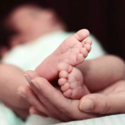
A high-resolution ultrasound device has shown great accuracy in detecting suspected meningitis in newborns and infants, potentially offering a non-invasive alternative to lumbar puncture, the traditional diagnostic method.
This is the main conclusion of an international study led by the Barcelona Institute for Global Health (ISGlobal), a centre supported by the ”la Caixa” Foundation, in collaboration with hospitals in Spain, Mozambique, and Morocco. The results have been published in the journal Pediatric Research.
Meningitis is an inflammation of the membranes surrounding the brain and spinal cord. When caused by bacteria or fungi, it can be fatal if not diagnosed and treated early. Even in cases where the disease is overcome, it can leave serious after-effects, such as neurological damage or cognitive disorders. Despite medical advances in recent decades, meningitis remains a major threat to child health, especially in low- and middle-income countries, where limited access to early diagnosis exacerbates its impact.
Current diagnosis: invasive and impractical
At present, diagnosing meningitis requires a lumbar puncture to collect cerebrospinal fluid, which is then analysed in the lab for signs of inflammation, such as elevated white blood cell counts.
This is an invasive technique, with associated risks and significant practical limitations. In high-income countries, it is performed routinely even in cases of low suspicion, resulting in a large number of “normal” procedures and therefore a low diagnostic yield. Conversely, in low-income countries, the lack of resources means the test is rarely performed, leading to significant underdiagnosis or, in many cases, empirical and often inaccurate diagnoses.
The alternative: an ultrasound device
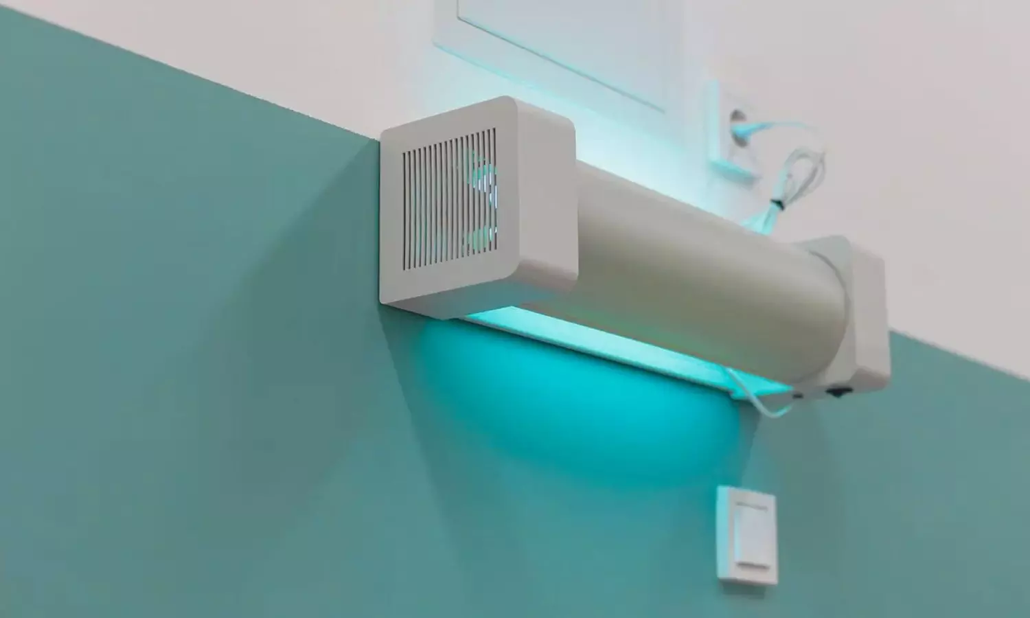
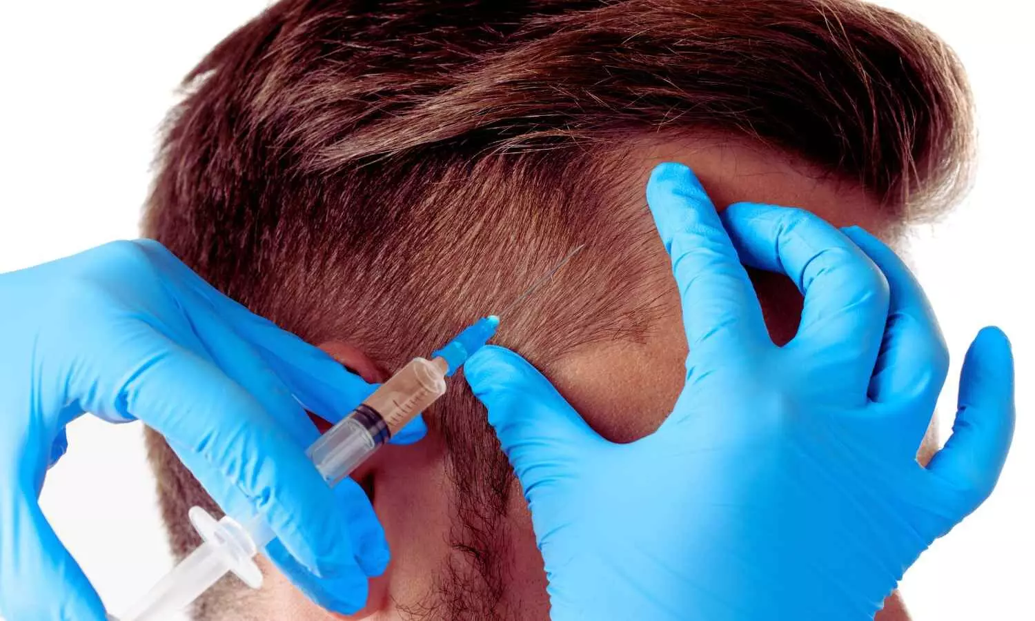
The aim of the study was to validate the NEOSONICS device, which uses high-frequency ultrasound applied through the baby’s open fontanelle —a membranous gap between the bones of the skull, which has not yet closed— to visualise and analyse cerebrospinal fluid. A deep learning algorithm interprets the images, identifies and counts the cells, and determines whether there are inflammatory signs consistent with meningitis.
The study, funded by the Bill and Melinda Gates Foundation, was conducted between 2020 and 2023 and included over 200 newborns aged up to 24 months from the Spanish hospitals Sant Joan de Déu, La Paz and Quironsalud, as well as Maputo Central Hospital (Mozambique) and the Hôpital d’Enfants de Rabat-Ibn Sina (Morocco). “The device was able to correctly classify 17 out of 18 meningitis cases and 55 out of 58 controls without meningitis,” explains Sara Ajanovic, researcher at ISGlobal and lead author of the study. “Specifically, it detected high white blood cell levels in cerebrospinal fluid with approximately 94% sensitivity and 95% specificity.”
The new device — cost-effective, portable and easy to use — could not only reduce the number of lumbar punctures, but also be used in clinically unstable patients where lumbar puncture is contraindicated. “Introducing a non-invasive tool could reduce unnecessary antibiotic use, prevent complications associated with lumbar puncture, and improve both early diagnosis and non-invasive monitoring of treatment response,” explains Quique Bassat, ISGlobal’s director general, ICREA researcher and senior author of the study.
Artificial Intelligence and the future of diagnostics
The validation of NEOSONICS marks a first step towards its future incorporation into clinical practice. At the same time, other studies coordinated by ISGlobal are exploring the potential of integrating Artificial Intelligence (AI) with ultrasound technology to enhance interpretation of results. Using advanced algorithms, AI enables image metrics to be analysed and texture patterns associated with inflammatory cells in cerebrospinal fluid to be detected, thus improving the system’s diagnostic capacity in cases of infant meningitis.
References: Ajanovic S, Jobst B, Jiménez J, Quesada R, Santos F, Carandell F, Lopez-Azorín M, Valverde E, Ybarra M, Bravo MC, Petrone P, Sial H, Muñoz D, Agut T, Salas B, Carreras N, Alarcón A, Iriondo M, Luaces C, Sidat M, Zandamela M, Rodrigues P, Graça D, Ngovene S, Bramugy J, Cossa A, Mucasse C, Buck WC, Arias S, El Abbass C, Tligi H, Barkat A, Ibáñez A, Parrilla M, Elvira L, Calvo C, Pellicer A, Cabañas F, Bassat Q; UNITED study group. Non-Invasive Meningitis Screening in Neonates and Infants: Multicentre International Study. Pediatr Res. 2025 Jul 23. https://doi.org/10.1038/s41390-025-04179-7
Sial, H., Carandell, F., Ajanovic, S., Jiménez, J., Quesada, R., Santos, F., Buck, W. C., Sidat, M., UNITED Study Consortium, Bassat, Q., Jobst, B., & Petrone, P. (2024). Novel AI-driven infant meningitis screening from high resolution ultrasound imaging. In medRxiv. https://doi.org/10.1101/2024.08.29.24312709
Powered by WPeMatico
Recent advancements in dermatology have highlighted the crucial role of dermal T cell immunity in regulating hair follicle regeneration and addressing immune-mediated alopecia. This emerging perspective offers promising insights into the mechanisms that govern hair growth, loss, and potential therapeutic interventions.
The intricate relationship between T cells and hair follicle regeneration is driven by key regulatory signaling pathways that dictate the activity of epithelial stem cells. Under normal conditions, these pathways maintain a balance between immune tolerance and hair follicle cycling, ensuring continuous hair renewal. However, when this balance is disrupted, immune-mediated disorders such as alopecia areata, androgenetic alopecia, and cicatricial alopecia can emerge, leading to varying degrees of hair loss.
At the core of this interaction, immune privilege of hair follicles serves as a protective mechanism, shielding hair follicles from immune attack. However, in conditions like alopecia areata, the collapse of this immune privilege triggers an autoimmune response, resulting in hair follicle destruction. The presence of CD8+ cytotoxic T cells, coupled with an overactive Th1/Th17 inflammatory response, exacerbates follicular damage, preventing normal hair growth.
Emerging research suggests that modulating T cell activity through immune-based therapies could pave the way for novel treatments. The ability of regulatory T cells (Tregs) to suppress excessive immune responses presents an opportunity to restore immune balance and promote hair regrowth. Furthermore, advances in cytokine-targeting therapies and JAK inhibitors hold significant potential in reversing immune-driven hair disorders.
Beyond alopecia, understanding the role of T cells in hair regeneration is reshaping the future of regenerative medicine. The influence of Tregs on stem cell activation, Wnt/β-catenin signaling, and epithelial-mesenchymal interactions underscores their broader application in tissue engineering and skin repair. These findings not only reinforce the importance of immune regulation in hair follicle biology but also open new avenues for developing precision therapies tailored to individual immune profiles.
With the increasing prevalence of hair loss disorders and the growing demand for effective treatments, immune-centered approaches represent a transformative shift in dermatology and regenerative medicine. By harnessing the power of dermal T cell immunity, the future of hair restoration is poised to move beyond conventional treatments, offering long-lasting solutions that target the root causes of hair follicle dysfunction.
Reference:
Nana Tao, Qingru Sun, Yuyuan Ying, Yitao Wang, Jianli Gao, Dermal T cell immunity and key regulatory signaling pathways: Implications in immune-mediated alopecia and hair regeneration, Genes & Diseases, https://doi.org/10.1016/j.gendis.2025.101518.
Powered by WPeMatico