Indian Case Study Supports Liquid Nitrogen as a Safe and Painless Gingival Depigmentation Technique

India: A recent case report published in the Cureus Journal has highlighted the effectiveness of liquid nitrogen cryotherapy as a novel approach for gingival depigmentation, offering promising cosmetic results with minimal discomfort. Gingival pigmentation, although often physiological, can be a major aesthetic concern for individuals, especially those with a gummy smile. To address this issue, various treatment modalities have been explored, including gingivectomy, grafts, laser therapy, and electrosurgery. However, cryosurgery using liquid nitrogen has emerged as a simple and cost-effective alternative.

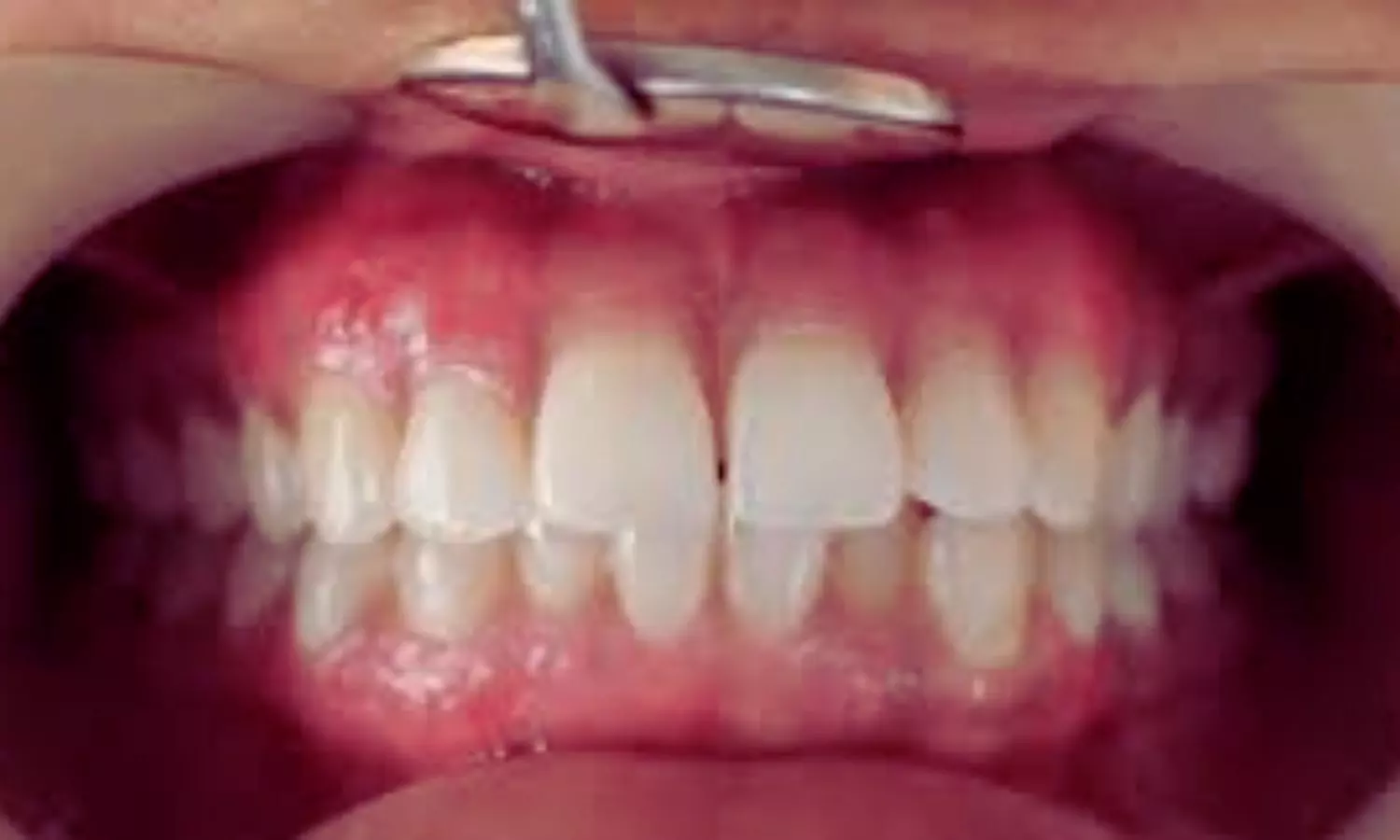
In the case, a 21-year-old female patient presented with the complaint of black gums, which had been present since childhood. Although the pigmentation was physiological, the patient sought treatment for cosmetic reasons. After a detailed discussion about various treatment options and potential repigmentation risks, liquid nitrogen cryotherapy was the preferred approach. The procedure was performed following thorough oral prophylaxis and patient consent.

During the procedure, the hyperpigmented areas of the maxillary and mandibular anterior gingiva were isolated using cotton rolls. A topical anesthetic spray was applied to minimize discomfort. Liquid nitrogen was administered using a pre-cooled cotton swab in a rolling motion for 30 seconds until blanching. Immediately after the procedure, slight erythema of the gingiva was noted, but the patient reported only mild discomfort without pain. No periodontal dressing was required postoperatively, and the patient was provided with oral hygiene instructions.

The healing process was uneventful, with the formation of a superficial necrotic layer that resolved within three to four weeks. The patient was monitored at one, three, and six months post-procedure, with no signs of recurrence. The gingiva remained pink, firm, and healthy, and the patient expressed high satisfaction with the aesthetic outcome.

This case emphasizes the potential of liquid nitrogen cryotherapy as an effective, safe, and minimally invasive method for gingival depigmentation. Unlike other procedures, it requires minimal equipment, does not induce bleeding, and does not necessitate anesthesia or dressing. Additionally, it provides excellent wound healing and long-term aesthetic stability. However, researchers emphasize the need for larger-scale studies and randomized clinical trials to further validate its long-term effectiveness and safety.

“With its simplicity and high patient acceptance, liquid nitrogen cryotherapy may offer a promising alternative for individuals seeking cosmetic improvement of gingival pigmentation. This technique holds great potential for clinical practice, providing a painless and efficient solution for managing gingival melanin pigmentation,” the authors concluded.

Reference:

A K K, Sankethguddad (January 27, 2025) Liquid Nitrogen as a Novel Treatment for Gingival Depigmentation: A Case Report. Cureus 17(1): e78099. DOI 10.7759/cureus.78099

Powered by WPeMatico

AI model can read ECGs to identify female patients at higher risk of heart disease: Study

A new AI model can flag female patients who are at higher risk of heart disease based on an electrocardiogram (ECG).

The researchers say the algorithm, designed specifically for female patients, could enable doctors to identify high-risk women earlier, enabling better treatment and care. Details are published today in Lancet Digital Health.

An ECG records the electrical activity of the heart and is one of the most common medical tests in the world. In their study, funded by the British Heart Foundation, the researchers used artificial intelligence to analyse over one million ECGs from 180,000 patients, of whom 98,000 were female.

In the latest study, the researchers developed a score that measures how closely an individual’s ECG matches ‘typical’ patterns of ECGs for men and women, and which showed a range of risk for each sex. Women whose ECGs more closely matched the typical ‘male’ pattern – such as having an increased size of the electrical signal – tended to have larger heart chambers and more muscle mass.

Crucially, these women were also found to have a significantly higher risk of cardiovascular disease, future heart failure, and heart attacks, compared to women with ECGs more closely matching the ‘typical female’ ECG.

Previous evidence has shown that men tend to be at higher risk of heart disease – more accurately called cardiovascular disease – which may be due to differences in hormone profiles and lifestyle factors. Because of this, healthcare professionals and the public believe that women’s risk of cardiovascular disease is low. This is even though the risk for women is also high, with women twice as likely to die of coronary heart disease, the main cause of heart attack, than from breast cancer in the UK. A recent consensus statement called cardiovascular disease the “number one killer” of women. The statement called for better diagnosis and treatment for women, as well as better female representation in clinical trials.

Dr Arunashis Sau, Academic Clinical Lecturer at Imperial College London’s National Heart and Lung Institute, and cardiology registrar at Imperial College Healthcare NHS Trust, led the research. He said:

“Our work has underlined that cardiovascular disease in females is far more complex than previously thought. In the clinic we use tests like ECGs to provide a snapshot of what’s going on but as a result this may involve grouping patients by sex in a way that doesn’t take into account their individual physiology. The AI enhanced ECGs give us a more nuanced understanding of female heart health – and we believe this could be used to improve outcomes for women at risk of heart disease.”

Dr Fu Siong Ng, Reader in Cardiac Electrophysiology at the National Heart & Lung Institute at Imperial College London and a consultant cardiologist at Imperial College Healthcare NHS Trust and Chelsea and Westminster Hospital NHS Foundation Trust, was the senior author of the study. He said: “Many of the women identified were in fact at even higher risk than the ‘average’ man. If it becomes used widely, over time the AI model may reduce gender differences in cardiac care, and improve outcomes for women at risk of heart disease.”

The research group recently published another paper on the related AI-ECG risk estimation model, known as AIRE, which can predict patients’ risk of developing and worsening disease from an ECG. Trials of AIRE in the NHS are already planned for late 2025. These will evaluate the benefits of implementing the model with real patients from hospitals across Imperial College Healthcare NHS Trust and Chelsea and Westminster Hospital NHS Foundation Trust. This model will be trialled in conjunction with AIRE.

Dr Sonya Babu-Narayan, Clinical Director at the British Heart Foundation, said: “Far too often, women are misdiagnosed or even dismissed by healthcare professionals, thanks to the myth that heart disease is ‘only a male’ issue. Even if they do receive the right diagnosis, evidence shows that women are less likely than men to receive recommended treatments.”

“This study has applied powerful AI technology to ECGs, a routine, cheap and widely available heart test. Harnessing the potential of this type of research could help better identify those patients at highest risk of future heart problems and reduce the gender gap in heart care outcomes. However, one test alone will not level the playing field. Ensuring every person gets the right heart care they need when they need it will require change in every part of our healthcare system.”

Powered by WPeMatico

Ultrasound may aid in rapid and accurate pulse assessment in CPR situations, reveals research

Recent study involved testing medical students’ ability to differentiate between ‘pulsation present’ and ‘no pulsation present’ using ultrasound videos of the common carotid artery (CCA) after watching a brief introductory video. The study aimed to assess the students’ accuracy in determining the presence of a pulse using different ultrasound modes (B-mode, M-mode, and Color Doppler).

Results and Conclusions

A total of 432 medical students participated in the study, evaluating 8640 decisions on CCA pulse detection. The results showed high accuracy in identifying pulsatile CCA using M-mode (96%) and Color Doppler (99.5%). B-mode exhibited lower accuracy in detecting pulsation (69%) but was highly reliable in identifying the absence of pulsation (99%). The study concluded that medical students could effectively detect the absence of a pulse using 2D ultrasound of the CCA, suggesting that a combination of Color Doppler and B-mode could be practical during CPR scenarios.

Challenges and Implications

The study highlighted the challenges in manually palpating pulses during CPR and emphasized the importance of prompt pulse checks. It also discussed the potential role of ultrasound in quickly assessing pulsation, especially when dealing with scenarios like reduced contractility post-resuscitation. Differentiating between pseudo-pulseless electrical activity (PEA) and true PEA using TEE or TTE echocardiography was mentioned, with a focus on the practicality of assessing CCA pulsation as a surrogate parameter.

Ultrasound in Pulse Detection

The research findings suggested that a simple dichotomous question of ‘CCA pulsation present’ or ‘no pulsation present’ could be effectively answered using ultrasound by medical students, even with limited experience. Color Doppler exhibited the highest sensitivity, while B-mode showed the highest specificity in pulse detection. The study indicated the potential for ultrasound to aid in rapid and accurate pulse assessment in CPR situations.

Study Limitations

The limitations of the study included the controlled setting and the use of pre-recorded ultrasound videos, which may not fully simulate real-world conditions. The need for further clinical studies to validate the technique in actual CPR settings was highlighted. Overall, the study underscored the efficacy of ultrasound in assisting healthcare providers in assessing pulsation during CPR and suggested areas for future research in this field.

Key Points

– The study focused on assessing medical students’ ability to differentiate between ‘pulsation present’ and ‘no pulsation present’ in the common carotid artery using ultrasound videos in various modes (B-mode, M-mode, and Color Doppler) after watching an introductory video.

– Results demonstrated high accuracy in identifying pulsatile common carotid artery using M-mode (96%) and Color Doppler (99.5%), with B-mode having lower accuracy in pulse detection (69%) but high reliability in identifying the absence of pulsation (99%).

– Medical students could effectively detect the absence of a pulse using 2D ultrasound of the common carotid artery, suggesting the practicality of utilizing a combination of Color Doppler and B-mode during CPR scenarios.

– Challenges in manually palpating pulses during CPR were noted, emphasizing the importance of prompt pulse checks. Ultrasound was discussed as a potential tool for quickly assessing pulsation, especially post-resuscitation scenarios with reduced contractility.

– Color Doppler showed the highest sensitivity, while B-mode exhibited the highest specificity in pulse detection, indicating the potential for ultrasound to aid in rapid and accurate pulse assessment in CPR situations.

– Study limitations included the controlled setting and the use of pre-recorded ultrasound videos, highlighting the need for further clinical studies to validate the technique in real CPR settings. The study emphasized the efficacy of ultrasound in assisting healthcare providers in pulse assessment during CPR and pointed out areas for future research in the field.

Reference –

B. Vojnar et al. (2024). Visual Detection Of Pulselessness By Carotid Artery Sonography – A Prospective Observational Study Among Medical Students.. *Resuscitation*, 110461 . https://doi.org/10.1016/j.resuscitation.2024.110461.

Powered by WPeMatico

Weight-loss surgery lowers risk of developing complications of liver disease in patients with cirrhosis and obesity: Study

A Cleveland Clinic study shows that patients with obesity and fatty liver-related cirrhosis who had bariatric (weight-loss) surgery significantly lowered their future risk of developing serious liver complications compared with patients who received medical therapy alone. The results were published in Nature Medicine journal.

Ali Aminian, M.D., director of Cleveland Clinic’s Bariatric & Metabolic Institute and lead investigator of the study, said results show that bariatric surgery should be considered as a treatment option in patients with cirrhosis and obesity. “Bariatric surgery was associated with a 72% lower risk of developing serious complications of liver disease and an 80% lower risk of progression to decompensated stage among patients with compensated cirrhosis and obesity.”

Obesity and diabetes are the leading causes of metabolic dysfunction-associated steatohepatitis (MASH), which is the most common form of chronic liver disease in the United States. Accumulation of fat within liver cells triggers a cascade of events ultimately leading to liver scarring. About 20% of people with MASH can progress to late-stage liver scarring which is called cirrhosis. Approximately three million people in the United States are estimated to have MASH-related cirrhosis.

Cirrhosis is categorized into two distinct stages: compensated and decompensated. In the compensated stage, despite presence of considerable damage, the liver maintains sufficient residual function to support the body’s needs and patients can appear relatively healthy. In the decompensated stage, severe and life-threatening complications occur reflecting the liver’s inability to sustain vital functions and liver transplantation becomes essential for survival.

“Patients with MASH-related cirrhosis have extremely limited treatment options. Currently, no therapeutic interventions have demonstrated efficacy in mitigating the risk of severe liver complications within this patient population,” said Sobia Laique, M.D., a transplant hepatologist and the study coinvestigator at Cleveland Clinic. “This underscores a critical unmet need for the development of effective therapies specifically targeting patients with compensated MASH-related cirrhosis.”

The aim of the SPECCIAL (Surgical Procedures Eliminate Compensated Cirrhosis In Advancing Long-term) study was to examine the long-term outcomes of bariatric surgery on the risk of developing major liver complications in patients with obesity and compensated MASH-related cirrhosis, compared with nonsurgical management.

A group of 62 Cleveland Clinic’s patients with compensated MASH-related cirrhosis and obesity who had bariatric surgery were compared with a control group of 106 nonsurgical patients and followed for 15 years. Study participants had similar characteristics such as severity of liver disease at their baseline liver biopsy.

Fifteen years after enrollment, study results show that 20.9% in the surgical group and 46.4% in the nonsurgical group developed one of the major complications of liver disease including liver cancer and death.

Over the course of 15 years, 15.6% in the surgical group and 30.7% in the nonsurgical group progressed from compensated cirrhosis to the decompensated stage. At 15 years, patients in the bariatric surgery group lost 26.6% (31.6 kg) of their weight and patients in the nonsurgical control group lost 9.8% (10.7 kg) of their weight.

Steven Nissen, M.D., Chief Academic Officer of the Heart, Vascular and Thoracic Institute at Cleveland Clinic and the study’s senior investigator, said: “Currently, lifestyle intervention is the only therapeutic recommendation for compensated MASH-related cirrhosis. However, lifestyle changes alone rarely provide the weight loss and metabolic changes needed to reduce the risk of liver complications in this patient population. The SPECCIAL study shows that bariatric surgery is an effective treatment that can influence the trajectory of cirrhosis progression in select patients.”

The authors note that the SPECCIAL study is the first to examine long-term clinical outcomes after bariatric surgery in patients with MASH-related cirrhosis. In 2021, the Cleveland Clinic-led SPLENDOR study suggested bariatric surgery as the first effective treatment for MASH without cirrhosis.

Future research is needed to study the new generation of anti-obesity medications and whether they can provide similar benefits in this patient population.  

Reference:

Aminian, A., Aljabri, A., Wang, S. et al. Long-term liver outcomes after metabolic surgery in compensated cirrhosis due to metabolic dysfunction-associated steatohepatitis. Nat Med (2025). https://doi.org/10.1038/s41591-024-03480-y

Powered by WPeMatico

Antimicrobial therapy for both partners found effective for women with bacterial vaginosis: NEJM

A new study published in The New England Journal of Medicine revealed that in addition to treating female patients, treating male companions with oral metronidazole and topical clindamycin decreased the prevalence of recurrent bacterial vaginosis after 12 weeks when compared to treating only women.

One-third of women of reproductive age have bacterial vaginosis, and recurrence is frequent. Treatment with a male partner may improve the chance of recovery, according to evidence of sexual exchange of organisms linked to bacterial vaginosis. In order to determine the impact of treating male and female partners independently on the prognosis of bacterial vaginosis, Lenka Vodstrcil and colleagues carried out this investigation.

The partners in which a woman experienced bacterial vaginosis and was monogamous with a male partner were included in an open-label, randomized, controlled study. In the partner-therapy group, the male partner got oral and topical antimicrobial treatment (two times a day for 7 days, 2% clindamycin cream applied to penile skin and 400 mg tablets of metronidazole) while the woman received first-line prescribed antibacterial medicines. The male partner in the control group received normal care, while the lady received first-line therapy. Recurrence of bacterial vaginosis within 12 weeks was the main result.

The partner-treatment group consisted of 81 couples, whereas the control group consisted of 83 couples. After 150 couples had finished the 12-week follow-up period, the data and safety monitoring board decided to end the experiment because the treatment of the woman alone was not as good as the treatment of the woman and her male companion.

Recurrence was observed in 24 out of 69 women (35%) in the partner-treatment group (recurrence rate: 1.6 per person-year; 95% CI: 1.1 to 2.4) and in 43 out of 68 women (63%) in the control group in the modified intention-to-treat population. This represents an absolute risk difference of −2.6 recurrences per person-year. Men receiving treatment had headaches, nausea, and a metallic taste as side effects.

Overall, when women were treated for bacterial vaginosis and their male partners received combination oral and topical antimicrobial medication, the risk of bacterial vaginosis recurrence after 12 weeks was lower than with conventional care.

Reference:

Vodstrcil, L. A., Plummer, E. L., Fairley, C. K., Hocking, J. S., Law, M. G., Petoumenos, K., Bateson, D., Murray, G. L., Donovan, B., Chow, E. P. F., Chen, M. Y., Kaldor, J., Bradshaw, C. S., & StepUp Team. (2025). Male-partner treatment to prevent recurrence of bacterial vaginosis. The New England Journal of Medicine, 392(10), 947–957. https://doi.org/10.1056/NEJMoa2405404

Powered by WPeMatico

Narrow Band Imaging Versus White Light: No Superiority in Detecting Recurrences After Chemo/Radiation for HNSCC

Netherlands: A recent randomized controlled trial compared white light (WL) and narrow-band imaging (NBI) using flexible laryngoscopy for detecting local recurrences after (chemo)radiation in patients with pharyngeal or laryngeal cancer. The study found that narrow-band imaging did not provide any advantage over white light in detecting local recurrences of head and neck squamous cell carcinoma after (chemo)radiotherapy.

“Detection rates were similar between the two methods (WL-NBI: 11.7%, WL: 10.0%), and NBI did not improve survival outcomes. Both overall survival and disease-free survival remained comparable, reinforcing WL as a reliable tool for routine post-treatment surveillance,” the researchers reported in Clinical Otolaryngology.

Early detection of local recurrences in head and neck squamous cell carcinoma (HNSCC) plays a vital role in improving long-term survival. Flexible laryngoscopy is widely used for post-treatment surveillance, with white light being the standard imaging technique. Narrow-band imaging (NBI), which enhances mucosal and vascular contrast, has been proposed as a potentially superior method for identifying recurrent tumors.

Against the above background, Constanze Scholman, Department of Otorhinolaryngology – Head & Neck Surgery, the University of Groningen, University of Medical Center Groningen, Groningen, the Netherlands, and colleagues aimed to assess the effectiveness of white light (WL) and narrow-band imaging (NBI) during flexible laryngoscopy in detecting local recurrences and to evaluate their impact on clinical outcomes in patients with HNSCC who underwent radiotherapy with or without chemotherapy ((C)RT).

For this purpose, the researchers conducted a prospective randomized controlled trial at a tertiary head and neck oncologic center to evaluate the effectiveness of WL and NBI in detecting local recurrences after (chemo)radiotherapy ((C)RT) for HNSCC. A total of 257 patients without residual disease post-treatment were enrolled and randomly assigned to either the WL group (n=120) or the WL-NBI group (n=137). These patients were closely monitored for 24 months to assess recurrence patterns and survival outcomes.

The primary focus was to compare local recurrence rates between the two groups, while additional analyses examined overall survival, disease-specific survival, disease-free survival, and local recurrence-free survival.

Key Findings

  • The detection rate of local recurrences was similar between both groups, with 11.7% in WL-NBI and 10.0% in WL.
  • Overall survival was comparable, with 88.3% in the WL group and 87.6% in the WL-NBI group.
  • Disease-specific survival rates were 86.7% for WL and 83.9% for WL-NBI, showing no significant difference.
  • Disease-free survival remained similar between groups, with 85.0% in WL and 83.2% in WL-NBI.
  • Local recurrence-free survival was 90.0% in the WL group and 89.1% in the WL-NBI group, with no observed superiority in the WL-NBI group.

The researchers found that the local recurrence rate in HNSCC patients without residual disease after (chemo)radiotherapy remained low during the first 24 months of follow-up. Their study demonstrated that flexible laryngoscopy using WL-NBI did not provide any added benefit in detecting local recurrences compared to standard white light imaging. Additionally, WL-NBI did not significantly impact clinical outcomes, reinforcing that white light remains an effective tool for routine surveillance.

“The findings suggest that there is currently no strong evidence to support the use of NBI as a superior imaging modality in post-treatment follow-up for HNSCC patients,” the researchers concluded.

Reference:

Scholman, C., Westra, J. M., Zwakenberg, M. A., Wedman, J., M. Steenbakkers, J. H., Oosting, S. F., Halmos, G. B., & C. Plaat, B. E. Comparison of White Light With Narrow Band Imaging Using Flexible Laryngoscopy for the Detection of Local Recurrences After (Chemo)Radiation for Pharyngeal or Laryngeal Cancer: A Randomised Controlled Trial. Clinical Otolaryngology. https://doi.org/10.1111/coa.14293

Powered by WPeMatico

Better Cardiovascular Health Linked to Lower Osteoporotic Fracture Risk, suggests study

Researchers have found in a new study that a high cardiovascular health score is associated with lower odds of osteoporotic fractures. Higher scores in Life’s Essential 8 metrics, such as physical activity, further reduce the risk. The cross-sectional study found that better cardiovascular health significantly decreases the likelihood of bone fractures related to osteoporosis. Further researchers emphasized the need for integrated public health strategies to improve both cardiovascular and bone health.

Osteoporotic fractures are a major public health concern, particularly among the aging population, as they significantly contribute to morbidity, mortality, and reduced quality of life. While cardiovascular health (CVH) has traditionally been linked to cardiovascular disease outcomes, emerging evidence suggests it may also influence bone health. This study investigates the association between CVH, as measured by the Life’s Essential 8 (LE8) score, and the prevalence of osteoporotic fractures in U.S. adults. This cross-sectional study utilized data from the National Health and Nutrition Examination Survey (NHANES) from 2005 to 2018. A total of 17,606 adults aged 20 and above were included in the analysis after excluding participants with missing data on CVH or osteoporotic fractures. CVH was assessed using the LE8 score, which incorporates eight modifiable cardiovascular health metrics: diet, physical activity, tobacco use, sleep, body mass index (BMI), lipid levels, blood glucose, and blood pressure. The primary outcome, osteoporotic fractures, was identified through self-reported data confirmed by a physician. Weighted multivariate logistic regression models were used to estimate the association between CVH and the prevalence of osteoporotic fractures, adjusting for demographic and health-related covariates. Participants with higher CVH scores had a lower prevalence of osteoporotic fractures. In the fully adjusted model, each 1-point increase in the LE8 score was associated with a 1% reduction in the odds of osteoporotic fractures (OR = 0.99, 95% CI: 0.98–0.99). Compared to participants with low CVH levels, those with moderate CVH had a 22% lower odds of osteoporotic fractures (OR = 0.78, 95% CI 0.70–0.87), and those with high CVH had a 34% lower odds (OR = 0.66, 95% CI 0.56–0.79). A significant linear trend was observed across different CVH levels (P for trend < 0.001). Subgroup analyses revealed that the inverse relationship between CVH and osteoporotic fractures was consistent across different demographic and health-related subgroups. This study highlights a significant inverse association between cardiovascular health and osteoporotic fractures in U.S. adults. These findings suggest that maintaining a high level of cardiovascular health, as measured by the LE8 score, may be important in reducing the risk of osteoporotic fractures. Public health strategies that integrate cardiovascular and bone health interventions may enhance overall health outcomes and reduce the societal burden of both cardiovascular diseases and osteoporosis.

Reference:

Ou, J., Wang, T., Lei, R. et al. Association between cardiovascular health and osteoporotic fractures: a national population-based study. Sci Rep 15, 3844 (2025). https://doi.org/10.1038/s41598-025-88020-5

Keywords:

Better, Cardiovascular, Health, Linked, lower, Osteoporotic, Fracture, Risk, suggests, study, Scientific Reports, Ou, J., Wang, T., Lei, R

Powered by WPeMatico

Mother’s use of acetaminophen linked to Child ADHD risk, reports research

Fetal acetaminophen exposure increases the likelihood that a child will develop attention-deficit/hyperactivity disorder (ADHD), according to a study published Feb. 6 in Nature Mental Health.

Prior research shows that upward of 70% of pregnant women use acetaminophen during pregnancy to control pain or reduce fever. The drug, which is the active ingredient of many pain-relief medications, is one of the few considered safe to take during pregnancy by the U.S. Food and Drug Administration.

The new findings suggest, however, that doctors should reconsider prescribing medications with acetaminophen to mothers during pregnancy, the researchers said.

“Most of the prior studies asked women to self-report whether they had taken Tylenol or anything that contained acetaminophen,” said lead author Brennan Baker, a researcher at Seattle Children’s Research Institute. Baker also works in the lab of Dr. Sheela Sathyanarayana, a UW Medicine pediatrician.

“This medication was also approved decades ago, and may need reevaluation by the FDA,” said Sathyanarayana, the paper’s senior author. “Acetaminophen was never evaluated for fetal exposures in relations to long-term neurodevelopmental impacts.”

Acetaminophen is widely used during pregnancy, with 41–70% of pregnant individuals in the United States, Europe and Asia reporting use. Despite acetaminophen’s classification as low risk by regulatory agencies such as the FDA, accumulating evidence suggests a potential link between prenatal acetaminophen exposure and adverse neurodevelopmental outcomes, including ADHD and ADHD autism spectrum disorder, the researchers noted.

This research tracked a cohort of 307 women from 2006 to 2011, who agreed to give blood samples during their pregnancy. The researchers tracked plasma biomarkers for acetaminophen in the samples.

The children born to these mothers were followed for 8 to 10 years. Among the women who did not use acetaminophen during pregnancy, the rate of ADHD was 9%, but for the women who used acetaminophen, the ADHD rate among their offspring was 18%.

Acetaminophen metabolites were detected in 20.2% of maternal plasma samples. Children whose mothers had these biomarkers present in their plasma had a 3.15 times higher likelihood of an ADHD diagnosis compared with those without detected exposure.

The association was stronger among daughters than sons, with the daughters of acetaminophen-exposed mothers showing a 6.16 times higher likelihood of ADHD while the association was weaker and nonsignificant in males. Researchers did not know why the association was stronger in females.

The investigators’ analysis used data from the Conditions Affecting Neurocognitive Development and Learning in Early Childhood (CANDLE) research cohort, which comprised 1,031 pregnant individuals in Memphis, Tenn., who were enrolled between 2006 and 2011. 

By happenstance, and not by design, the study cohort included only Black women, Baker said, adding that the results could be generalized to woman and children of any race or ethnicity.

Mothers often are advised to turn to acetaminophen, the primary agent in Tylenol, rather than ibuprofen, which is more likely to adversely affect the fetal kidney or heart, Baker said.

“(Acetaminophen) is really the only option to control fever or pain during pregnancy,” he said.

So, what is a mother to do?

“There is obviously more work that needs to be done in this area,” he said. “And we need to continually update our guidance.”

For example, he suggested, during prenatal visits, patients should discuss the dosage of a drug that contains acetaminophen or talk about what pain it is intended to help manage, he said. Another drug class, such as triptans, is safe and effective for managing migraines, he added.

More work needs to be done to find out if some people can tolerate acetaminophen during pregnancy with no ill effects on the fetus while others cannot, he said.

He added that research findings on the effects of the drug and its potential risks during pregnancy have not been consistent.

One study recently released in Sweden, showed no link between maternal acetaminophen use and ADHD in their children; while another study out of Norway, did in fact find a link. The study out of Sweden, however, relied on self-reported data, Baker noted.

“The study out of Sweden, however, reported that only 7% of pregnant individuals used acetaminophen,” Baker noted. “And that study could have underestimated the exposure.

“I think it goes back to how the data was collected,” he added. “The conflicting results means that more research is needed.”

Medical societies and the FDA should update guidance on the use of acetaminophen as safety data emerges, Sathyanarayana said. 

Reference:

Baker, B.H., Bammler, T.K., Barrett, E.S. et al. Associations of maternal blood biomarkers of prenatal APAP exposure with placental gene expression and child attention deficit hyperactivity disorder. Nat. Mental Health (2025). https://doi.org/10.1038/s44220-025-00387-6

Powered by WPeMatico

Lung abnormalities seen in children and teens with long COVID: Study

An advanced type of MRI uncovers significant lung abnormalities in children and adolescents with long COVID, according to a new study published today in Radiology, a journal of the Radiological Society of North America (RSNA).

Post-COVID-19 condition, commonly known as long COVID, can affect individuals of all ages and is diagnosed when symptoms persist for more than 12 weeks after an initial COVID-19 infection. Children and adolescents typically experience a milder form of the condition, but common symptoms such as chronic fatigue, headaches and poor concentration can negatively impact school performance and social activities.

While chest CT is frequently used to diagnose and monitor lung function of adults with long COVID, it is not typically recommended in children because it exposes the patient to ionizing radiation and may require injection of a contrast agent.

Young patients with suspected long COVID are typically evaluated with pulmonary function tests, echocardiography and reviews of medical history. Unfortunately, conventional pulmonary tests often show normal lung and cardiac function, even in symptomatic patients.

“Parents should understand that their children’s persistent symptoms after COVID-19 may have a measurable physiological basis, even when standard medical tests appear normal,” said lead study author Gesa H. Pöhler, M.D., a senior physician in the Department of Diagnostic and Interventional Radiology at Hannover Medical School in Germany.

The researchers employed phase-resolved functional lung (PREFUL) MRI. This advanced MRI technology can analyze lung ventilation (air movement in and out of the lungs) and perfusion (blood flow through the lungs). PREFUL MRI doesn’t require the use of radiation or intravenous contrast agents and can be done while the patient breathes freely, making it a suitable procedure for children.

“Our research provides the first comprehensive evidence of measurable regional lung perfusion abnormalities in pediatric post-COVID-19 condition using radiation-free, contrast-free lung imaging,” Dr. Pöhler said.

For the prospective study, conducted between April 2022 and 2023, the researchers enrolled 54 patients ranging in age from 11 to 17 years. Half of the patients were diagnosed with long COVID, and the other half were healthy controls. A self-reported assessment called the bell score was used to assess symptom severity in patients with long COVID.

Compared to healthy controls, children and adolescents with long COVID had significantly reduced blood flow in the lungs. A reduction in blood flow patterns in organs or other areas of the body can result in a lack of sufficient oxygen and nutrients.

The most prevalent symptom of fatigue affected all but one patient with long COVID.

“Importantly, the severity of fatigue symptoms correlated with these blood flow changes, suggesting a possible biological basis for the patients’ ongoing symptoms,” Dr. Pöhler said.

In addition to poor blood flow, a subgroup of long COVID patients with cardiopulmonary symptoms, such as shortness of breath, also showed a reduction of air movement and reach in the lungs.

The researchers suggest that continuous monitoring of lung abnormalities in children with long COVID at various stages of the condition could help guide therapeutic interventions and monitoring strategies.

“Quantitative lung MRI establishes a potential imaging biomarker profiling and helps to enable disease severity follow-up for this complex condition in the future,” Dr. Pöhler said.

Powered by WPeMatico

Adults with T2DM have high odds of periodontitis: Study

Adults with T2DM have high odds of periodontitis, and serum PLP may play a modulatory effect in this association, as suggested by a new study published in BMC Oral Health.

Periodontitis is a chronic inflammatory disease that seriously affects the quality of patients’ lives. Diabetes mellitus (DM) is an independent risk factor of periodontitis. The association between vitamin B6 and several inflammatory diseases have been reported in previous studies. However, the effect of vitamin B6 on the association of T2DM and periodontitis remains unclear. This study aimed to explore the effect of vitamin B6 [evaluated by serum pyridoxal 5’-phosphate (PLP)] on the association of T2DM and periodontitis in Athe merican population. Data of this cross-sectional study were extracted from the National Health and Nutrients Examination Survey (NHANES) 2009–2010. Serum PLP level was the indicator of vitamin B6 status in vivo and measured by enzymatic assay. Covariates included demographic information, physical examination, lifestyle characteristics, laboratory parameters and complications. The weighted univariate and multivariate logistics regression models were conducted to explore the association of PLP, T2DM and periodontitis, with the odds ratios (ORs) and 95% confidence intervals (CIs). Subgroup analyses were further performed to explore these associations based on age, body mass index (BMI), cardiovascular disease (CVD) and dental decay. Results: Finally, 3,491 eligible adults with the information of periodontitis measurement, T2DM diagnosis and PLP detection were included. Among them, 1,999 (57.26%) had periodontitis. After adjusted confounders, we found adults with T2DM had high odds of periodontitis (OR = 1.45, 95%CI: 1.04–2.02); while no significant association between PLP and periodontitis was observed. Adults with low PLP level (< 67.20 nmol/L) and combined with T2DM had high odds of periodontitis (OR = 1.82, 95%CI: 1.29–2.55), no significant association was found between T2DM and periodontitis in adults with high PLP level (≥ 67.20 nmol/L). These results suggested that serum PLP levels may have the modulatory effect on the association of T2DM and periodontitis. This modulatory effect remains robust in subgroup analysis, especially in adults aged ≥ 60 years (OR = 4.54, 95%CI: 2.15–9.62), with obese (OR = 3.06, 95%CI: 1.31–7.18), without the history of CVD (OR = 2.25, 95%CI: 1.06–4.79) and without dental decay (OR = 2.93, 95%CI: 1.51–5.68) (all P < 0.05). The study suggested that adults with T2DM had the high odds of periodontitis, and serum PLP may plays a modulatory effect in this association. T2DM patients maintaining a higher intake of vitamin B6 may have potential benefits in reducing the periodontitis risk.

Reference:

Zhu, J., Xu, W., Wu, S. et al. Vitamin B6 status, type 2 diabetes mellitus, and periodontitis: evidence from the NHANES database 2009–2010. BMC Oral Health 25, 299 (2025). https://doi.org/10.1186/s12903-025-05597-z

Keywords:

Vitamin B6, Pyridoxal 5′-phosphate (PLP), Type 2 diabetes mellitus (T2DM), Periodontitis, National health and nutrition examination surveys (NHANES) database, Zhu, J., Xu, W., Wu, S

Powered by WPeMatico