Karnataka Health Minister writes to Centre seeking supply of anti-TB drugs

Bengaluru: Karnataka Health Minister Dinesh Gundu Rao has accused the Central government of impeding the supply of anti-tuberculosis drugs in the state, endangering the lives of thousands of TB patients. He has written to the Centre to take immediate action, seeking a supply of anti-TB drugs.

In his post on X, Dinesh Gundu Rao mentioned that critical medications are not available in sufficient quantities. 

“It is deeply concerning that the Union Government’s frequent interruptions in the supply of Anti-TB drugs are jeopardising the health and well-being of over 80,000 TB patients annually in Karnataka. Despite our relentless efforts to ensure continuous treatment, the recent directives have only exacerbated the situation,” he said.

Also Read:Renowned Ophthalmologist Dr Atul Kamath bags Ophthalmic Heroes of India Award

“It is unacceptable that critical medications are not available in sufficient quantities, especially when the lives of thousands of TB patients are at stake. The Union Government’s delayed communication and issuance of directives during the model code of conduct have severely hampered our efforts to procure essential drugs promptly,” he added. 

Further, he urged the Central government to immediately take action and solve the situation.

According to ANI, “Neglecting people’s health under the pretext of the election code of conduct is unjustifiable. I urge the Union Government to take immediate action to rectify this situation,” the Karnataka health minister added.

Furthermore, Deputy Chief Minister DK Shivakumar criticized the Centre for what he termed as “stepmotherly treatment” towards Karnataka, especially concerning funding for various development projects amid drought conditions.

In an exclusive conversation with ANI, when asked about his claims of paucity of funds for projects, DK Shivakumar said, “…In Karnataka, there was a double-engine government. Chief Minister Bommai, when he was in power himself, admitted on record in the assembly that we would have to fight for our share.”

He accused the Centre of not giving an extra 50 days of work in drought-hit Taluks.

“I am not talking about big things, there is MGNREGA programme, in which every drought time they have to permit us 150 days, but they are giving only 100 days, they have to give 50 more days. This is the law of the country, this is mandatory. We have 200 Taluks under drought, but not a single rupee has been given funds for drought. My Chief Minister and ministers all met the Prime Minister, Home Minister and other Union Ministers– requested all of them. But nothing has been given,” the Congress leader said.

Powered by WPeMatico

Low-fat vegan diet improves insulin sensitivity and glycemic control in type 1 diabetes patients, finds new study

Researchers have found in a groundbreaking new study that low-fat vegan diet reduces insulin requirements and improves insulin sensitivity for people with type 1 diabetes.

A low-fat vegan diet rich in fruits, vegetables, grains, and beans reduces insulin needs and improves insulin sensitivity and glycemic control in people with type 1 diabetes, according to a first-of-its-kind study by the Physicians Committee for Responsible Medicine published in Clinical Diabetes. The study also found that a vegan diet led to improvements in cholesterol levels, kidney function, and weight.

Type 1 diabetes is thought to be caused by an autoimmune reaction that destroys the beta cells in the pancreas that make insulin. Insulin is a hormone that helps move glucose (sugar) from the blood into muscle and liver cells to be used as energy. People with type 1 diabetes must take insulin because their body doesn’t produce enough. Some people with type 1 diabetes may also have insulin resistance, which is a condition in which cells don’t respond well to insulin and glucose remains in the blood. Insulin resistance is strongly influenced by dietary fat, which can inhibit glucose from entering the cells. Over time, high blood glucose levels can lead to health complications.

In the 12-week study, which is the first randomized clinical trial to look at a vegan diet in people with type 1 diabetes, 58 adults with type 1 diabetes were randomly assigned to either a low-fat vegan group with no limits on calories or carbohydrates, or a portion-controlled group that reduced daily calorie intake for overweight participants and kept carbohydrate intake stable over time.

Those in the low-fat vegan diet group reduced the amount of insulin they needed to take by 28% and increased insulin sensitivity (how well the body responds to insulin) by 127%, compared with those following the portion-controlled diet. This was associated with changes in body weight. Body weight decreased by about 11 pounds on average in the vegan group, compared with a nonsignificant change in body weight in the portion-controlled group. Changes in insulin sensitivity were also associated with increased carbohydrate and fiber intake. Previous research shows that reducing fat and protein intake is also associated reduced insulin requirements and improved insulin sensitivity in people with type 1 diabetes.

In the vegan group, total cholesterol decreased by 32.3 mg/dL compared to 10.9 mg/dL in the portion-controlled group. LDL cholesterol decreased by approximately 18.6 mg/dL in the vegan group and did not change significantly in the portion-controlled group.

Type 1 diabetes is associated with an increased risk of cardiovascular disease and death. In this study, the reduction in insulin use on the vegan diet corresponds to a 9% reduction in cardiovascular risk; the decrease in HbA1c corresponds to a 12% and 8.8-12% reduced risk of heart attack and cardiovascular disease, respectively; and the reduction in LDL cholesterol corresponds to an approximate 20% reduced risk for a major cardiac event, including heart attack and stroke.

Approximately 40,000 new cases of type 1 diabetes are diagnosed each year. Recent analyses project up to a 107% increase in prevalence of type 1 diabetes by 2040. The annual cost of type 1 diabetes care increased by more than 50% from 2012 and 2016, primarily due to rising costs of insulin and diabetes monitoring equipment.

“With the cost of insulin remaining a concern for many, our groundbreaking research shows that a low-fat vegan diet that doesn’t restrict carbs may be the prescription for reducing insulin needs, managing blood sugar levels, and improving heart health in people with type 1 diabetes,” says Hana Kahleova, MD, PhD, the lead author of the study and director of clinical research at the Physicians Committee for Responsible Medicine.

Reference:

Hana Kahleova, Tatiana Znayenko-Miller, Karen Smith, Cyrus Khambatta, Robby Barbaro, Macy Sutton, Danielle N. Holtz, Mark Sklar, Desiree Pineda, Richard Holubkov, Neal D. Barnard; Effect of a Dietary Intervention on Insulin Requirements and Glycemic Control in Type 1 Diabetes: A 12-Week Randomized Clinical Trial. Clin Diabetes 2024; cd230086. https://doi.org/10.2337/cd23-0086.

Powered by WPeMatico

Customized Healing Abutments for Immediately Placed Implants tied to Favorable Peri-Implant Soft Tissue Outcomes: Study

Customized Healing Abutments for Immediately Placed Implants are tied to Favorable Peri-Implant Soft Tissue Outcomes suggests a study published in The International Journal of Oral & Maxillofacial Implants. 

A study was done to evaluate the peri-implant outcomes of customized healing abutments (CstHA) placed on dental implants inserted in fresh tooth extraction sockets. Materials and Methods: The study was registered on PROSPERO: CRD42022304320. A systematic search in PubMed, Scopus, and Web of Science was conducted between April 2022 and October 2022 to identify clinical studies involving immediate implant surgery associated with CstHA placement. The Joanna Briggs Institute Critical Appraisal and RoB2 tool were performed for the risk of bias analysis. Results: A total of 12 studies were included, most of them with low risk of bias. Four studies compared CstHA vs conventional healing abutments (CnvtHA), two compared CstHA vs cover screw and collagen matrix (CMa), and six were clinical case series. For the CstHA vs CnvtHA comparison, favorable results were observed for CstHA considering papilla maintenance and probing depth, yet the mean marginal bone level was statistically similar between CstHA and CnvtHA. CstHA showed advantages when compared to CMa for total bone volume, papilla height, and midfacial mucosa maintenance. Significantly less horizonal bone loss was reported when using CstHA compared with CMa. Horizontal and vertical bone loss was observed in a few (or no) sites in the case series using CstHA. Conclusions: CstHA provides favorable peri-implant response because in general it does not result in a significant loss of soft and hard tissues.

Reference:

Chokaree P, Poovarodom P, Chaijareenont P, Rungsiyakull P. Effect of Customized and Prefabricated Healing Abutments on Peri-Implant Soft Tissue and Bone in Immediate Implant Sites: A Randomized Controlled Trial. J Clin Med. 2024 Feb 2;13(3):886. doi: 10.3390/jcm13030886. PMID: 38337580; PMCID: PMC10856257.

Keywords:

Customized Healing, Abutments, Immediately, Placed, Implants, Favorable, Peri-Implant, Soft Tissue Outcomes, The International Journal of Oral & Maxillofacial Implants, soft tissue–implant interactions, prosthodontics, wound healing, Chokaree P, Poovarodom P, Chaijareenont P, Rungsiyakull

Powered by WPeMatico

Febuxostat Use Among Gout Patients Linked to Increased Cardiovascular Risk in new study

Us of Febuxostat linked to increased CV risk among Asians suggests a new study published in the Clinical and Translational Science.

The cardiovascular (CV) safety of febuxostat compared to allopurinol for the treatment of hyperuricemia among Asian patients is uncertain. In this study, we conducted a systematic review and meta-analysis to compare the CV safety profiles of febuxostat with allopurinol in Asian patients with hyperuricemia. A total of 13 studies were included. On the basis of the pooled results of cohort studies, febuxostat users were at a significantly higher risk for acute coronary syndrome (ACS; hazard ratio [HR]: 1.06, 95% confidence interval [CI]: 1.03-1.09, p < 0.01), atrial fibrillation (HR: 1.19, 95% CI: 1.05-1.35, p < 0.01) than allopurinol users, whereas no significant difference between febuxostat and allopurinol existed for urgent coronary revascularization (HR: 1.07, 95% CI: 0.98-1.16, p = 0.13), and stroke (HR: 0.96, 95% CI: 0.91-1.01, p = 0.13).

Nevertheless, that difference in results of acute decompensated heart failure (ADHF; HR: 0.73, 95% CI: 0.35-1.53, p = 0.40) and all-cause death (HR = 0.86, 95% CI: 0.49-1.51, p = 0.60) was not significant based on randomized controlled trials. In the Chinese subgroup, febuxostat could increase the risk of ADHF (HR: 1.22, 95% CI: 1.01-1.48, p < 0.05), CV death (HR: 1.25, 95% CI: 1.03-1.50, p < 0.05), and all-cause mortality (HR: 1.07, 95% CI: 1.01-1.14, p < 0.05) compared to allopurinol. In conclusion, the use of febuxostat, compared with allopurinol among Asian patients, was associated with a significantly increased risk of adverse CV events.

Reference:

Deng JH, Lai PH, Xie LS, Qiu SS, Qiu DS, Zhang JX. Cardiovascular safety of febuxostat versus allopurinol among the Asian patients with or without gout: A systematic review and meta-analysis. Clin Transl Sci. 2024;17(3):e13757. doi:10.1111/cts.13757

Powered by WPeMatico

Twice-daily PPI dosage improves outcomes among patients of eosinophilic esophagitis: Study

A recent study by Mayssan Muftah and colleagues explored the most effective dosing strategies for treating Eosinophilic Esophagitis (EoE) which is a chronic immune system disease that affects the esophagus. The key findings of this study were published in the American Journal of Gastroenterology.

Eosinophilic Esophagitis is marked by the accumulation of eosinophils in the esophagus that leads to inflammation, difficulty swallowing and food impaction. This comprehensive analysis involved a total of 305 newly diagnosed EoE patients and compared their histologic response rates among various dosing regimens of proton pump inhibitors (PPIs), a common treatment for the condition.

This study meticulously evaluated the effects of different PPI dosages including standard (omeprazole 20 mg daily), moderate (either 40 mg once daily or 20 mg twice daily) and high (40 mg twice daily) doses were administered over a period of at least 8 weeks. Remarkably, these findings reveal a significant disparity in the efficacy of treatment which highlights the superiority of twice-daily dosing over once-daily regimens.

About 42.3% of the patients achieved a histologic response to their PPI treatment with response rates that were markedly higher in the twice-daily groups. Also, 52.8% of the patients on the moderate twice-daily dose and 54.3% of the patients on the high twice-daily dose showed histologic improvement. In stark contrast, the once-daily dosing regimens (standard and moderate) had significantly lower response rates of 11.8% and 10%, respectively.

The multivariable analysis further illuminated the advantage of the twice-daily dosing schedule. The moderate twice-daily dose was associated with an adjusted odds ratio of 6.75 for achieving a histologic response. The high twice-daily dose had an even more pronounced effect with an adjusted odds ratio of 12.8. These statistics outcome suggest that patients who were receiving the twice-daily regimens were significantly more likely to express histological improvement by making a compelling case for revisiting current EoE treatment protocols.

This research illuminates the path forward for more effective management of Eosinophilic Esophagitis and also underlines the importance of dosage timing in the treatment regimen. Overall, these findings direct us to the potential of PPI for improved quality of life for EoE patients that offers promise for the individuals with this condition.

Reference:

Muftah, M., Goldin, A. H., Barshop, K., Hsu Blatman, K., Hamilton, M. J., Lo, W.-K., Hornick, J. L., & Chan, W. W. (2024). Twice-Daily Proton Pump Inhibitor Induces Higher Remission Rate in Eosinophilic Esophagitis Than Once-Daily Regimen Regardless of Total Daily Dose. In American Journal of Gastroenterology. Ovid Technologies (Wolters Kluwer Health). https://doi.org/10.14309/ajg.0000000000002712

Powered by WPeMatico

Social Isolation Accelerates Biological Aging and Increases Mortality

A recent study conducted by researchers at Mayo Clinic sheds light on the detrimental effects of social isolation, revealing its association with accelerated biological aging and increased mortality risk. These findings underscore the importance of social connections in maintaining overall health and well-being. This study was published in the journal JACC Advances. The study was conducted by Rajai and colleagues.

Social isolation has long been recognized as a risk factor for various health issues, but its impact on biological aging and mortality risk has not been fully understood. This study aimed to examine the relationship between social contact and biological aging using advanced AI technology.

Researchers analyzed data from 280,000 adults aged 18 years and older who underwent outpatient care between June 2019 and March 2022. The study utilized an AI-enabled electrocardiogram (AI-ECG) to determine participants’ predicted biological age and assessed social isolation using the Social Network Index.

The key findings of the study were:

  • The study revealed that social isolation is associated with accelerated biological aging, as evidenced by a significant age gap between chronological and biological age.

  • Participants with lower Social Network Index scores, indicating greater social isolation, had a larger difference between chronological and biological age.

  • Social isolation was also linked to a higher risk of all-cause mortality, with participants who had low social index scores experiencing a significantly greater mortality risk.

  • Racial disparities were observed, with non-white participants showing a greater difference between chronological and biological age compared to white participants.

  • Comorbidities such as hypertension, hyperlipidemia, and diabetes were more prevalent among individuals with lower Social Network Index scores.

The study highlights the critical role of social connections in mitigating the effects of biological aging and reducing mortality risk. Addressing social isolation through interventions aimed at promoting social interaction and support networks could have significant implications for public health.

Social isolation is not only detrimental to mental well-being but also accelerates biological aging and increases mortality risk. Healthcare providers should consider screening for social isolation and implementing interventions to foster social connections, ultimately improving overall health outcomes and longevity.

Reference:

Rajai, N, Medina-Inojosa, J, Lewis, B. et al. Association Between Social Isolation With Age-Gap Determined by Artificial Intelligence-Enabled Electrocardiography. JACC Adv. null2024, 0 (0) .https://doi.org/10.1016/j.jacadv.2024.100890

Powered by WPeMatico

Study suggests protective effect of ergocalciferol on beta cells in new-onset type 1 diabetes

USA: A secondary analysis of a randomized clinical trial (RCT) published as a research letter in JAMA Network Open has shed light on the effect of ergocalciferol, an inactivated vitamin D analog, on β-cell function in new-onset type 1 diabetes (T1D).

The researchers showed that high-dose ergocalciferol, also known as vitamin D2, may help preserve β-cell function in kids with new-onset T1D and prolong the partial remission phase. 

In the study, ergocalciferol significantly reduced the ratio of fasting proinsulin to C-peptide (PI: C) and slowed the decrease in the percent change from baseline in the area under the curve (%∆AUC) of C-peptide among youths with type 1 diabetes.

The results suggest a protective action of ergocalciferol on β cells and possible mechanisms of action to prolong the partial remission (PR) phase of T1D. Ergocalciferol’s ∆ effect size for β-cell protection (15%) is comparable to that of verapamil, imatinib, and other agents (15%-19.4%). Thus, vitamin D may be combined with other treatments (eg, baricitinib and teplizumab) to prolong PR.

About 30% to 50% residual β-cell function may remain at the time of T1D diagnosis, and this may persist for months or years. A prolonged partial remission phase of T1D leads to decreased long-term complications and improved glycemic control. Previous studies have shown a significant decrease with ergocalciferol, in circulating tumor necrosis factor (TNF)-α and temporal trends in both hemoglobin A1c (HbA1c) and insulin dose–adjusted A1c (IDAA1c), a marker of PR, compared with placebo.

Benjamin Udoka Nwosu, Cohen Children’s Medical Center, New Hyde Park, New York, and colleagues report the effect size of high-dose ergocalciferol (50 000 IU/wk for two months, then biweekly for ten months) versus placebo on β-cell function, denoted by the ratio of fasting proinsulin to C-peptide and the percent change from baseline in the area under the curve of C-peptide.

For this purpose, the researchers conducted a post hoc secondary analysis of a double-blind, single-center, placebo-controlled, parallel-group randomized clinical trial of ergocalciferol vs placebo in youths (aged 10-21 years) with newly diagnosed T1D. They obtained written informed consent from adults and parents and assent from youths (aged <18 years). The study followed the CONSORT reporting guideline.

Exclusion and inclusion criteria included fasting C-peptide (>0.1 nmol/L) or stimulated C-peptide (≥0.2 nmol/L) and a positive diabetes-associated autoantibody profile. Participants entered a run-in phase of 1 to 2 months, maintained a treat-to-target insulin regimen, and were randomized to ergocalciferol or placebo. Participants completed mixed-meal tolerance tests to estimate fasting glucose, C-peptide, and proinsulin (normal range, 3.6-22 pmol/L) at 0, 3, 6, 9, and 12 months.

Of 48 youths with type 1 diabetes eligible for the 12-month trial, 36 were randomized to ergocalciferol or placebo. Their mean age was 13.5 years.

Based on the analysis, the researchers reported the following findings:

  • Ergocalciferol significantly decreased fasting PI: C vs placebo (mean [SE], −0.0009 versus 0.0011) for the monthly overall difference in trends.
  • · The mean decrease in %∆AUC C-peptide was similar for both groups in the first three months (−10.9 versus −8.2) but subsequently decreased more slowly with ergocalciferol vs placebo (−28.4 versus −41.5), with a significant reduction in monthly overall temporal trends (mean [SE], −2.8% vs −4.7 %).

“Although this RCT was limited by its single-center setting, the results suggest a protective action of ergocalciferol on β cells and possible mechanisms of action to prolong PR,” the researchers concluded.

Reference:

Nwosu BU, Parajuli S, Sharma RB, Lee AF. Effect of Ergocalciferol on β-Cell Function in New-Onset Type 1 Diabetes: A Secondary Analysis of a Randomized Clinical Trial. JAMA Netw Open. 2024;7(3):e241155. doi:10.1001/jamanetworkopen.2024.1155

Powered by WPeMatico

Sweeteners don’t increase appetite, suggests new evidence from randomised controlled trial

Replacing sugar with artificial and natural sweeteners in foods does not make people hungrier-and also helps to reduce blood sugar levels, a significant new study has found.

The double blind randomised controlled trial found that consuming food containing sweeteners produced a similar reduction in appetite sensations and appetite-related hormone responses as sugary foods – and provides some benefits such as lowering blood sugar, which may be particularly important in people at risk of developing type 2 diabetes.

The use of sweeteners in place of sugar in foods can be controversial due to conflicting reports about their potential to increase appetite. Previous studies have been carried out but did not provide robust evidence.

However, the researchers say their study, which meets the gold standard level of proof in scientific investigation, provides very strong evidence that sweeteners and sweetness enhancers do not negatively impact appetite and are beneficial for reducing sugar intake.

The trial was led by the University of Leeds in collaboration with the The Rhône-Alpes Research Center for Human Nutrition. It is the latest study to be published by the SWEET consortium of 29 European research, consumer and industry partners which is working to develop and review evidence on long term benefits and potential risks involved in switching over to sweeteners and sweetness enhancers in the context of public health and safety, obesity, and sustainability. It was funded by Horizon Europe.

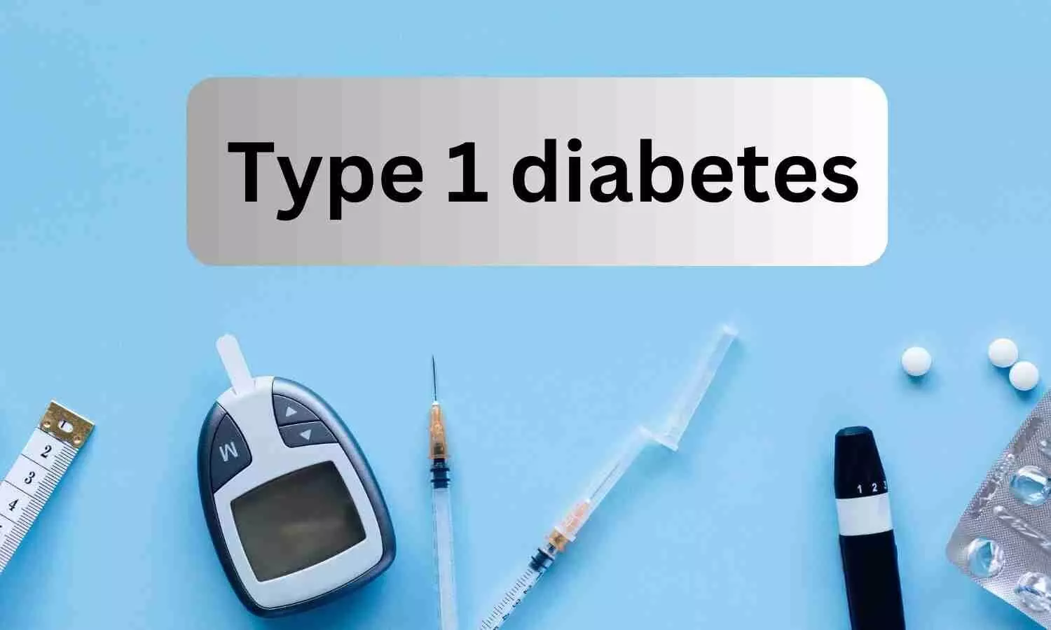
Lead author Catherine Gibbons, Associate Professor in the University of Leeds’ School of Psychology, said: “Reducing sugar consumption has become a key public health target in the fight to reduce the rising burden of obesity-related metabolic diseases such as type 2 diabetes.

“Simply restricting sugar from foods without substitution may negatively impact its taste or increase sweet cravings, resulting in difficulties sticking to a low-sugar diet. Replacing sugars with sweeteners and sweetness enhancers in food products is one of the most widely used dietary and food manufacturing strategies to reduce sugar intake and improve the nutritional profile of commercial foods and beverages.”

Principal investigator Graham Finlayson, Professor of Psychobiology in the University of Leeds’ School of Psychology, said: “The use of sweeteners and sweetness enhancers has received a lot of negative attention, including high profile publications linking their consumption with impaired glycaemic response, toxicological damage to DNA and increased risk of heart attack and stroke. These reports contribute to the current befuddlement concerning the safety of sweeteners and sweetness enhancers among the general public and especially people at risk of metabolic diseases.

“Our study provides crucial evidence supporting the day-to-day use of sweeteners and sweetness enhancers for body weight and blood sugar control.”

The study, which is the first of its kind, looked at the effects of consuming biscuits containing either sugar or two types of food sweetener: natural sugar substitute Stevia, or artificial sweetener Neotame on 53 adult men and women with overweight or obesity.

Until now, virtually all studies of the effects of sweeteners and sweetness enhancers on appetite and glycaemia have been conducted using beverages as the vehicle. Few studies include volunteers with overweight or obesity and few have included volunteers of both sexes.

Most studies have only compared a single sweetener, mostly aspartame, with a control, and very few studies have examined the effect of repeated daily intake of a known sweetener or sweetness enhancer in the normal diet.

The new trial took place at the University of Leeds and the Rhône-Alpes Research Center for Human Nutrition (CRNH-RA), France between 2021 and 2022. Participants were all aged 18 to 60, with overweight or obesity.

The trial consisted of three two-week consumption periods, where participants consumed biscuits with either fruit filling containing sugar; natural sugar substitute Stevia, or artificial sweetener Neotame, each separated by a break of 14-21 days. Day 1 and day 14 of the consumption periods took place in the lab.

Participants were instructed to arrive in the lab after an overnight fast, a blood sample was taken to establish baseline levels of glucose, insulin and appetite-related hormones. They were also asked to rate their appetite and food preferences.

After consuming the biscuits, they were asked to rate how full they felt over several hours. Glucose and insulin levels were measured, as were ghrelin, glucagon-like peptide 1 and pancreatic polypeptide – hormones associated with the consumption of food.

The results from the two sweetener types showed no differences in appetite or endocrine responses compared to sugar, but insulin levels measured over two hours after eating were reduced, as were blood sugar levels.

SWEET project joint co-ordinator Professor Anne Raben, from the University of Copenhagen, Denmark, said: “The findings show that sweeteners are a helpful tool to reduce intake of added sugar without leading to a compensatory increase in appetite or energy intake, thereby supporting the usefulness of sweeteners for appetite, energy and weight management.” 

Reference:

Catherine Gibbons, Kristine Beaulieu, Eva Almiron-Roig, Santiago Navas-Carretero, J. Alfredo Martínez l, Beverley O’Hara, Acute and two-week effects of neotame, stevia rebaudioside M and sucrose-sweetened biscuits on postprandial appetite and endocrine response in adults with overweight/obesity—a randomised crossover trial from the SWEET consortium, EBioMedicine, DOI:https://doi.org/10.1016/j.ebiom.2024.105005.

Powered by WPeMatico

Study reveals bidirectional link between premenstrual disorders and perinatal depression

A study from Karolinska Institutet published in the journal PLOS Medicine has revealed a bidirectional link between premenstrual disorders and perinatal depression.

Acccording to the  study,  Women with premenstrual syndrome (PMS) or premenstrual dysphoric disorder (PMDD) have a higher risk of perinatal depression. Conversely, women with perinatal depression have a higher risk of developing premenstrual disorders.

Premenstrual disorders like PMS or PMDD and perinatal depression are similar in the way that symptoms appear in connection with hormonal changes. This fact has given rise to the hypothesis that the disorders share both causes and risk factors. Now a study by researchers at Karolinska Institutet shows a bidirectional association between premenstrual disorders and perinatal depression.

“We can show that women with PMS or PMDD have a higher risk of developing perinatal depression and vice versa,” says Qian Yang, affiliated researcher at the Institute of Environmental Medicine, Karolinska Institutet and one of the main authors of the paper. “The results support the hypothesis that the diseases might have common causes.”

Between 2001 and 2018, approximately 1,800,000 pregnancies were registered in the Swedish Medical Birth Register. Among these, the researchers were able to identify nearly 85,000 women who suffered from perinatal depression. Additional national registers, such as the patient register and the drug register, were also used to identify women diagnosed with PMS or PMDD. These were then compared to a control group of nearly 850,000 birthing women who did not develop perinatal depression during the same period.

The results showed that women with premenstrual disorders were five times more likely to experience perinatal depression. Conversely, women who experienced perinatal depression were twice as likely to develop premenstrual disorders.

The bidirectional association was noted for both prenatal and postnatal depression, regardless of history of psychiatric disorders.

“It is important that healthcare professionals who meet with women during pregnancy are aware of the link between premenstrual disorders and perinatal depression in order to provide well-informed advice,” says Donghao Lu, Associate Professor at the Institute of Environmental Medicine, Karolinska Institutet and last author of the paper.

The authors emphasize that more research is needed to understand the biological link between premenstrual disorders and perinatal depression. In addition, more information is needed to clarify whether the association also applies to women with mild PMS or perinatal depression.

Reference:

Qian Yang ,Emma Bränn,Elizabeth R. Bertone- Johnson,Arvid Sjölander,Fang Fang,Anna Sara Oberg,Unnur A. Valdimarsdóttir,Donghao Lu, The bidirectional association between premenstrual disorders and perinatal depression: A nationwide register-based study from Sweden, PLoS Medicine, https://doi.org/10.1371/journal.pmed.1004363.

Powered by WPeMatico

Bone scintigraphy combined with integrated SPECT/CT advantageous in differentiating prosthetic infection from loosening: study

Prosthetic loosening and infection are still common complications after joint replacement. Over the past few years, single-photon emission computed tomography–computed tomography (SPECT/CT) was widely used and showed unique value based on the combination of anatomic and metabolic information of foci. However, its performance in differentiating between prosthetic loosening and periprosthetic infection after joint replacement is still the focus of clinicians and deserves further investigation.

Yaxin Tian et al conducted a retrospective study to determine whether bone scintigraphy combined with SPECT/CT still can differentiate prosthetic infection from loosening in patients after joint replacement. The differential efficacy in hip and knee prosthesis was also analyzed. Blood biomarkers for the diagnosis of periprosthetic infection were also evaluated.

The study was conducted at Ningxia Medical University, Yinchuan, China. It was published in “Indian Journal of Orthopaedics.”

Data sets of 74 prosthetic joints (including knees and hips), with suspected prosthetic loosening or infection were evaluated. Besides the results of nuclear imaging, X-ray images and serum biomarker were also recorded. Telephone follow-up and revision surgery after SPECT/CT were used as a gold standard. The sensitivity and accuracy of different imaging modalities were calculated by Chi-square test. The diagnostic efficacy of imaging methods and serum biomarkers were then analyzed by the area under curve (receiver operating characteristic curves, ROC) in SPSS 26.

The key findings of the study were:

• In all, 47 joints (14 knees and 33 hips) were confirmed as aseptic loosening, while 25 joints (18 knees and 7 hips) were confirmed as infection.

• The sensitivity and accuracy of SPECT combined with SPECT/CT imaging were the highest (92.86% and 87.84%, respectively).

• The differential efficacy of bone scintigraphy combined with SPECT/CT imaging was also better than any other single imaging modality.

• In the analysis of involved prosthesis, prosthetic loosening occurred more in hip prosthesis and knee prosthesis was easily infected (P<0.05).

• The sensitivity of ESR and CRP were 80% and 84%, respectively.

The authors concluded – “Bone scintigraphy with hybrid SPECT/CT remains encouraging in differentiating prosthetic infection from loosening after joint replacement. The diagnostic efficacy of differentiation in hip prosthesis was better than knee. Serum biomarkers cannot be used alone to differentiate prosthetic infection from loosening.”

Further reading:

Can99mTc MDP–SPECT/CT Differentiate Loosening and Infection After Hip and Knee Replacements?

Yaxin Tian, Yanghongyan Jiang et al

Indian Journal of Orthopaedics (2024) 58:316–322

https://doi.org/10.1007/s43465-024-01095-6

Powered by WPeMatico