No increased risk of stroke in older adults who received COVID-19 bivalent vaccine: JAMA

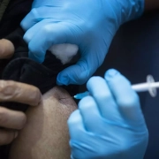
USA: In a self-controlled case series of 11,000 Medicare beneficiaries age 65 or older, the primary analysis showed no evidence of a significantly increased risk of stroke during the days immediately following the administration of either brand of the COVID-19 bivalent vaccine.

“In the Medicare beneficiaries who experienced stroke following the administration of either brand of the COVID-19 bivalent vaccine, the stroke risk was not significantly increased during the 1- to 21-day or 22- to 42-day risk window after vaccination compared with the 43- to 90-day control window,” the researchers reported. The findings were published online in the Journal of the American Medical Association (JAMA) on March 19, 2024.

In January 2023, the US Food and Drug Administration and the US Centers for Disease Control and Prevention raised a safety concern for ischemic stroke among adults aged 65 years or above who received the Pfizer-BioNTech BNT162b2; WT/OMI BA.4/BA.5 COVID-19 bivalent vaccine.

Against the above background, Yun Lu, Center for Biologics Evaluation and Research, US Food and Drug Administration, Silver Spring, Maryland, and colleagues aimed to evaluate stroke risk after administering(1) either brand of the COVID-19 bivalent vaccine, (2) either brand of the COVID-19 bivalent plus a high-dose or adjuvanted influenza vaccine on the same day (concomitant administration) and (3) a high-dose or adjuvanted influenza vaccine.

For this purpose, the researchers conducted a self-controlled series including 11 001 Medicare beneficiaries aged 65 years or older who experienced stroke after receiving either brand of the COVID-19 bivalent vaccine (among 5 397 278 vaccinated individuals). The study period was 2022 through 2023.

The main outcomes were stroke risk (transient ischemic attack, non-hemorrhagic stroke, combined outcome of transient ischemic attack or non-hemorrhagic stroke, or hemorrhagic stroke) during the 1- to 21-day or 22- to 42-day risk window following vaccination versus the 43- to 90-day control window.

The researchers reported the following findings:

  • 5 397 278 Medicare beneficiaries received either brand of the COVID-19 bivalent vaccine (median age, 74 years; 56% were women).
  • Among the 11 001 beneficiaries who experienced a stroke after receiving either brand of the COVID-19 bivalent vaccine, there were no statistically significant associations between either brand of the COVID-19 bivalent vaccine and the outcomes of transient ischemic attack, non-hemorrhagic stroke, non-hemorrhagic stroke or transient ischemic attack, or hemorrhagic stroke during the 1- to 21-day or 22- to 42-day risk window vs the 43- to 90-day control window.
  • Among the 4596 beneficiaries who experienced a stroke after concomitant administration of either brand of the COVID-19 bivalent vaccine plus a high-dose or adjuvanted influenza vaccine, there was a statistically significant association between vaccination and non-hemorrhagic stroke during the 22- to 42-day risk window for the fizer-BioNTech BNT162b2; WT/OMI BA.4/BA.5 COVID-19 bivalent vaccine and a statistically significant association between vaccination and transient ischemic attack during the 1- to 21-day risk window for the Moderna mRNA-1273.222 COVID-19 bivalent vaccine.
  • Among the 21 345 beneficiaries who experienced stroke after administration of a high-dose or adjuvanted influenza vaccine, there was a statistically significant association between vaccination and non-hemorrhagic stroke during the 22- to 42-day risk window.

In conclusion, there was no evidence of a significantly increased risk for stroke during the days immediately after vaccination among Medicare beneficiaries aged 65 years or older who experienced stroke after receiving either brand of the COVID-19 bivalent vaccine.

Reference:

Lu Y, Matuska K, Nadimpalli G, et al. Stroke Risk After COVID-19 Bivalent Vaccination Among US Older Adults. JAMA. 2024;331(11):938–950. doi:10.1001/jama.2024.1059

Powered by WPeMatico

Obesity in childhood may double risk of developing multiple sclerosis in early adulthood, shows study

New research to be presented at this year’s European Congress on Obesity in Venice, Italy (12-15 May) shows that having obesity in childhood is associated with a more than doubling of the risk of later developing multiple sclerosis. The study is by Professor Claude Marcus and Associate Professor Emilia Hagman, Karolinska Institutet, Stockholm, Sweden, and colleagues.

Emerging evidence implies a link between high BMI in adolescence and an increased risk of Multiple Sclerosis (MS). Yet, most studies evaluating this association are cross-sectional, have retrospective design with self-reported data, have used solely genetic correlations, or use paediatric weight data before the obesity epidemic. Therefore, to authors aimed to prospectively evaluate the risk of developing MS in a large cohort of patients with paediatric obesity compared with the general population.

They included patients aged 2 – 19 years with obesity enrolled in the Swedish Childhood Obesity Treatment Register (BORIS) between 1995 – 2020, and a matched comparison group from the general population. Matching criteria included sex, year of birth, and residential area. Exclusion criteria were secondary obesity (eg brain tumours such as craniopharyngioma), and genetic syndromes (eg Prader Willi, morbus Down), and MS diagnosis before 15 years of age (that is, already developing in childhood). MS was identified through Sweden’s National Patient Register. Individuals were followed from obesity treatment initiation, or from 15 years of age if treatment was initiated earlier, until MS diagnosis, death, emigration, or August 2023, whichever came first. The authors computer and statistical modelling to calculate any potential association. Due to previous reported genetic associations of MS, the authors also assessed levels of parental MS which was present in 0.99% in the obesity cohort and 0.68% in the general population comparators.

The data included 21 661 patients (54% boys) from the paediatric obesity cohort with a median age of obesity treatment initiation (behaviour and lifestyle modification) of 11.4 years (years and 102 230 general population comparators. The median follow-up time was 5.6 years, corresponding to median age of 20.8 years in the follow-up population (and 50% of the population were aged between 18 and 25 at the point analysis, with the highest age in the cohort 45 years).

During follow-up, 0.13% [n=28, 18 (64%) female, 10 (36%) male] developed MS in the obesity cohort, whereas the corresponding number in the general population was 0.06% [n=58, 38 (66%) female, 20 (34%) male]. The mean (SD) age of MS diagnosis was comparable between the groups; 23.4 years in the obesity cohort versus 22.8 years in the general population comparators. (see graph in full abstract). The small numbers who developed MS so far means that the study was not sufficiently statistically powered to state the increased risk to females developing MS – however the results follow the general increased risk to females (the estimate ratio of female: male affected by MS in the general population is 4:1).

The crude incidence rate of MS per 100 000 person years was 19.3 in the obesity cohort and 8.3 in the general population cohort. Analyses adjusted for presence of parental MS (heredity) (which was more prevalent in the obesity cohort, as above) revealed that the risk of developing MS was more 2.3 times higher than in the paediatric obesity cohort, with both these findings statistically significant.

The authors say: “Despite the limited follow-up time, our findings highlight that obesity in childhood is associated with an increased susceptibility of early-onset MS more than two-fold. Given that paediatric obesity is prevalent, it is likely to serve as a critical etiological contributor to the escalating prevalence of MS. Paediatric obesity is associated with several autoimmune diseases and the leading hypothesis is that the persistent low-grade inflammatory state, typically observed in obesity, is mediating the association. Understanding these pathways is crucial for developing targeted prevention and intervention strategies to normalise the risk for MS in children and adolescents with obesity.”

They add: “There are several studies showing that MS has increased over several decades and obesity is believed to be one major driver for this increase. Thanks to our prospective study design, we can confirm this theory.”*

“Even though the risk for MS is more than double among children and adolescence with obesity, the absolute risk for MS remains lower than for many other comorbidities associated with obesity. Nevertheless, our study adds to the evidence that obesity in early life increases the risk for a plethora of diseases including MS, and not only the well-known cardiometabolic conditions such as heart disease and diabetes.”

Reference:

Study shows obesity in childhood associated with a more than doubling of risk of developing multiple sclerosis in early adulthood, European Association for the Study of Obesity, Meeting: European Congress on Obesity (ECO2024).

Powered by WPeMatico

Passive smoking during pregnancy tied to increased risk of atopic dermatitis in offspring: Study

China: A recent meta-analysis published in the Journal of the European Academy of Dermatology and Venereology has shown an association between passive smoking during pregnancy and an increased risk of eczema development in offspring.

Atopic dermatitis (eczema), a condition that causes dry, itchy, and inflamed skin, is common worldwide. Several studies have explored the modifiable factors of atopic dermatitis (AD), including smoking. The adverse effects of smoking on asthma have been well documented in adolescents, however, the implications of smoking on atopic dermatitis are controversial.

The findings are contradictory and could be due to disparities in study design and covariables adjusted for in the analysis. For instance, the results of a few prospective cohort studies, which reported non-significant or negative correlations between atopic dermatitis and passive smoking, may have exhibited selection bias due to the inclusion of restricted residential regions and a lack of compliance with the survey or involving a small study population. Additionally, because AD generally develops in young individuals and it is rare for children to smoke, the focus of the majority of studies has been the effects of passive smoking on atopic dermatitis in children

Against the above background, Wenjie Ren, School of Public Health, Xinxiang Medical University, Xinxiang, China, and colleagues examine the evidence of the relationship between active smoking or passive smoking during pregnancy and atopic dermatitis (AD) in offspring.

For this purpose, the protocol was written following the PRISMA Checklist and was registered in the PROSPERO database. The researchers searched online databases to identify all potentially related articles from inception through 1 December 2022. Cohort and case–control studies were assessed using the Newcastle–Ottawa Scale (NOS), and the Joanna Briggs Institute (JBI) critical appraisal tool was used to assess the quality of cross-sectional studies.

Heterogeneity was determined using Cochrane Q tests and I2 statistics. Additionally, the reasons for the heterogeneity were analyzed according to the population source, research design, and population size. Fifteen observational studies were included in the analysis.

“Our meta-analysis suggests that atopic dermatitis in offspring is not associated with active smoking during pregnancy (pooled OR, 0.96), however, it is related to passive smoking (OR, 1.52),” the researchers reported.

“Passive smoking during pregnancy is associated with an increased risk of eczema development in offspring.”

“More research is required to explore the risk of active smoking and atopic dermatitis development in offspring, particularly the association between measurements of pregnancy cotinine levels in maternal body fluids and AD in offspring,” they concluded.

Reference:

Chao, L., Liang, W., Zhao, X., Liang, Z., Wu, W., Song, J., & Ren, W. Maternal tobacco exposure during pregnancy and atopic dermatitis in offspring: A systematic review and meta-analysis. Journal of the European Academy of Dermatology and Venereology. https://doi.org/10.1111/jdv.19958

Powered by WPeMatico

Surgical nerve decompression tied to reduced pain in patients with diabetic neuropathy: Study

USA: A recent five-year study published in the Annals of Surgery has shed light on the effect of lower extremity nerve decompression in patients with painful diabetic peripheral neuropathy (DPN). 

“Although nerve decompression was tied to reduced pain, the benefit of surgical decompression requires further investigation since a placebo effect may be responsible for part or all of these effects,” the researchers wrote. 

Surgical nerve decompression, used to treat conditions such as carpal tunnel syndrome and sciatica, could play a role in relieving the pain of diabetic neuropathy patients, researchers at UT Southwestern Medical Center found.

The five-year study, published in the Annals of Surgery, was the first randomized control trial to assess the effectiveness of lower extremity nerve decompression surgery on patients with diabetic peripheral neuropathy.

Approximately 20 million Americans suffer from diabetic neuropathy, a progressive condition that damages the nerves, primarily in the legs and feet. The primary treatment today is the use of temporary pain-relieving medications, but many patients find them ineffective after prolonged use.

“Diabetic neuropathy can be debilitating, leading to a lack of mobility and a severe reduction in quality of life,” said study leader Shai Rozen, M.D., Professor and Vice Chair of Plastic Surgery. “It’s believed that roughly one-third of those with neuropathy pain have nerve compression – where there is direct and chronic pressure on a peripheral nerve – due to physiological changes brought on by diabetes. Our research suggests that nerve decompression surgery to release compressed nerves from surrounding tissue could offer lasting relief for those individuals.”

In diabetic neuropathy, nerves can swell and eventually be compressed by surrounding semirigid tissue, causing pain, muscle weakness, or both. In the surgery, the semirigid tissue is removed from the swollen nerve, allowing for improved blood flow to relieve symptoms.

The study followed 78 patients at UT Southwestern and Parkland Health who were randomly placed into two groups-one receiving surgery and one serving as an observation group who remained on medication only. Those selected for surgery also had one leg randomly chosen for “sham surgery”-when the surgeon makes incisions to mimic the procedure but without principal therapeutic actions (i.e., nerve release)-while the other leg underwent nerve decompression.

Patients agreed to being blinded about which leg underwent sham surgery and which had decompression surgery. Additionally, the evaluators were blinded regarding which group the patients were in. “This masking of patients and evaluators further increases the reliability of the results,” Dr. Rozen said.

During follow-up visits, patients completed standard pain and lifestyle questionnaires. At the 12-month visit, the patients who underwent surgical intervention reported significantly less pain in both legs, while the observation group’s pain rankings were unchanged.

At 56 months, under the same patient- and evaluator-blinded conditions, the surgical group reported even greater pain reduction, while the observation group had worse pain. Unlike the 12-month follow-up, however, surgical patients reported far more improvement in their decompressed legs than in their sham surgery legs.

“The one-year reports of pain improvement in both legs could mean that there is a placebo effect taking place, but the five-year results suggest that the procedure actually does have a positive long-term impact on pain,” Dr. Rozen said. “There is still much debate in the medical community about the value of decompression surgery in treating diabetic neuropathy, and while this study doesn’t settle the issue, it should help expand the discourse among stakeholders and hopefully lead to even more research. The goal is to better understand the efficacy of nerve decompression surgery on diabetic neuropathy and improve our ability to identify patients who are likely to respond to surgical intervention.” 

Reference:

Rozen, Shai M. MD*; Wolfe, Gil I. MD†; Vernino, Steven MD, PhD‡; Raskin, Philip MD§; Hynan, Linda S. PhD∥,¶; Wyne, Kathleen MD, PhD#; Fulmer, Rita FNP-BC**; Pandian, Geetha MD**; Sharma, Shiv K. MD††; Mohanty, Ahneesh J. MD‡‡; Sanchez, Cristina V. BS*; Hembd, Austin MD*; Gorman, April PhD∥. Effect of Lower Extremity Nerve Decompression in Patients with Painful Diabetic Peripheral Neuropathy: The DNND Randomized, Observation Group- and Placebo Surgery-controlled Clinical Trial. Annals of Surgery ():10.1097/SLA.0000000000006228, February 8, 2024. | DOI: 10.1097/SLA.0000000000006228

Powered by WPeMatico

Study identifies high BP and triglycerides as risk factors for IgA nephropathy

China: A recent Mendelian randomization (MR) study published in Frontiers in Medicine has shed light on the causal effect of high blood pressure (BP) and triglyceride (TG) on IgA nephropathy.

The study revealed high BP and triglyceride as risk factors causally connected with immunoglobulin A nephropathy (IgAN), of which high BP was strongly related to IgAN onset.

Results from previous research indicate that both TG and high BP have strong correlations with IgAN. However, there is no clarity if there is a direct causal effect of triglycerides and high blood pressure on IgAN.

High blood pressure and hyperlipidemia are common complications of renal dysfunction in IgAN. Although its exact cause is unknown, recent research indicates that high BP and triglycerides may play a causal role in IgAN.

Against the above background, Yijun Yan, First Affiliated Hospital of Xi’an Jiaotong University, Xi’an, China, and colleagues aimed to explore the causalities between HBP and TG, and IgAN based on MR analysis.

For this purpose, the researchers sourced from genome-wide association study (GWAS) summary data of IgAN (GCST90018866) and two exposure factors, TG (ukb-d-30870_raw) and HBP (ukb-a-437), from the GWAS Catalog and Integrative Epidemiology Unit (IEU) OpenGWAS databases, respectively.

Five methods were utilized to perform MR analysis after taking out single nucleotide polymorphisms (SNPs) as instrumental variables, including MR-Egger, weighted mode, weighted median, simple mode, and inverse variance weighted (IVW), followed by the sensitivity analysis containing the heterogeneity, horizontal pleiotropy test, and leave-one-out (LOO) analysis. Finally, the researchers performed the enrichment analysis and interaction network construction of genes corresponding to SNPs of high BP and TG.

The study led to the following findings:

  • The univariate MR results revealed that high BP and TG regarded as risk factors were causally related to IgAN based on random-effect IVM method, of which TG had a weaker impact.
  • The reliability of these univariate MR results was certified by the sensitivity analysis, in which there was no horizontal pleiotropy and exaggerated influence of each SNP.
  • High BP was markedly causally related to IgAN with the help of multivariate MR analysis, rather than TG. Therefore, when HBP and TG occur simultaneously, HBP is a direct influencing factor on IgAN.
  • A total of 208 and 153 genes separately corresponding to SNPs of TG and HBP were included in enrichment analysis, and thereinto, genes relevant to TG were mainly enriched in lipid homeostasis and cholesterol metabolism, while genes concerned with HBP played their roles in the regulation of cell growth, aldosterone synthesis and secretion and so forth.

In conclusion, the results showed a causal effect of triglyceride and high BP on IgAN, both of them are risk factors, and HBP is the direct influencing factor of IgAN.

“Since the sample data used in this study originated in East Asia and Europe, the causality currently applies only to these regions. Enrichment analysis of genes corresponding to SNPs of TG and high BP might spark interest in further molecular biological mechanism investigation in IgAN,” the researchers wrote.

Reference:

Yang, Y., Li, Y., Feng, X., Ding, C., Zhang, J., & Liu, Z. (2024). The causal effect of triglyceride and high blood pressure on IgA nephropathy: A Mendelian randomization study. Frontiers in Medicine, 11, 1338462. https://doi.org/10.3389/fmed.2024.1338462

Powered by WPeMatico

Early intervention after first seizure prevents long-term epilepsy and associated cognitive deficits: Study

Only a very small percentage of neurons show changes after an epileptic seizure in mice, but these alterations can be permanent and trigger future seizures that can affect the whole brain and lead to impaired cognition, like memory and learning, according to new research from the Perelman School of Medicine at the University of Pennsylvania. The researchers identified an experimental treatment that, if provided within the first 48 hours after the first seizure, can prevent these long-term changes. The findings, which were published recently in The Journal of Clinical Investigation, suggest a promising target for developing treatments for epilepsy and preventing downstream effects of seizures.

Epilepsy is characterized by excessive activity of brain cells-neurons-which generate seizures. Research is increasingly showing that the development of epilepsy involves changes of synapses, which are structures that connect one neuron to another. While an estimated 3.4 million people in the United States live with some form of epilepsy, it is still unknown what causes it, and there is no cure. Further, half of individuals with epilepsy experience cognitive impairment, such as problems with memory, or with emotional regulation, but it remains unclear why or how epilepsy changes brain cells to cause this. What’s more, epilepsy is common in children with autism and individuals with dementia.

“It is clear that there is some connection between an epileptic brain, impaired memory and trouble controlling emotions and how we act on those feelings, but we don’t understand the underlying mechanisms,” said Frances E. Jensen, MD, chair of the Department of Neurology, and senior author of the study. “Existing treatments for epilepsy only help manage seizures. This research gives us a promising starting point for developing therapies that prevent them from happening.”

In this study, the researchers used a method that “tagged” neurons in the hippocampus-an area commonly affected by epilepsy, and critical for memory-of mice that were activated by epileptic activity. The researchers were able to monitor those activated neurons over time and observe how they responded to subsequent seizures. They found that only about twenty percent of neurons in the hippocampus were activated by seizures. Over time, the overactivity of these neurons diminished their ability to make connections with other neurons, called synapses, which is necessary for learning.

“The overactive neurons lose their ability to build the strong synapses necessary for learning, which may explain why some people with epilepsy have trouble with learning and with memory,” said Jensen. “If we can stop these neurons from undergoing changes after being activated by seizures, our hope is that we can also prevent not only the progression of epilepsy, but also avoid these cognitive deficits individuals experience long-term.”

To see if they could prevent neurons from becoming permanently epileptic, the researchers used an experimental glutamate receptor-blocker, called IEM-1460, which has been shown to reduce neuron hyperexcitability in models of mice with epilepsy. They found when they treated mice with this blocker in the first 48 hours after their very first seizure the neurons did not become permanently activated, and the subjects did not experience future seizures or the associated effects, like impaired cognition and trouble learning.

“Now that we have identified the subgroup of neurons that are impacted by epilepsy, we can investigate what makes these cells vulnerable to becoming epileptic, and whether that is something we can develop a therapy to stop,” said Jensen. “We are also eager to determine whether there is a glutamate receptor-blocker that works similarly to IEM-1460 in humans, which could be given to people after their first seizure, and prevent the lifelong struggles associated with epilepsy.”

Reference:

Bo Xing, Aaron J. Barbour, Joseph Vithayathil, Xiaofan Li, Sierra Dutko, Jessica Fawcett-Patel, Eunjoo Lancaster, Delia M. Talos, and Frances E. Jensen, Reversible synaptic adaptations in a subpopulation of murine hippocampal neurons following early-life seizures, Journal of Clinical Investigation, DOI:10.1172/JCI175167.

Powered by WPeMatico

SGLT2 inhibitors clinically beneficial in patients with gout, suggests review

USA: Sodium-glucose cotransporter type 2 (SGLT2) inhibitors may provide improved outcomes in treating gout and its common comorbidities, a recent review has shown. SGLT2 inhibitors are a popular drug class in the management of cardiometabolic and diabetic conditions.

The review, published in Nature Reviews Rheumatology, suggested that the mechanism of SGLT2 inhibition in patients with arthritic disease could provide anti-inflammatory benefits while reducing the risk of renal and cardiovascular outcomes in such patients—as the drug class is already indicated to treat.

Chio Yokose, Massachusetts General Hospital, Boston, MA, USA, and colleagues described the characteristics of gout and its most common comorbidities.

They noted that gout is the most common form of inflammatory arthritis worldwide, which is characterized by painful recurrent flares of inflammatory arthritis and is associated with a transiently increased risk of adverse cardiovascular events. Furthermore, gout is associated with various cardiometabolic–renal comorbidities such as chronic kidney disease (CKD), type 2 diabetes, and cardiovascular disease (CVD). These comorbidities, potentially combined with gout flare-related inflammation, contribute to persistent premature mortality in gout, independently of traditional cardiovascular risk factors and serum urate concentrations.

The researchers add, “Although better implementation of standard goud care could improve gout outcomes, there is a need for deliberate efforts to address the CV risk in gout patients to reduce mortality.”

SGLT2 inhibitors are approved for multiple indications owing to their ability to lower the risk of hospitalizations for heart failure and CKD, all-cause, and cardiovascular death, making them an attractive treatment option for gout. These medications have been also shown to reduce serum urate concentrations, the causal culprit in gout risk, and are linked with a reduced risk of incident and recurrent gout, potentially due to their anti-inflammatory effects. Thus, SGLT2 inhibition could simultaneously address both gout symptoms and its comorbidities.

According to the authors, this is not the first analysis to consider a potential role in gout management for the SGLT2 inhibitor class.

A study presented during the European Congress on Rheumatology (EULAR) 2023 meeting in May 2023 showed initiating an SGLT2 inhibitor significantly lowered the risk of incident gout among at-risk patients with type 2 diabetes, compared to other drug classes, including GLP-1 receptor agonists (HR, 0.39), DPP-4 inhibitors (hazard ratio [HR], 0.54), and sulfonylurea (HR, 0.61).

These promising clinical data come with the disclosure that researchers are still seeking to understand the anti-gout and urate-lowering properties of SGLT2 inhibitors, Yokose, and colleagues noted. They suggested such mechanisms may be due to enhanced uricosuria and anti-inflammatory pathways in SGLT2 inhibition.

“Although additional research is needed to determine SGLT2 inhibitors’ role in gout management, available evidence indicates that these drugs have the potential to improve outcomes among gout patients,” the researchers concluded.

Reference:

Yokose, C., McCormick, N., Abhishek, A., Dalbeth, N., Pascart, T., Lioté, F., Gaffo, A., FitzGerald, J., Terkeltaub, R., Sise, M. E., Januzzi, J. L., Wexler, D. J., & Choi, H. K. (2024). The clinical benefits of sodium–glucose cotransporter type 2 inhibitors in people with gout. Nature Reviews Rheumatology, 20(4), 216-231. https://doi.org/10.1038/s41584-024-01092-x

Powered by WPeMatico

FDA clears rapid PCR test for sore throat and respiratory infections

The US Food and Drug Administration has granted 510(k) clearance to bioMerieux for a rapid respiratory/sore throat panel. The test will help clinicians make more informed decisions about antibiotic prescribing.

The new PCR test is capable of detecting upto 15 most common bacteria, viruses, and viral subtypes that cause respiratory or sore throat infections in about 15 minutes from samples taken from nasopharyngeal and throat swabs.

The COVID-19 pandemic has demonstrated the need for healthcare professionals to have diagnostic tests available as close as possible to the patient, providing actionable results quickly. A fast and innovative syndromic testing range, BIOFIRE  SPOTFIRE  perfectly matches these new medical needs worldwide.

The BIOFIRE  SPOTFIRE  R/ST Panel is a unique multiplex PCR1 test capable of detecting and identifying nucleic acids from up to 15 of the most common bacteria, viruses, and viral subtypes responsible for respiratory or sore throat infections in about 15 minutes. Samples can be taken from a nasopharyngeal swab when a respiratory tract infection is suspected, or from a throat swab in case of a pharyngitis syndrome.

“To prescribe or not to prescribe antimicrobials is the age-old question for outpatient upper respiratory infections. The flexibility of this syndromic panel allows healthcare professionals to test for multiple pathogens with overlapping signs and symptoms, ultimately allowing the diagnostic to drive informed decision-making during the outpatient visit. These results further empower the advancement of antimicrobial stewardship and modernize patient care.” declared Dr Charles K. Cooper, Executive Vice-President, Chief Medical Officer, bioMérieux

The BIOFIRE SPOTFIRE R/ST Panel is the third panel to receive FDA clearance for use on the BIOFIRE® SPOTFIRE® System. The two other panels available for use on this system are the BIOFIRE  SPOTFIRE  Respiratory (R) Panel and BIOFIRE® SPOTFIRE® Respiratory (R) Panel Mini, detecting 15 and 5 of the most common respiratory pathogens respectively.

A CLIA-waiver allows the BIOFIRE  SPOTFIRE  System and its so-authorized panels to be used by non-lab professionals and in any clinical setting where patients seek care including an urgent care, physician office, local pharmacy, student health clinic, or an emergency department.

“Receiving FDA clearance for the BIOFIRE  SPOTFIRE  R/ST Panel, just one year after the successful launch of the BIOFIRE  SPOTFIRE  solution, marks another milestone in our mission to move testing closer to the patient,” said Jennifer Zinn, Executive Vice President, Clinical Operations, bioMérieux. “With our innovative approach, we are committed to enhancing patient care by providing healthcare professionals with the tools they need to deliver expedited and effective diagnoses. Together, we are revolutionizing the landscape of healthcare, one diagnosis at a time.”

The BIOFIRE  SPOTFIRE  R/ST Panel is currently CE-marked under IVDD (In Vitro Diagnostic Directive) and has been submitted for CE-marking under IVDR (In Vitro Diagnostic Regulation).

bioMérieux will also submit the BIOFIRE  SPOTFIRE  Respiratory / Sore Throat (R/ST) Panel Mini to the FDA for review for a 510(k) clearance. This panel is intended to detect and identify nucleic acids from 5 of the most common viral and bacterial causes of respiratory tract infections from either a nasopharyngeal or throat swab respectively. The BIOFIRE  SPOTFIRE  R/ST Panel Mini is not yet available for sale.

Powered by WPeMatico

Prices Of Essential Medicines To Rise Slightly From April 1

New Delhi: A slight hike in the price of essential medicines, such as painkillers, antibiotics, and anti-infectives, is set to take place on April 1st, in line with the change observed in the Wholesale Price Index (WPI) between 2022 and 2023.

Through a recent notification, the National Pharmaceutical Pricing Authority (NPPA) has declared to permit the pharmaceutical sector to increase the Maximum Retail Price (MRP) of scheduled formulations by approximately 0.00551% at the start of the fiscal year 2024-25, considering the change observed in the Wholesale Price Index (WPI) between 2022 and 2023.

As a consequence, beginning on April 1, there may be a slight spike in the cost of essential medicines such as painkillers, antibiotics, and others.

The notice stated,

‘Based on the Wholesale Price Index (WPI) data provided by the office of the Economic Advisor, Department of Industry and Internal Trade Ministry of Commerce and Industry, the annual change in WPI works out as (+) 0.00551% during the calendar year 2023 over the corresponding period in 2022.”

In continuation, it is worth notifying that in accordance with Paragraph 16(2) of the Drug Price Control Order (DPCO) 2013, the manufacturers may increase the maximum retail price (MRP) of scheduled formulations once a year, in the month of April, on the basis of the wholesale price index with respect to previous calendar year and no prior approval of the Government in this regard shall be required.

Therefore in light of the above, the notice further added,

“As per provisions of Paragraph 16(2) of Drug Price Control order (DPCO) 2013, the manufacturers may increase the Maximum Retail Price (MRP) of scheduled formulations on the basis of this WPI and no prior approval of the government in this regard shall be required.”

The previous year, the Medical Dialogues Team reported that based on the Wholesale Price Index (WPI) data provided by the Office of the Economic Advisor, Department of Industry and Internal Trade, Ministry of Commerce and Industry, the annual change in the WPI worked out as 12.1218% during the calendar year 2022 over the corresponding period in 2021.

To view the official notice, click the link below:

Powered by WPeMatico

Hair-straightening products linked to acute kidney failure risk: Research

In the pursuit of beauty, there can be unexpected risks. Recent research has uncovered a concerning connection between hair-straightening products and kidney health. French researchers have unveiled a disturbing link between these popular cosmetic items and acute kidney failure, shedding light on a potentially overlooked health hazard. Their findings, published in The New England Journal of Medicine, reveal a troubling association between glyoxylic acid—a common ingredient in these products—and the formation of calcium oxalate crystals within the kidneys.

Lead researcher Dr. Emmanuel Letavernier of Tenon Hospital in Paris, France, expressed alarm at the rapid onset of severe kidney failure observed in animal models within a mere 24 hours of exposure to the suspect straightening cream. These findings have prompted urgent calls to action, with researchers urging the removal of products containing glyoxylic acid from the market and notifying relevant regulatory agencies.

Reference: Kidney Injury and Hair-Straightening Products Containing Glyoxylic Acid,The New England Journal of Medicine, DOI: 10.1056/NEJMc2400528

Powered by WPeMatico