Successful Breastfeeding Initiation Linked to Lower Risk of Postpartum Depression, Study Finds

Canada: A recent study published in the Journal of Obstetrics and Gynaecology Canada has found that successfully initiating breastfeeding is strongly linked to a reduced risk of postpartum depression (PPD).

Postpartum depression is a mood disorder that affects approximately 10-20% of new mothers, manifesting within the first year after childbirth. Symptoms include persistent sadness, fatigue, irritability, and difficulty bonding with the baby. The causes of PPD are multifaceted, involving hormonal, psychological, and environmental factors. However, recent findings have pointed to breastfeeding initiation as a potential protective factor against the development of PPD.

Against the above background, Anne-Sophie Roy and Nils Chaillet from Obstetrics and Gynecology, Faculty of Medicine, Laval University, QC, Canada, and colleagues aimed to assess the impact of successful breastfeeding initiation on postpartum depression among women who gave birth in Quebec.

For this purpose, the researchers conducted a secondary analysis of the “Quality of Care, Obstetrics Risk Management, and Mode of Delivery” (QUARISMA) trial, which took place in Quebec from April 1, 2008, to October 31, 2011. The trial aimed to reduce cesarean delivery rates in the region. The study included all women aged 18 and older who gave birth to a single baby at 37 weeks or later. To assess the effect of successful breastfeeding initiation on PPD rates, logistic regression was used. The results were reported using adjusted odds ratios (ORs).

The study led to the following findings:

  • The study included 151,708 women, of whom 21,525 had unsuccessful breastfeeding initiation and 130,183 had successful breastfeeding initiation.
  • The analysis revealed a significant link between successful breastfeeding initiation and a lower rate of postpartum depression, with 0.16% of women who successfully initiated breastfeeding experiencing PPD, compared to 0.29% in those who did not.
  • The odds of developing PPD were 43% lower for women with successful breastfeeding initiation (OR 0.57), and this association was statistically significant.

The findings revealed that successful initiation of breastfeeding is strongly linked to a reduced risk of postpartum depression. The researchers suggest that, due to this association, breastfeeding initiation could serve as a potential preventive strategy for postpartum depression. As a result, health professionals are encouraged to incorporate discussions about breastfeeding initiation into their counseling with new mothers, emphasizing its potential benefits for maternal mental health.

“The success of breastfeeding initiation appears to offer more than just physical benefits for infants—it may also serve as an important factor in supporting maternal mental health. As research continues, healthcare providers must offer guidance and encouragement to mothers in the early postpartum period, ensuring that they have the resources and emotional support necessary to navigate the challenges of both breastfeeding and mental health,” the researchers concluded.

Reference:

Roy, A., & Chaillet, N. (2024). Relation Between Initiation of Breastfeeding Success and Postpartum Depression. Journal of Obstetrics and Gynaecology Canada, 46(11), 102666. https://doi.org/10.1016/j.jogc.2024.102666

Powered by WPeMatico

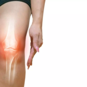
Blood Flow Restriction in Knee OA Exercise Yields Superior Pain Relief and Strength Gains: Study

Researchers have discovered that exercise programs combined with blood flow restriction (BFR) provide better outcomes for patients who are suffering from knee osteoarthritis (KOA). The authors of a recent study at Ghent University Hospital, found that BFR-added exercise resulted in a larger short-term reduction of pain and increase in strength. This study was conducted by Jacobs E. and colleagues and published in the Annals of the Rheumatic Diseases.

Knee osteoarthritis is one of the most significant causes of disability globally. Exercise is very commonly prescribed, but typically, standard programs alone will only result in limited relief. This study aimed to assess whether BFR could complement exercise and enhance its effects on reducing KOA symptoms, muscle strength, and overall functionality.

The randomized controlled trial known as the Vascular Occlusion for optimizing Functional Improvement in patients with Knee Osteoarthritis, or VO-FIKOA, included 120 KOA patients. The randomization was made into a standard exercise program versus BFR-enhanced programs and conducted over 24 supervised sessions over 12 weeks. Outcomes were measured at baseline, 6 weeks, 12 weeks, and after 3 months post-intervention.

Main outcome measurements included the Knee Injury and Osteoarthritis Outcome Score pain subscale; secondary outcome measures were also assessed using quadriceps strength, the Pain Catastrophizing Scale, as well as functional performance-based measurements. Intention-to-treat principles will be followed for analysis.

  • Pain subscale of KOOS significantly improved in the BFR group with an effect size of 0.58 at 12 weeks (p=0.0009).

  • These improvements remained statistically significant at the 3-month follow-up with slightly decreased effect size of 0.55 (p=0.0008).

  • The BFR group significantly increased the strength of quadriceps with an ES of 0.81 compared to the control group, considerably improved (p<0.0001).

  • Reducing strain in knee joints is an essential step towards alleviating symptoms of KOA.

  • Functional performance, as measured by KOOS functional tests, was improved significantly with the BFR group across the 12-week intervention period and retained its advantages at the 3-month post-intervention time.

  • The improvement in quality of life was observed in the BFR group; hence, exercise with BFR can significantly impact daily life and overall well being.

The results of this study demonstrate that exercise plus BFR leads to larger improvements in pain, strength, and functional measures as compared to exercise alone. It will allow patients to increase their strength and diminish pain with less joint stress. BFR-supported programs may encourage a more active lifestyle, thereby improving muscle function and quality of life even after the end of the intervention.

Reference:

Jacobs, E., Stroobant, L., Victor, J., Elewaut, D., Tampere, T., Wallaert, S., Witvrouw, E., Schuermans, J., & Wezenbeek, E. (2024). Vascular occlusion for optimising the functional improvement in patients with knee osteoarthritis: a randomised controlled trial. Annals of the Rheumatic Diseases, ard-2024-226579. https://doi.org/10.1136/ard-2024-226579

Powered by WPeMatico
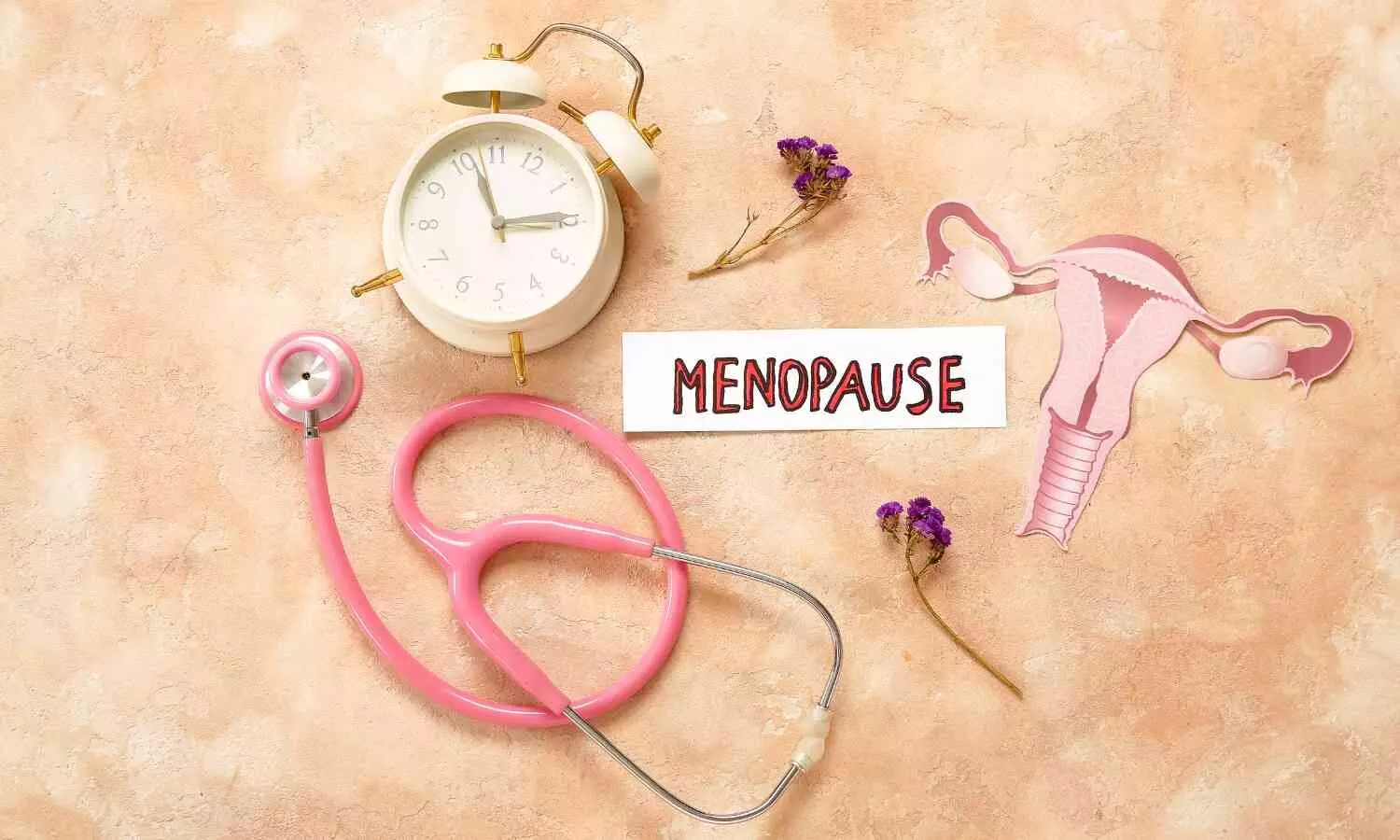
Women entering menopause later in life at higher risk of developing asthma: Study

Many studies suggest that an earlier age at menopause is more detrimental to a woman’s health, leading to an increased risk for adverse health conditions such as heart disease, diabetes, osteoporosis, and depression, among others. However, a new study is linking a later age at natural menopause with a greater risk for asthma. Results of the study are published online today in Menopause, the journal of The Menopause Society.

Asthma is a common, chronic disease affecting more than 300 million people worldwide. The prevalence of asthma has been increasing over recent years, creating a substantial economic impact because it is one of the highest diseases for healthcare use. Adult-onset asthma is typically more severe and more difficult to treat than childhood asthma.

Multiple studies have suggested a possible link between asthma and sex hormones. Most notable is the fact that adult-onset asthma is more common in women than men. In childhood, asthma is more prevalent in boys. After puberty, however, asthma occurs more often in girls. Women also tend to have more severe asthma and are less likely to have remission of the disease.

Some studies have found a peak incidence of asthma at around age 40 years, which is commonly the age of the menopause transition, whereas other studies found a peak at the average age at menopause, which is 51 years. Both natural estrogen and synthetic estrogen, such as used in hormone therapy, offer similar risk profiles. Women using hormone therapy were shown to have a 63% increased risk of asthma, whereas women who stopped hormone therapy were two times more likely to quit asthma treatment. Higher body mass index also is shown to be a risk factor for women, but not men, because fat produces estrogen.

Unfortunately, research on the association between menopause and asthma incidence is limited and has yielded conflicting results. That is why this newest study, based on 10 years of follow-up data from more than 14,000 postmenopausal women, was designed to investigate the association between the age at natural menopause and incidence of asthma in nonsmoking postmenopausal women. The study researchers found that women with early menopause (which occurs between 40 and 44 years of age) are at a reduced risk of asthma, which led them to suggest a role of estrogen with asthma risk.

Study results are published in the article “The association between age at natural menopause and risk of asthma among postmenopausal women from the Canadian Longitudinal Study on Aging.”

“This study highlights sex-based differences in asthma, with women at a greater risk for asthma than men in adulthood. It also showed that women with later onset of menopause are at greater risk than those with early onset of menopause. Clinicians should be aware of this link and should monitor women with later age at natural menopause for asthma symptoms,” says Dr. Stephanie Faubion, medical director for The Menopause Society.

Reference:

Kesibi, Durmalouk MA; Rotondi, Michael PhD; Edgell, Heather PhD; Tamim, Hala PhD. The association between age at natural menopause and risk of asthma among postmenopausal women from the Canadian Longitudinal Study on Aging. Menopause ():10.1097/GME.0000000000002443, October 29, 2024. | DOI: 10.1097/GME.0000000000002443.

Powered by WPeMatico

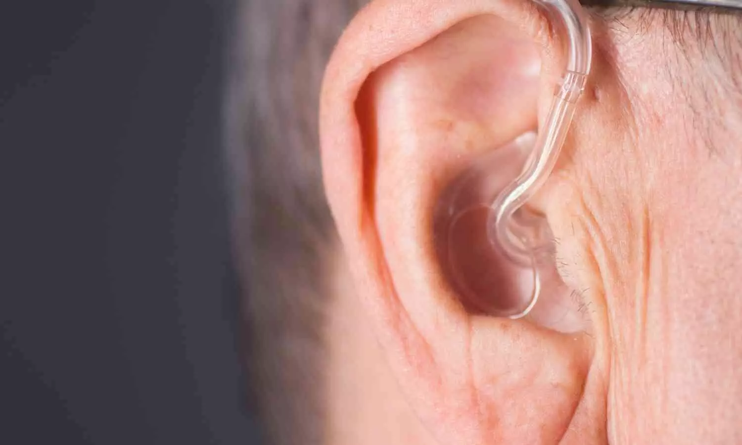
Hearing Loss may be Independent risk factor for Parkinson’s Disease – JAMA study

Recent research published in JAMA Neurology  has unveiled a compelling link between hearing loss and Parkinson’s disease, marking a significant advance in our understanding of preventable risk factors for this debilitating neurological condition. The study, analyzing data from over 3.5 million U.S. veterans through their electronic health records, provides significant evidence that hearing impairment may precede Parkinson’s disease development.

The findings revealed a 26% increased risk of developing Parkinson’s disease in individuals with hearing loss compared to those with normal hearing. More significantly, the research demonstrated a dose-dependent relationship – the more severe the hearing loss, the higher the risk of developing Parkinson’s disease.

Using objective audiometric data rather than relying on subjective reports or clinical judgments, the study followed patients for an average of 7.6 years, with some followed up to 20 years. The study found that patients who received hearing aids within two years of their hearing loss diagnosis showed a significantly reduced risk of developing Parkinson’s disease. Statistical analysis revealed that providing hearing aids to 462 people could prevent one case of Parkinson’s disease over a ten-year period.

The research team also discovered that hearing loss interacts synergistically with known prodromal symptoms of Parkinson’s disease, such as constipation, depression, anxiety, sleep disorders, and loss of smell. When hearing loss occurred alongside these symptoms, the risk of developing Parkinson’s was higher than would be expected from either condition alone.

While the exact mechanism linking hearing loss to Parkinson’s disease remains unclear, several possibilities exist. Hearing loss might directly contribute to neurodegeneration, as evidenced by studies showing elevated tau levels and accelerated brain volume atrophy in individuals with hearing impairment. Alternatively, hearing loss might work indirectly by limiting social engagement or increasing cognitive load, both known risk factors for neurological disorders.

The study’s findings have significant implications for clinical practice. They suggest that hearing screening should be implemented at the primary care level, even when patients haven’t expressed hearing concerns. These results are particularly relevant as we face an aging global population and an expected increase in Parkinson’s disease cases. The World Health Organization projects that neurological disorders, including Parkinson’s, will become leading causes of disability worldwide in coming decades.

The study’s implications are clear: hearing loss should not be dismissed as an inevitable part of aging. Regular hearing screening should be incorporated into routine healthcare, especially for individuals over 40 or those with other risk factors for Parkinson’s disease. Early intervention with hearing aids might serve as a practical, non-invasive strategy to potentially reduce the risk of this serious neurological condition.

This research opens new avenues for prevention and highlights the importance of considering sensory health in the context of neurodegenerative disease risk.

Reference

Neilson LE, Reavis KM, Wiedrick J, Scott GD. Hearing Loss, Incident Parkinson Disease, and Treatment With Hearing Aids. JAMA Neurol. Published online October 21, 2024. doi:10.1001/jamaneurol.2024.3568

Powered by WPeMatico

New AI tool “game changer” in improving outcome predictions for kidney transplant patients, claims research

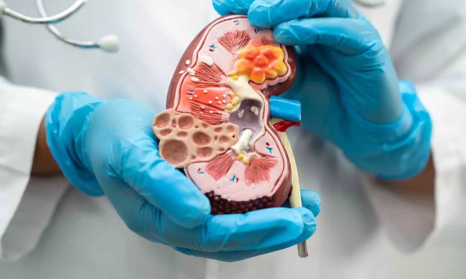
A new advanced artificial intelligence (AI) tool, developed by renal doctors internationally, represents a significant step forward in predicting and potentially improving outcomes for UK kidney transplant patients.

For patients with late-stage renal failure, a kidney transplant can be life-changing, offering the promise of improved survival and a better quality of life compared to other treatment options. But in the UK alone, around 5,000 people are on the waiting list for a kidney transplant, with an average wait time of 2-3 years for a deceased donor organ.

A team of experts from hospitals across the US and UK have created new software, which could ultimately pave the way for updated kidney allocation policies – potentially leading to better patient outcomes and the more efficient use of this precious resource.

The tool, entitled the ‘UK Deceased Donor Kidney Transplant Outcome Prediction’ (UK-DTOP), uses advanced AI. It was developed using data from nearly 30,000 transplant cases across 15 years.

Dr Hatem Ali, a renal specialist at University Hospitals Coventry and Warwickshire NHS Trust, leads a new study to report on the tool’s capabilities in the peer-reviewed journal Renal Failure.

He explains how his research team believe the model “promises to be a game changer in kidney transplantation”.

“The UK-DTOP offers hope for more efficient organ allocation and improved outcomes for patients in need,” Dr Ali says.

“By harnessing the power of AI and machine learning, we’ve created a more accurate and reliable decision-support system, which could lead to improved donor selection, transplant strategies, and ultimately, better outcomes for kidney transplant patients.”

“As an author of this study, I am enthusiastic about the potential impacts of the UK-DTOP tool on kidney transplantation. This AI-enabled model enhances our predictive capabilities and helps refine our approach to donor-recipient matching. By improving how we allocate organs, we can ensure better outcomes for transplant recipients. It is my hope that this tool will be embraced globally, leading to significant advancements in patient care and the efficient use of critical health resources.”

A kidney transplant carries inherent risks and with the demand for organs far outstripping supply, it is crucial to ensure that every donated kidney is used in the most effective way.

However, existing predictive models, such as the widely used Kidney Donor Risk Index (KDRI), have shown limitations in accurately forecasting patient outcomes-highlighting the urgent need for more sophisticated tools that can better guide clinical decision-making.

Using the data from 29,713 transplant cases recorded in the UK Transplant Registry (UKTR) between 2008 and 2022, the expert term evaluated the predictive performance of three advanced machine learning techniques, considering various donor, recipient, and transplant factors. The UK-DTOP emerged as the superior model with a predictive power of 0.74, significantly outperforming the KDRI (0.57) and its UK counterpart, the UK-KDRI (0.62).

“The UK-DTOP is a versatile tool for assessing deceased donor kidney transplantation outcomes. It refines pre-transplant decision-making while recognising that the final decision to accept an organ rests with the recipient and their risk tolerance,” adds co-author Dr Miklos Molnar, from Division of Nephrology & Hypertension at University of Utah.

“Our findings advocate for a shift toward the adoption of advanced, data-driven tools across healthcare systems worldwide, potentially revolutionising donor-recipient matching and organ allocation, improving transplant success rates and saving lives.”

The researchers also used unsupervised machine learning techniques to identify five distinct groups of kidney transplant patients with varying survival rates. Ultimately, this approach could enable more personalised risk assessments to inform decisions about whether or not to proceed with a transplant.

While the UK-DTOP represents a significant advancement, the team also acknowledge this decision-support system has certain limitations that could affect its predictions. These include variability in reported data, missing information on some donor characteristics, and the absence of certain factors that may influence long-term outcomes, such as specific antibodies and certain biological markers. 

Reference:

Hatem Ali,Arun Shroff,Karim Soliman,Miklos Z. Molnar,Adnan Sharif,Bernard Burke, Improved survival prediction for kidney transplant outcomes using artificial intelligence-based models: development of the UK Deceased Donor Kidney Transplant Outcome Prediction (UK-DTOP) Tool, Renal Failure, https://doi.org/10.1080/0886022X.2024.2373273.

Powered by WPeMatico

Smoking significantly increases risk of early implant failure, particularly in maxilla: Study

Smoking significantly increases risk of early implant failure, particularly in maxilla suggests a study published in the Journal of Dentistry.

A study was done to assess the association between smoking and early dental implant failure by conducting a systematic review and meta-analysis of observational studies. Thirty-two observational clinical studies published between 1994 and 2024 were included, with a total of 59,246 implants at implant level and 14,115 patients at individual level. At implant level, a meta-analysis of 21 included cohort studies showed that smoking was associated with increased risk of early dental implant failure compared with non-smoking (odds ratio [OR], 2.59; 95 % confidence interval [CI], 2.08–3.23). Three included studies reported that smoking was associated with higher maxillary early dental implant failure risk (OR, 5.90; 95 %CI, 2.38–14.66) than that of mandible (OR, 3.76; 95 %CI, 1.19–11.87). At individual level, meta-analysis of thirty cohort studies indicated that risk of early implant failure in smokers was 100 % higher than in non-smokers (OR, 2.00; 95 %CI, 1.43–2.80). Three case-control studies found that risk of early implant failure of smokers was 59 % higher than that of non-smokers (OR, 1.59; 95 %CI, 1.28–1.97). Smoking was significantly associated with early dental implant failure, particularly at the maxillary location, at both implant and individual level. These findings suggest smoking cessation is a crucial factor in reducing risk of early dental implant failure. There is uncertainty about the extent to which smoking influences early dental implant failure, our meta-analysis of findings emphasize smoking was significantly associated with early dental implant failure, particularly at the maxillary location.

Reference:

Ying-Ying Fan, Shu Li, Ye-Jun Cai, Tai Wei, Peng Ye. Smoking in relation to early dental implant failure: A systematic review and meta-analysis. Journal of Dentistry, Volume 151, 2024, 105396, ISSN 0300-5712. https://doi.org/10.1016/j.jdent.2024.105396. (https://www.sciencedirect.com/science/article/pii/S0300571224005669)

Keywords:

Smoking, significantly, increases, risk, early, implant failure, particularly, maxilla, Study, Ying-Ying Fan, Shu Li, Ye-Jun Cai, Tai Wei, Peng Ye

Powered by WPeMatico

Alcohol and Inflight Hypoxia Significantly Worsen Sleep and Oxygen Levels, reveals study

Researchers have demonstrated that alcohol consumed during the preparation for sleep in an environment of low pressure such as in an airplane cabin significantly lowers the quality of sleep, promotes elevated heart rate, and prolongs hypoxia time. A recent study was published in the journal Thorax conducted by Trammer and colleagues.

Long-haul flights subject passengers to inadequate oxygen due to a lower pressure in the cabin which can impact sleep. Moreover, alcohol consumption from the list of additives may enhance these effects and lengthen periods characterized by low SpO2 and high heart rates with decreased deep sleep. Low air pressure can decrease the amount of oxygen in blood. It is one of the predominant causes of hypoxia in cabins of flights. This study was conducted to research the combined effect of alcohol consumption and hypobaric hypoxia on sleep patterns, oxygen saturation, and heart rate.

The subjects were two groups of healthy participants followed up during sleep in a normal sleep laboratory or in a hypobaric chamber simulating the reduced air pressure found at an altitude of 2,438 meters (about 8,000 feet). They had a 4-hour sleep opportunity from 00:00 to 04:00 hours and were given alcohol before one of the nights, with a mean blood alcohol concentration of 0.043%. Both nights with and without alcohol were counterbalanced in the order for the groups. The sleep stages were recorded using polysomnography, and oxygen saturation and heart rate were measured all through the night. To ensure a full recovery of participants, two full recovery nights of sleep separated the study conditions.

The study developed a number of important findings on the effects of alcohol and inflight hypoxia on sleep and levels of oxygen:

  • Under both conditions combined (alcohol and hypobaric hypoxia), SpO2 fell to a median value during sleep to 85.32% compared to 88.07% without alcohol under the condition of hypobaric hypoxia, whereas in the alcohol-only condition, it was still 94.97%. In the sleep laboratory (ordinary atmospheric pressure), without alcohol, SpO2 reached 95.88 %.

  • It increased heart rate to a significant extent in the combined condition with a median of 87.73 bpm as compared with 72.90 bpm in the non-alcohol hypobaric condition, 76.97 bpm in the alcohol condition, and 63.74 bpm in the sleep laboratory without alcohol.

  • The time below the clinical threshold of 90% SpO2 median was reached to be 201.18 minutes within the combined condition. For the non-alcohol hypobaric condition, median time below this threshold reached 173.28 minutes. In the sleep laboratory, participants showed no exposure time to the level below 90% SpO2, regardless of alcohol consumption.

  • Deep sleep was reduced to 46.50 minutes with the combined condition, as compared to 84.00 minutes while alcohol was used in the sleep laboratory and 67.50 minutes without alcohol.

A combination of alcohol consumption and inflight hypobaric hypoxia severely impairs sleep quality with a further increase in heart rate and prolongs the duration of hypoxemia (SpO2 <90%) in healthy volunteers. It is worth remembering these risks when consuming alcohol for long flights, because the carelessness of alcohol consumption might deteriorate people’s overall well-being and recovery while traveling and after arriving at the destination.

Reference:

Trammer, R. A., Rooney, D., Benderoth, S., Wittkowski, M., Wenzel, J., & Elmenhorst, E.-M. (2024). Effects of moderate alcohol consumption and hypobaric hypoxia: implications for passengers’ sleep, oxygen saturation and heart rate on long-haul flights. Thorax, 79(10), 970–978. https://doi.org/10.1136/thorax-2023-220998

Powered by WPeMatico

Iron supplements efficacious for children living with HIV, reveals research

A University of Minnesota Medical School research team has found that giving iron supplements to children living with human immunodeficiency virus (HIV) in sub-Saharan Africa could be an important first step in optimizing brain development.

The research, published in the journal Lancet HIV, demonstrates that iron, while often withheld from children with HIV due to fear of increasing infection risk, is in fact beneficial. This finding paves the way for future research examining iron’s role in neurodevelopmental outcomes in children with HIV.

“With the success and widespread availability of antiretroviral therapy (ART), children with HIV in sub-Saharan Africa are living longer, and optimizing their brain development is a new public health imperative,” said Sarah Cusick, PhD, associate professor at the U of M Medical School and a member of the Masonic Institute for the Developing Brain.

Between May 2018 and November 2019, researchers enrolled 200 children with HIV and anemia who had received ART for at least six months. The study participants were randomly chosen to receive either iron supplements or a placebo for three months. Children who received iron had higher hemoglobin concentrations and better markers of iron nutrition than those who received the placebo. There also was no evidence of increased risk of infection.A of time.

Reference:

Anne E P Frosch, Victor Musiime, Christopher Staley, Andrea L Conroy, Safety and efficacy of iron supplementation with 3 months of daily ferrous sulphate in children living with HIV and mild-to-moderate anaemia in Uganda: a double-blind, randomised, placebo-controlled trial, The Lancet HIV, DOI:10.1016/S2352-3018(24)00238-8. 

Powered by WPeMatico

No end to doctor violence! Cardiologist considers quitting profession after mob attack

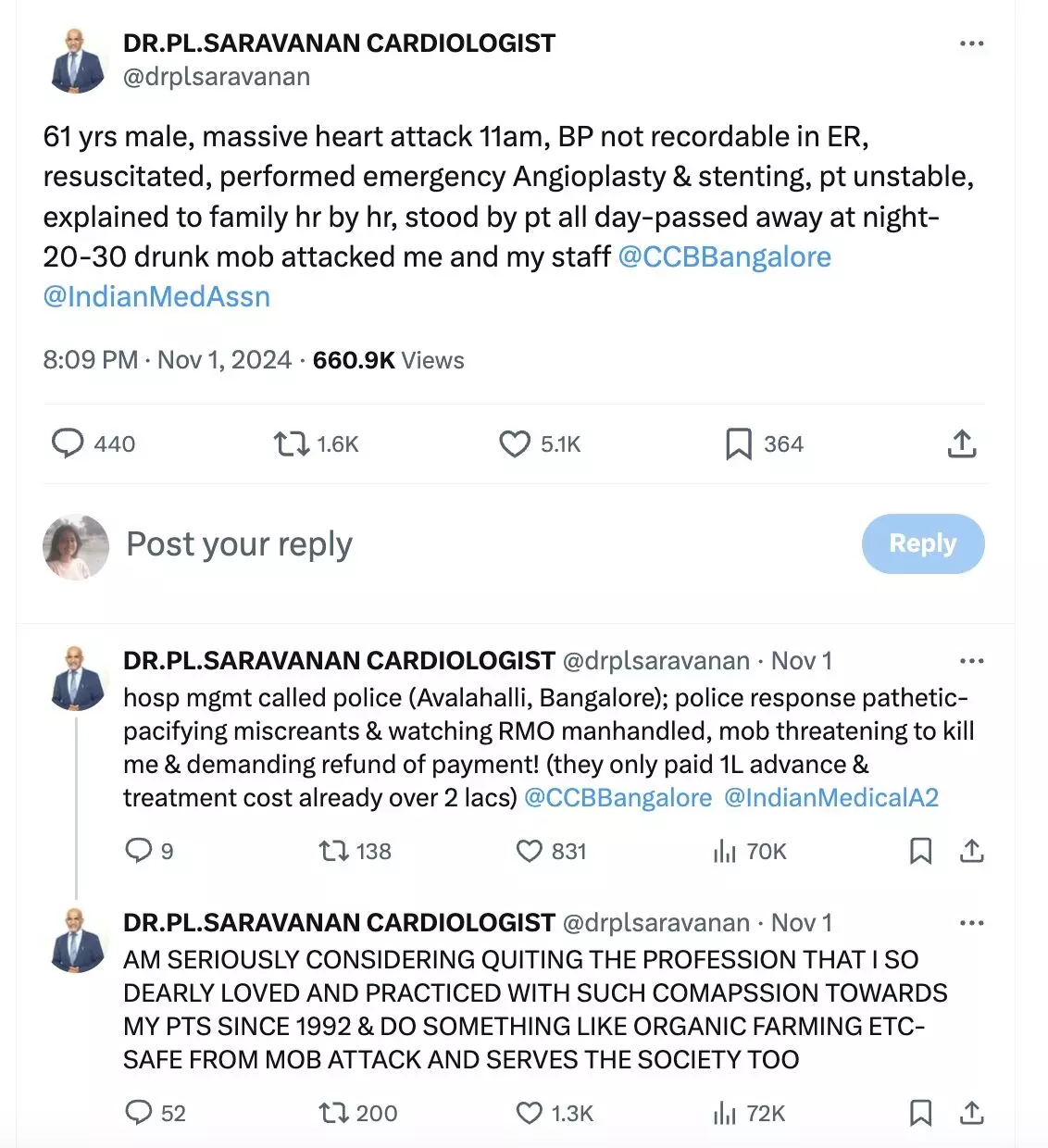
New Delhi: A recent X (formerly Twitter) post by a senior cardiologist went viral as he narrated how he was attacked by a mob of 20 to 30 people after providing all-day long treatment to a patient, who ultimately passed away.

It has been alleged that when the doctor reached out to the police, the response was “pathetic” and the Inspector of Police at Avalahalli Police Station allegedly pressurised the doctor to delete the post and not to register an FIR!

In another X post, the doctor mentioned that he was considering quitting the medical profession and doing something such as organic farming—to serve society and not face any mob attack. Since the post was posted, around 6,61,000 people have viewed it, and thousands of people have shared it on their social media handles.

The doctor made the first X post on the evening of November 1st, 2024. He mentioned how a 61-year-old male, having a massive heart attack at 11 am came to him for treatment and how after staying by the side of the patient for the whole day, a 20-30 drunk mob attacked him and his staff.

Also Read: Violence against Doctors: Punjab Govt doctors prepare for agitation over lack of security measures

“61 yrs male, massive heart attack 11am, BP not recordable in ER, resuscitated, performed emergency Angioplasty & stenting, pt unstable, explained to family hr by hr, stood by pt all day-passed away at night- 20-30 drunk mob attacked me and my staff,” the doctor’s X post read.

violence against doctors

He further alleged that when the hospital management called the police, the response was pathetic and added, “hosp mgmt called police (Avalahalli, Bangalore); police response pathetic- pacifying miscreants & watching RMO manhandled, mob threatening to kill me & demanding refund of payment! (they only paid 1L advance & treatment cost already over 2 lacs).”

Expressing how he was considering leaving the medical profession, the doctor, who has been practising since 1992, further mentioned in the post, “AM SERIOUSLY CONSIDERING QUITING THE PROFESSION THAT I SO DEARLY LOVED AND PRACTICED WITH SUCH COMPASSION TOWARDS MY PTS SINCE 1992 & DO SOMETHING LIKE ORGANIC FARMING ETC- SAFE FROM MOB ATTACK AND SERVES THE SOCIETY TOO.”

“CAN YOU BELEIVE THIS – Inspector of police- Avalahalli police station- pressurising to delete post in X and not to register an FIR!! Only he knows the reason- but I am led to beleive this is standard police tactic to avoid the hassle of inv and arrrest etc,” he further alleged.

In another post, the doctor claimed that the law to protect the doctor only exists on pen and paper. Later, sharing updates regarding the incident, the doctor claimed that due to the power of the social media, police finally registered his complaint and the commissioner of health services asked for a full report. He also informed how the Indian Medical Association (IMA) and CSI i.e. the Cardiologists Society of India took the matter seriously and how they plan to take it up to the Karnataka Home Minister.

However, in another X post yesterday, he informed that till now no action has been taken even though the RMO at the hospital gave a detailed complaint along with CCTV footages.

Meanwhile, in response to the X post, another user mentioned that people are angry how hospitals looted them during COVID 19 pandemic in the name of treatment and how people lost everything they had including their loved ones. The post further mentioned how during the pandemic, dead people were kept on ventilators to inflate the bill and obnoxious rates were charged for meds and how people have now lost their patience.

Responding to this, the doctor mentioned, “I feel this is so inappropriate. Just because of some people being unethical, U can’t paint majority of sincere doctors toiling to save a life on Diwali day (also happened to be my Birthday & I could have happily enjoyed the festivities with family& friends) with the same brush.”

Further addressing the comments that referred to corporate culture in medical field these days, the doctor mentioned that he had not anything wrong but they were upset with the outcome because the patient had died despite best efforts and how those people manhandled 6 persons altogether.

“The excuses put forward r “they were emotional” “they were inebriated” “they r illiterate”- does any right thinking person agree that any of the above justifies beating up 6 health care professionals doing their duty on Diwali day!” he added in another X post.

Also Read: Orthopaedic Surgeon attacked at Trichy ESI Hospital after traffic dispute, 5 booked

Powered by WPeMatico

Kundapur Govt Hospital Doctor on emergency duty verbally abused, threatened with death by patient’s attendant

Udupi: A case has been registered against an attendant of a female patient who allegedly verbally abused an on-duty medical officer at Kundapur government hospital and threatened him with death over a dispute regarding the patient’s admission process. 

According to the complaint, the attendant is the patient’s husband who allegedly in a fit of rage verbally assaulted the doctor, snatched his wife’s medical records, and tore official documents on the doctor’s table.

The incident occurred around 11:15 p.m. on Sunday when the doctor who has served as an emergency care doctor at the hospital for over two years, was treating a female patient who came in complaining of severe stomach pain with the assistance of another woman. 

Also read- Orthopaedic Surgeon attacked at Trichy ESI Hospital after traffic dispute, 5 booked

In his complaint, the doctor claimed that he examined the patient along with her medical records and recommended her admission for further observation in the presence of a nurse. During the process, the patient’s husband arrived at the hospital and he was asked to complete the admission process. 

However, shortly afterwards, the couple unexpectedly left the hospital without completing the formalities. The doctor said that he was in the middle of examining the patient when they left the hospital. 

Within half an hour, at approximately 11:45 p.m., the husband in a rage returned to the hospital’s emergency room. It was claimed that he barged in and verbally assaulted the doctor, snatched the patient’s medical records, and tore official documents on the doctor’s table, disrupting the emergency room.

As per DH news report, the situation further escalated when the husband accompanied by another woman, allegedly issued death threats to the doctor apart from continuing to verbally abuse him. 

Following this, the doctor filed a complaint at the police station. Acting on the complaint, the police registered a case against the husband under relevant sections of  BNS. An investigation in this regard is underway. 

Also read- TN Nurse attacked, sexually assaulted by youth while returning from hospital

Powered by WPeMatico