KNRUHS Notifies On 2nd Phase of Web-Based Counselling for Nursing Courses 2024

Telangana- Through a notification, Kaloji Narayana Rao University of Health Sciences (KNRUHS) has notified regarding the conduct of the 2nd phase of web-based counselling under the Competent authority Quota for vacant seats in B.Sc. (Nursing) 4 YDC course for 2024-25 in affiliated Government, Private Nursing Colleges after 1st phase of counselling. Also, the conduct of the 2nd phase of web-based counselling under the Competent Authority Quota for vacant seats in the Post Basic B.Sc. (Nursing) 2 YDC course for 2024-25 in affiliated Government and Private Nursing Colleges after the 1st phase of counselling.

As per the notification, Candidates whose certificates were uploaded at the time of online registration and provisionally verified by the staff of concerned departments and whose names are notified as eligible candidates in the provisional final merit list for B.Sc. (Nursing) 4 YDC and P.B. B.Sc. (Nursing) 2YDC courses 2024-25 on the KNRUHS official website are eligible to exercise web – options online from the computer with internet connection.

All the eligible candidates whose names are displayed in the Provisional final merit list including PWD candidates on the KNRUHS website can exercise web options for admission into B.Sc (Nursing) 4 YDC and P.B. BSc (Nursing) 4 YDC course till 08th November 2024 upto 3.00 PM.

UNIVERSITY FEES

Allotted Candidates have to pay the University fee of Rs. 6000/- through a payment gateway by using an online payment method such as a Debit Card / Credit Card / Internet Banking and download the allotment letter. Candidates who have paid University fees in the first phase need not pay again. The University Fee and other fees once paid shall not be refunded under any circumstances.

Meanwhile, candidates are hereby informed that final verification of original certificates will be conducted at the allotted college and in case of any discrepancy, the provisional allotment will be cancelled and action may be initiated as per University regulations. Therefore, original certificates will not be returned to candidates unless the candidate discontinues from the course.

To view the notifications, click the link below

Powered by WPeMatico

Carbohydrate-Restricted Diet may Improve Beta-Cell Function in Type 2 Diabetes Patients: Study

Researchers have been able to demonstrate that type 2 diabetic patients, when placed on a carbohydrate-restricted diet, are capable of experiencing short-term recovery of beta-cell function, which produces insulin. Researchers in the Journal of Clinical Endocrinology & Metabolism proposed that an eating regimen carbohydrate-restricted helps to rejuvenate the beta-cell function, and therefore one may utilize such an eating habit to potentially improve glycemic control, independent of weight reduction.

Beta-cell dysfunction is an important determinant in the mechanism of developing type 2 diabetes by losing first-phase insulin response. Led by Marian Yurchishin, MS, a PhD student at the University of Alabama at Birmingham, a new study aimed to determine if a carbohydrate-restricted diet could enhance beta-cell function over a short period of intervention. The researchers’ hypothesis was based on the belief that decreasing glucose exposure-to what is considered “glucose toxicity” for the beta cell-may allow for repair and restoration of cell function.

The study consisted of 57 patients with type 2 diabetes who were not on insulin therapy. Ninety-three participants were randomized to a carbohydrate-restricted diet (9% of energy from carbohydrates, 65% from fats) or a higher-carbohydrate diet (55% energy from carbohydrates, 20% from fats). The carbohydrate-restricted group included 5 men and 22 women; mean age 53 years, 17 Black and 10 white. The higher-carbohydrate group included 8 men and 22 women; mean age 55 years, 20 Black and 10 white.

At 12 weeks, the authors noted marked increases in beta-cell function in the carbohydrate-restricted diet group:

• The acute C-peptide response was twice as large in the carbohydrate-restricted group compared with the higher-carbohydrate group (p < 0.01).

• The maximal C-peptide response, an index of the beta-cell’s ability to produce insulin, was 22% larger in the carbohydrate-restricted group (p < 0.05).

• Disposition index, a marker of insulin sensitivity and secretion, was 32% higher in the carbohydrate-restricted group compared with the oral glucose tolerance test (p< 0.05).

• The study also noted race differences in beta-cell function. Maximum C-peptide response was 48% higher in white patients on the carbohydrate-restricted diet compared with those on the higher-carbohydrate diet (p< 0.01), but not in Black patients. This suggests that mechanisms that lead to beta-cell failure may vary by race.

Carbohydrate restriction ameliorates the function of white subjects’ beta cells, particularly of type 2 diabetic subjects. Such a dietary intervention may potentially enhance glycemic control and improve insulin secretions at least in the absence of weight loss. However, the racial differences in beta-cell recovery bode well for further research in the mechanisms that underlie such disparities.

Reference:

Swain, E. (2024, October 22). Effects of a Carbohydrate Restricted Diet on Beta-cell Response in Adults with Type 2 Diabetes, Journal of Clinical Endocrinology & Metabolism (2024). dx.doi.org/10.1210/clinem/dgae670

Powered by WPeMatico

Researchers Highlight Coronary Artery Calcium Scoring as New Gatekeeper for Evaluating Stable Chest Pain

USA: A recent editorial comment in the Journal of the American College of Cardiology: Cardiovascular Imaging highlights the evolving role of coronary artery calcium (CAC) scoring as a critical tool for evaluating patients with stable chest pain. Traditionally, clinicians relied on the Diamond-Forrester model, which estimates the likelihood of obstructive coronary artery disease (CAD) based on age, sex, and angina characteristics. However, contemporary analyses suggest that this model may overestimate CAD probabilities and fail to incorporate newer testing methods.

The editorial references current guidelines, including the 2021 American Heart Association/American College of Cardiology Multisociety Chest Pain Guidelines, which advocate for integrating cardiovascular risk factors and CAC scores to refine the pretest probability (PTP) of CAD in patients with chest pain. According to the editorial authors David E. Winchester, and Mahmoud Al Rifai (both from USA), this is particularly relevant given that stable chest pain can indicate varying levels of underlying cardiovascular issues.

In the featured study by Rasmussen et al., which included 4,371 patients undergoing anatomical assessment for CAD, the researchers assessed the effectiveness of different PTP models. The findings indicated that both risk factor-weighted clinical likelihood (RF-CL) and CAC score-weighted clinical likelihood (CACS-CL) models provided significantly improved predictions for hemodynamically obstructive CAD compared to the basic PTP model. Notably, CAC scoring demonstrated a 98.9% negative predictive value for obstructive CAD, making it an effective tool for ruling out serious conditions.

The editorial emphasizes that while CAC scoring alone can serve as a valuable screening tool, it is crucial to consider individual clinical characteristics to minimize misclassification of CAD status. With nearly half of patients presenting with stable chest pain potentially having a CAC score of zero, the absence of coronary artery calcium suggests that further testing may be unnecessary.

Future recommendations from the 2023 Appropriate Use Criteria for multimodality imaging in chronic coronary disease reinforce this perspective. These guidelines suggest that for patients with low to intermediate PTP for CAD, a negative CAC score could warrant deferring further testing. Conversely, the presence of CAC might indicate the need for additional assessments, such as coronary computed tomography angiography or myocardial perfusion imaging.

Despite its promise, challenges remain in determining which patients would benefit most from CAC scoring. The editorial underscores the need for ongoing research to evaluate various PTP strategies and refine how CAC can be used effectively in clinical practice. Ultimately, the integration of CAC scoring into routine assessments could lead to more accurate and safer management of patients with stable chest pain, improving overall cardiovascular care.

Reference:

Winchester DE, Al Rifai M. Coronary Artery Calcium as a Gatekeeper for Patients With Stable Chest Pain. JACC Cardiovasc Imaging. 2024 Oct;17(10):1211-1213. doi: 10.1016/j.jcmg.2024.05.025. Epub 2024 Aug 7. PMID: 39115500.

Powered by WPeMatico

Landiolol Effectively Controls Heart Rate in Septic Shock Patients Without Increasing Vasopressor Needs: Landi-SEP Trial

Germany: A recent multicenter randomized clinical trial (Landi-SEP) has revealed promising results for the use of Landiolol, an ultra-short-acting beta-blocker, in managing heart rate in patients experiencing septic shock and persistent tachycardia. The findings suggest that landiolol can effectively reduce heart rate without increasing the need for vasopressors after 24 hours of treatment. 

“Landiolol effectively lowers and sustains heart rate for 24 hours in patients with septic shock and persistent tachycardia, all while avoiding an increased requirement for vasopressors.,” the researchers wrote in Intensive Care Medicine.

Septic shock is a severe condition characterized by a significant drop in blood pressure due to infection, leading to organ dysfunction. Patients often present with persistent tachycardia, making heart rate management a crucial aspect of their treatment. Traditional therapies for controlling heart rate have had mixed results, highlighting the need for effective alternatives.

Against the above background, Sebastian Rehberg, University Hospital of Bielefeld, Bielefeld, Germany, and colleagues examined whether heart rate (HR) control could be achieved without raising the need for vasopressors by using the titratable, highly selective ultra-short-acting β1-blocker landiolol.

For this purpose, the researchers conducted a randomized, open-label, controlled trial across 20 sites in seven European countries from 2018 to 2022. They evaluated the efficacy and safety of landiolol in adult patients experiencing septic shock and persistent tachycardia. Participants were randomly assigned to receive either landiolol combined with standard treatment (n = 99) or standard treatment alone (n = 101).

The primary endpoint focused on achieving a heart rate within the range of 80 to 94 beats per minute and maintaining that rate without increasing the need for vasopressors during the first 24 hours following the start of treatment. Key secondary endpoints included 28-day mortality rates and the occurrence of adverse events.

The findings are summarized as follows:

  • The study included a total of 196 septic shock patients, with 98 receiving standard treatment combined with landiolol and 98 receiving standard treatment alone.
  • A significantly higher percentage of patients in the landiolol group achieved the combined primary endpoint compared to the control group:
    • Landiolol group: 39.8% (39 out of 98 patients)
    • Control group: 23.5% (23 out of 98 patients)
  • The between-group difference in achieving the primary endpoint was 16.5%.
  • There were no statistically significant differences between the two groups in secondary outcomes or adverse events.

The study revealed that the ultra-short-acting beta-blocker landiolol effectively reduced and maintained heart rate in patients with septic shock and persistent tachycardia without increasing the need for vasopressors after 24 hours. However, there were no significant differences in adverse events or clinical outcomes, including 28-day mortality, compared to standard care.

“These findings suggest that a strategy focused on strict heart rate reduction to below 95 beats per minute may not be appropriate for all patients with septic shock and persistent tachycardia. Further research is necessary to identify specific patient phenotypes within this population that may clinically benefit from heart rate control,” the researchers concluded.

Reference:

Rehberg, S., Frank, S., Černý, V. et al. Landiolol for heart rate control in patients with septic shock and persistent tachycardia. A multicenter randomized clinical trial (Landi-SEP). Intensive Care Med 50, 1622–1634 (2024). https://doi.org/10.1007/s00134-024-07587-1

Powered by WPeMatico

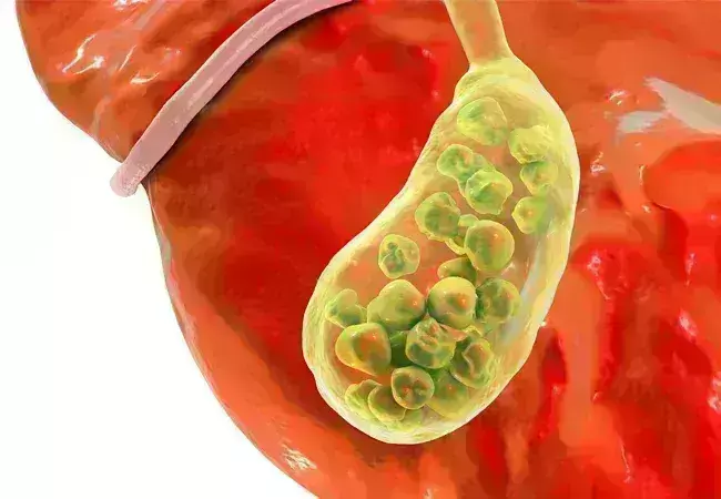
Neutrophil-to-HDL Cholesterol Ratio and associated with Gallstone Disease Risk: Insights from NHANES 2017-2020 Data

A recent study revealed that the neutrophil-to-high-density
lipoprotein cholesterol ratio (NHR) could be a useful tool to identify individuals
at risk of developing gallstone disease (GSD). NHR can be used as an easy and
cost-effective tool for the early detection of individuals at GSD as per a
study published in the journal Lipids in Health and Disease.

Gallstone disease (GSD) is one of the most frequent and expensive
gastrointestinal disorders caused by cholesterol, bile pigment, and mixed stones,
with cholesterol stones outranking the three. They are caused by the oversaturation
of calcium salts and cholesterol in bile, leading to cholesterol deposits. Gallstones
have an increased risk of cancer. The neutrophil to high-density lipoprotein
cholesterol ratio (NHR) is a novel comprehensive marker of inflammatory
responses and lipid metabolism that can detect cardiovascular disease,
depression, acute biliary pancreatitis, and hepatocellular carcinoma. Previous research
has shown that inflammatory reactions can promote gallstone formation by
altering protein and lipid metabolic pathways and increasing bile acid salt
concentrations by involving neutrophils and lipids. As there is ambiguity on
the association between NHR and gallstones, researchers from Zhejiang Chinese
Medical University, China conducted a study on the relationship between
gallstone disease (GSD) and the neutrophil-to-high-density lipoprotein
cholesterol ratio (NHR) in American patients with gallstones.

The study was conducted by collecting data from the National
Health and Nutrition Examination Survey (NHANES) between 2017–2020. Data was collected
through interviews, health exams, and lab tests. Out of 15,560 participants,
7,894 were included in the study. NHR calculation was done by dividing the neutrophil
count by HDL-C levels and the participants were divided into 3 NHR categories
based on the count. Gallstone data was reported by the participants based on
the physician’s diagnosis. Unadjusted and adjusted logistic regression models
were used to evaluate the association between NHR and GSD.

Key findings:

  • Among the 7894 participants analyzed in this
    study, the prevalence of GSD was 10.98%, and the average NHR value was 3.41 ±
    0.06.
  • An obvious positive association between NHR and
    the likelihood of GSD was found as per the fully adjusted multivariable
    logistic regression analysis (OR = 1.09, 95% CI: 1.01, 1.16; P = 0.0197).
  • Subset analyses confirmed the consistency of
    this association and interaction tests across various subgroups, including
    those categorized by smoking status and asthma.
  • Furthermore, smoothed curve fitting and
    threshold effect analyses revealed a nonlinear relationship with a threshold of
    2.86.

Thus, the study concluded that a higher NHR is associated
with a greater likelihood of having GSD and NHR can be used as a low-cost
method to screen patients at risk of GSD. NHR can also be used in early
diagnosis so that preventive measures can be taken to reduce the risk of gallstones.

Further reading: Ma
D, Ma H, Li Y, Yang L. Association between Neutrophil-to-high-density
lipoprotein-cholesterol ratio and gallstones: insights from the national health
and nutrition examination survey (2017-2020). Lipids Health Dis.
2024;23(1):355. Published 2024 Oct 31. doi:10.1186/s12944-024-02349-w.

Powered by WPeMatico

Which is better TTE or TEE for Grading RV Diastolic Dysfunction?

Assessing right ventricular diastolic function is crucial in cardiac surgery, as it significantly impacts long-term prognosis. Typically, RV diastolic dysfunction is evaluated using criteria from the American Society of Echocardiography (ASE) or the British Society of Echocardiography (BSE) via transthoracic echocardiography (TTE). Nevertheless, transesophageal echocardiography (TEE) is the primary method for perioperative evaluation during cardiac surgery. Recent study aimed to assess the agreement between transthoracic echocardiography (TTE) and transesophageal echocardiography (TEE) in grading right ventricular diastolic dysfunction (RVDD) using criteria from the American Society of Echocardiography (ASE) and the British Society of Echocardiography (BSE).

The study included 81 patients undergoing cardiac surgery. TTE and TEE measurements were taken within 10 minutes of each other, under similar hemodynamic, anesthetic, and ventilatory conditions. RVDD grading was performed separately using TTE, TEE mid-esophageal right ventricular inflow-outflow (MERVIO) view, and TEE deep transgastric right ventricular inflow-outflow (DTGRVIO) view, based on both ASE and BSE criteria. According to the ASE criteria, disagreement of ≥1 RVDD grade was seen in 53.1% of patients when comparing TTE and TEE-MERVIO, and in 39.5% when comparing TTE and TEE-DTGRVIO. The weighted kappa was 0.14 and 0.3, respectively, indicating poor to fair agreement. Using the BSE criteria, disagreement occurred in 11.1% and 14.8% of patients when comparing TTE with TEE-MERVIO and TEE-DTGRVIO, respectively, with unweighted kappa of 0.25 and 0.26, indicating fair agreement.

Limits of Agreement and Rater Consistency

The study found wide limits of agreement between individual 2D and Doppler parameters measured by TTE and TEE. There was almost perfect agreement between independent raters for RVDD grading using both TTE and TEE. In conclusion, the study revealed at best only fair agreement between TTE and TEE in grading RVDD. The authors recommend further research to develop a TEE-based algorithm for grading RVDD and to evaluate the prognostic effectiveness of perioperative TEE for predicting adverse clinical outcomes associated with RVDD.

Key Points

1. The study aimed to assess the agreement between transthoracic echocardiography (TTE) and transesophageal echocardiography (TEE) in grading right ventricular diastolic dysfunction (RVDD) using criteria from the American Society of Echocardiography (ASE) and the British Society of Echocardiography (BSE).

2. The study included 81 patients undergoing cardiac surgery, with TTE and TEE measurements taken within 10 minutes of each other under similar hemodynamic, anesthetic, and ventilatory conditions. RVDD grading was performed separately using TTE, TEE mid-esophageal right ventricular inflow-outflow (MERVIO) view, and TEE deep transgastric right ventricular inflow-outflow (DTGRVIO) view, based on both ASE and BSE criteria.

3. Using the ASE criteria, there was a disagreement of ≥1 RVDD grade in 53.1% of patients when comparing TTE and TEE-MERVIO, and in 39.5% when comparing TTE and TEE-DTGRVIO, with poor to fair agreement as indicated by the weighted kappa values.

4. Using the BSE criteria, disagreement occurred in 11.1% and 14.8% of patients when comparing TTE with TEE-MERVIO and TEE-DTGRVIO, respectively, with fair agreement as indicated by the unweighted kappa values.

5. The study found wide limits of agreement between individual 2D and Doppler parameters measured by TTE and TEE, but almost perfect agreement between independent raters for RVDD grading using both TTE and TEE.

6. The authors conclude that the study revealed at best only fair agreement between TTE and TEE in grading RVDD, and they recommend further research to develop a TEE-based algorithm for grading RVDD and to evaluate the prognostic effectiveness of perioperative TEE for predicting adverse clinical outcomes associated with RVDD.

Reference –

Singh N C, Biswas I, Kumar B, et al. (October 07, 2024) The Agreement Between Transthoracic and Transesophageal Echocardiography in the Assessment of Right Ventricular Diastolic Dysfunction Grades in Adult Patients Undergoing Cardiac Surgery: A Prospective Observational Study.

Cureus 16(10): e70976. DOI 10.7759/cureus.70976

Powered by WPeMatico

Add on Estradiol to 2% minoxidil offers no additional advantage in Female pattern hair loss, suggests study

A new study published in the Clinical and Experimental Dermatology journal showed that estradiol with minoxidil (EMX) may raise the risk of irregular menstruation in individuals with female pattern hair loss (FPHL) which is also known as female androgenetic alopecia (AGA). The variables that influence miniature follicle development and the pathophysiology of FPHL are yet unknown. There is proof that genetic, hormonal, and environmental factors also play a part in male AGA, in addition to the most obvious impact of androgens. Women who have thick, healthy hair are more likely to feel secure, confident, and capable of change. They are also more likely to engage with others.

The prevalence of female pattern hair loss is high and has a detrimental effect on one’s quality of life. The most effective treatment for FPHL is minoxidil, however it is more difficult to treat than male pattern hair loss. Several trials treated FPHL with 17α-estradiol solution, either by alone or in combination with minoxidil, with varying degrees of success. Thus, this study evaluated the safety and effectiveness of topical 17α-estradiol 0.01% with minoxidil 2% against minoxidil 2% in the treatment of FPHL, by Nermeen Ibrahim Bedair and colleagues.

For this study, 43 women with FPHL were instructed to apply a spray-on solution comprising either 17α-estradiol 0.01% plus minoxidil 2% (EMX group) or minoxidil 2% alone (MX group) blindfolded twice a day. The course of treatment lasted six months. After baseline and after the conclusion of the therapy, trichoscopic and clinical evaluations were conducted.

Both groups displayed indicators of improvement at the conclusion of the therapy session. The EMX group outperformed the MX group in terms of improvement metrics, however, this difference was not statistically significant. Menstrual abnormalities were more common among the patients of EMX group. Overall, the findings of this study suggest that a 0.01% 17α-estradiol and 2% minoxidil solution does not appear to provide a statistically significant benefit over minoxidil alone, and there may be a greater chance of related menstrual abnormalities in  the treatment of FPHL.

Reference:

Bedair, N. I., EL-Komy, M. H. M., Mohamed, R. E., Shamma, R. N., & Amer, M. A. (2024). Efficacy and safety of combined topical ethinylestradiol with minoxidil versus topical minoxidil in female pattern hair loss. A trichoscopic randomized controlled clinical study. In Clinical and Experimental Dermatology. Oxford University Press (OUP). https://doi.org/10.1093/ced/llae436

Powered by WPeMatico

Elective Single-Embryo and Transfer Mechanisms leading to embryonic splitting and its risk factors, explains study

Multiple gestations are the most frequent complications
associated with assisted reproductive techniques (ART). To decrease the rate of
twin and higher order multiple gestations, the elective single-embryo transfer
(eSET) is currently encouraged. However, blastocyst stage SET was still found
to be associated with multiple gestations. The present data revealed that the
frequency of embryo splitting post-eSET is 1.36% with the rate of monozygotic
twin and triplet pregnancies being 0.9-3.1% and 0.048%, respectively. However,
dizygotic twin and triplet pregnancies can also be encountered post-eSET. Both
monozygotic and dizygotic pregnancies can occur in the setting of fresh embryo
transfers, in natural or modified natural frozen embryo transfers (FET), and
rarely in medicated FET cycles. Such events challenge the twinning dogma
proposed by Corner.

Incidence of Zygotic Splitting after SET

Single-embryo transfer is becoming the most favored method
of ART conclusion in recent years as it decreased the complications associated
with multiple gestations. However, multiple gestations in the form of mono- or
dizygotic twinning are still encountered. The classic definition of when one
embryo undergoes fission into 2 or more genetically identical embryos is called
monozygotic splitting, while when 2 different embryos implant, a dizygotic
pregnancy ensures.

The findings of monochorionic and multiple chorionic
pregnancies in blastocyst eSET confirmed the fact that embryo splitting took
place after the transfer. However, not all pregnancies are monozygotic. As per
Osianlis et al., the calculated dizygotic rate in their paper was 0.5% with an
overall Di-Di birth rate of 1%. Based on these numbers, they concluded that 50%
of the multizygotic pregnancies are due to actual embryo splitting while the
other 50% could be explained by concomitant natural conception at the same time
of the ART conception. On another level, the Japanese ART national registry
database along with a survey done by Yamashita et al. documented 122 triplet
pregnancies, of which 46 were single gestational sac pregnancies, 18 were
double gestational sac pregnancies, and 59 were with 3 gestational sacs. It is
worth mentioning that the trichorionic pregnancies had zero fetuses in 9 cases,
1 fetus in 12 cases, 2 fetuses in 9 cases, and three fetuses in 29 cases. One
quadruplet case was also documented.

Chorionicity

Chorionicity refers to the placenta the origin of which can
be determined accurately. Zygosity on the other hand, which is the origin of
the fetus, can be predicted in half of the cases as multiple gestation
pregnancies can originate from one or multiple embryos especially when the sex
of the babies is discordant. Given this fact, same-sex twins or triplets could
be true monozygotic or dizygotic in origin. The only way to accurately diagnose
the zygosity is to do DNA fingerprinting which is expensive and thus not
performed in daily practice.

In contrast, monochorionic multiple gestations are always
monozygotic. Originally, it was thought that the earlier the embryonic
division, the more separate and independent the fetuses were. In other words,
cleavage stage divisions were believed to result in dichorionic diamniotic
pregnancies while blastocyst stage divisions resulted in monochorionic
monoamniotic pregnancies. According to Konno et al., dichorionic pregnancies
were found to be more common with ART. As such, authors can conclude that SET
can result in monozygotic (monochorionic and multichorionic) as well as
dizygotic pregnancies (multichorionic pregnancies).

Risk Factors

Naturally occurring twinning, especially the dizygotic form,
is believed to be linked to a genetic predisposition most commonly located on
chromosome 3. Some ethnicities were found to be more predisposed to dizygotic
twinning where the rate reached 50/1000 in Nigeria. This contrasts with the
naturally occurring monozygotic twinning which was found to be nonaffected by
the ethnicity or the genetic makeup of the couple.

ART on the other hand has increased the incidence of
monozygotic twining. It has been shown that the patient’s young age might
predispose to zygotic twinning while unexplained infertility was found to be
protective. It was proposed that ART associated embryo manipulations such as
FET per se, blastocyst culture, and assisted hatching could be risk factors for
zygotic splitting while the zona manipulation of the oocyte in the form of
intracytoplasmic sperm insemination (ICSI) was not. Interestingly, there was no
difference in the splitting rate neither between the cleavage stage and the
blastocyst stage transfers nor between fresh and frozen embryo transfer cycles.

Another risk factor for splitting is a lower inner cell mass
(ICM) grading of B or C. It is thought that loose intercellular connections may
induce the ICM fission. This has been documented through the time-lapse imaging.
The quality of the culture media is also thought to stimulate zygotic
splitting. An increase in the free radicals’ concentration due to increased
glucose concentration in the culture media used for prolonged culture could
lead to ICM splitting at the site of glucose-induced apoptosis of certain
regions of the ICM.

The new sequential culture systems with antioxidant activity
might explain the lack of increase of the rate of embryo splitting despite the
major increase in the number of IVF cycles and embryo transfers worldwide. When
coupled with the improvement in the embryologists training and experience, the
rate of splitting associated with a blastocyst transfer has been found to
decrease. The OR for embryo splitting decreased from 2.2 to 1.7 when comparing
the periods of 2007 to 2010 and 2010 to 2014. Embryo biopsy on the other hand
was not found to increase the risk of embryo splitting contrary to what was
believed before.

Suggested Mechanisms of Division

It has been shown that blastomeres from a 4-cell stage
embryo can develop into an ICM and trophectoderm; hence, any division after
this stage could give rise to 2 or more embryos with an implantation potential.
Of the suggested mechanisms, abnormal cellular axis formation and cytoplasm
folding in the secondary oocyte prior to fertilization or during the actual
fertilization lead to duplication. It is speculated that gonadotropin
stimulation might disrupt the fine balance and gradients of signalling
molecules affecting the polarity of the oocyte. This is thought to lead to the
formation of 2 cells referred to as daughter cells or tertiary oocytes that
could be fertilized. This is speculated to be caused by the displacement of the
meiotic spindle due to oocyte aging postovulation. This disruption might lead
to the duplication of the axes and formation of 2 embryos upon fertilization or
the fission of the ICM into 2 at the blastocyst stage. This theory would be
replaced later on by the formation of 2 zygotes postfertilization of the
secondary oocyte and not 2 blastomeres.

Other studies advocated the fission to happen closer to the
cleavage stage, and thus, the sequence of events happening during hatching
would then explain the type of the twin gestation. If both blastocysts were
released at the same time, then the resultant pregnancy would be a dichorionic
diamniotic twin gestation.

If on the other hand the blastocysts fused with the
conservation of 2 separate ICMs prior to hatching, then monochorionic
diamniotic twins would appear. If complete fusion of the trophectoderm and the
ICM happened, then monochorionic monoamniotic twins would be created.

Another suggestion was that the ICM would split due to
mechanical compression during hatching through the manipulated zona pellucida
of the embryo which is also referred to as atypical hatching. This atypical
hatching is referred to as 8-shaped hatching, which usually takes place when
the embryo is squeezing out through the hatch of the hardened zona pellucida
due to prolonged culture to blastocyst stage and in cryopreserved-thawed
blastocysts especially with the application of the day 3 prehatching protocol.
It is speculated that this phenomenon might also be the culprit for the
monozygotic triplet gestations that have been documented post-SET.

Another possible explanation to dual or even more ICM is the
nature of human blastomere plasticity. Studies have shown that isolated
trophectoderm cells when cultivated could give rise to a whole new embryo with
an implantation potential. As such, if a blastomere gets separated from the
trophectoderm into the blastocele due to low-grade compaction of the
trophectoderm, this blastomere could give rise to an ICM. Theoretically, each
ICM should give rise to a separate fetus with the surrounding amnion while the
chorion develops during the implantation. The mechanism of chorionic
differentiation between mono and higher order chorionicity in monozygotic
pregnancies is still unknown.

Triplets: Possible Explanation

The explanation of embryo splitting into three is
challenging since triplets after SET is a very rare event. What is known so far
is that to have implantation, an embryo with an intact ICM should be present.
The number of the ICM that the embryo has will define the number of fetuses
that will be seen on the pregnancy ultrasound.

The chorionicity of the pregnancy will depend on the number
of zygotes present at the time of implantation. In theory, the chorion should
rise from trophectoderm cells; thus, it would be logical to consider that the
higher the order of the chorionicity, the higher the number of separate embryos
available for implantation.

In the setting of monochorionic triplets, it is believed
that the blastocyst harbors three distinct ICMs. The mechanism of their
creation might be similar to the ones suggested for the monochorionic twin
gestation. The trigger factor for the splitting into 3 and not into 2 is still
unknown. In the setting of the multichorionic triplets, a suggested explanation
might be the complete division of the hatching embryos resulting in 3 and not
only 1 fully hatched embryo.

Another possible explanation for this is if the origin of
the sister ICMs is a trophectoderm blastomere. Since the implantation potential
of reconstructed embryos cannot be tested at this point due to ethical reasons,
one can only postulate that such cellular plasticity might confer to the newly
formed ICM the whole genetic makeup necessary for a successful implantation and
healthy fetal development. The question that arises here is that whether the
embryo initially had multiple ICM followed by trophectoderm splitting upon
hatching or the splitting of the ICM took place during hatching due to the
mechanical pressure exerted by the hardened zona pellucida. The latter might
explain the high incidence of blighted ova in triplet pregnancies. Due to the
abnormal cell division in the embryo(s) as well as increased cellular stress,
the ICM fails to continue itsdivision resulting in a blighted ovum.

Zygotic splitting is a well-described event in ART, yet the
complete mechanism of these events is not completely elucidated. The hypotheses
that authors have so far remain unproven due to the rarity of zygotic splitting
as well as the ethicolegal considerations of human embryo research. The
presence of such incidents necessitates extensive counselling of couples
undergoing SET.

Source: Mokhamad Zhaffal,1 Rania Al Jafari,2 and Anastasia Salame;Hindawi Journal of Pregnancy Volume 2024, Article ID 2686128, 4 pages https://doi.

Powered by WPeMatico

Prolonged fasting for multiple orthopedic surgeries linked to malnutrition, worse outcomes: Study

People who have multiple orthopedic surgeries during the same hospital stay are more likely to suffer malnutrition due to repeated or prolonged fasting, which can slow recovery and increase the risk of death, according to a study of more than 28 million patients presented at the ANESTHESIOLOGY® 2024 annual meeting.

Because food or liquid retained in the stomach increases the risk of regurgitation and aspiration in the airway and lungs during general anesthesia and deep sedation, most patients are directed to fast for at least eight hours before surgery. This is generally accomplished by not eating after midnight. It is also recommended that only clear liquids may be consumed up to two hours before the procedure. Patients having multiple surgeries while in the hospital, especially those clustered together over several days, are required to fast repeatedly or for cumulative prolonged periods.

“Our research determined that repeated fasting in hospitalized patients having multiple orthopedic surgeries over days or weeks increases the risk for protein-calorie malnutrition, leading to longer hospital stays, slower recovery and higher health care costs,” said Ivie Izekor, B.S., lead author of the study and a fourth-year medical student at Texas A&M College of Medicine, Bryan. “While fasting is a crucial part of ensuring patients’ safety during surgery, our findings suggest modifications to clinical practice should be considered for patients who are at risk for malnourishment, such as those who are older than 65, have a chronic illness like diabetes or congestive heart failure or have limited access to adequate nutrition prior to surgery for socioeconomic reasons. Patients who have frequent surgeries or hospital stays and those with conditions that impair nutrition absorption also are likely at higher risk.”

For the study, researchers analyzed the National Inpatient Sample database between 2016 and 2019. They identified 28,475,485 patients who had orthopedic surgery of any type in the hospital, 1,853,360 (6.5%) of whom were diagnosed with malnutrition after admission. Patients were grouped based on the number of surgeries they had, all of which were performed during a single hospitalization. Patients who were diagnosed with malnutrition had an average of 2.31 surgeries, while those who were not malnourished had an average of 1.57 surgeries.

Researchers found malnourished patients:

  • were at least 15% more likely to die (and the risk increased with more surgeries),
  • had higher hospital costs (an average of $98,000 vs. $48,000) and
  • had longer hospital stays (an average of 9.07 days vs. 4.34 days).

The cause of death in malnourished patients typically was related to infection, complications from poor wound healing or general frailty exacerbated by malnutrition. Researchers chose orthopedic surgeries because they do not directly involve the gastrointestinal system, which could complicate the findings. Patients who have multiple orthopedic surgeries include those with chronic joint conditions such as osteoarthritis, traumatic injuries that require several stages of repair and those needing revisions of initial surgeries.

To prevent malnutrition, researchers suggest that patients undergoing multiple surgeries receive personalized nutritional support during their hospital stay. This support may include dietary assessments by a dietitian, nutritional supplementation, and monitoring nutritional status to help facilitate faster recovery and reduce complications.

“The combined effects of repeated fasting and surgical stress can compromise nutritional status, regardless of the type of surgery, and it is likely our findings would hold true for patients undergoing multiple surgeries of any type,” said George Williams, M.D., FASA, FCCM, FCCP, senior author of the study and professor and vice chair of critical care medicine in the Department of Anesthesiology and Pain Medicine at McGovern Medical School, UT Health, Houston. “However, it may be more challenging to assess the cause-and-effect relationship in gastrointestinal surgeries due to their direct impact on the digestive system.”

Reference:

Prolonged fasting for multiple orthopedic surgeries raises risk of malnutrition, leading to worse outcomes, American Society of Anesthesiologists, Meeting: Anesthesiology 2024.

Powered by WPeMatico

Lipid Pneumonia Presents as Lung Cancer in Breast Cancer Survivor: A Rare Case Study

Taiwan: A recent case study published in the Annals of Thoracic and Cardiovascular Surgery has described a rare case report of lipid pneumonia mimicking lung cancer in a middle-aged woman.

In a remarkable case from a recent medical study, doctors have successfully diagnosed and treated a rare condition known as exogenous lipid pneumonia (ELP) that closely resembled lung cancer in a middle-aged woman. This unusual presentation highlights the importance of accurate diagnosis in differentiating between lipid pneumonia and malignancies.

Lipid pneumonia is a rare lung inflammation caused by the accumulation of fatty substances. It is classified into two types: exogenous and endogenous, with exogenous lipid pneumonia resulting from the inhalation or aspiration of fats. The specific pathogenesis of ELP is still unclear, and only a few case reports exist.

The case described involved a 49-year-old woman who worked as a waitress and had a history of left breast cancer. After undergoing a left modified radical mastectomy at 38, she received chemotherapy and regional radiotherapy. Initially, her chest radiography appeared normal, and she had no reported history of oil ingestion.

Seven years later, the patient experienced general malaise. Chest X-rays revealed increased infiltration in the left lung field, and a CT scan showed regional ground-glass opacity (GGO) in the left lower lung. Over the next year, she developed a dry cough and chest discomfort, with follow-up scans indicating an increase in the size of the GGO. Given these findings, malignancy, specifically lobar bronchioloalveolar carcinoma, was suspected.

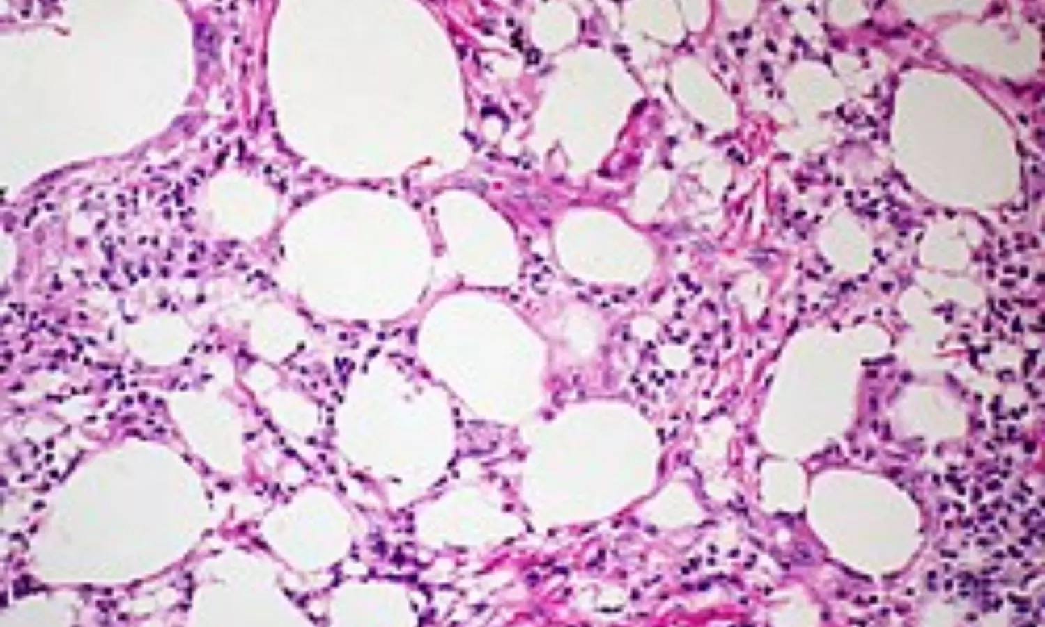
To investigate further, she underwent a thoracoscopic lobectomy of the left lower lung. Pathological examination of the removed tissue revealed chronic inflammatory cell infiltration and alveolar spaces filled with CD163+ foamy histiocytes and proteinaceous exudates, confirming the diagnosis of lipid pneumonia.

One year after the surgery, the patient was asymptomatic and underwent a chest CT scan as part of her postoperative surveillance. This scan revealed new areas of mixed consolidation and GGO in the right middle lung. Ongoing monitoring and an optimal postoperative surveillance strategy are planned for her care.

This case highlights the importance of considering ELP in patients with a history of lung abnormalities, especially when previous malignancies or treatments may contribute to lung complications. Continued research and documentation of such cases will enhance understanding of ELP and inform better management strategies for affected individuals.

“We share our experience managing a rare case of exogenous lipid pneumonia that initially presented as lung cancer. The condition was successfully diagnosed and treated through thoracoscopic lobectomy. In specific clinical situations, CT-guided or transbronchial biopsy may be valuable pre-operative diagnostic tools. Notably, the patient had received cyclophosphamide, an antineoplastic agent, seven years earlier, suggesting a potential link to the development of ELP,” Jiun-Chang Wu, Department of Medicine, MacKay Medical College, New Taipei, Taiwan, and colleagues wrote.

“This case highlights the need for further research to establish effective management guidelines for ELP moving forward,” they concluded.

Reference:

Wu JC, Chen TY, Wen-Chien H, Lee JJ, Chen CH. Lipid Pneumonia Mimicking Lung Cancer in a Middle-Age Woman. Ann Thorac Cardiovasc Surg. 2024;30(1):24-00117. doi: 10.5761/atcs.cr.24-00117. PMID: 39477502; PMCID: PMC11524679.

Powered by WPeMatico