TVS assessment of cervix better predictor of successful labor induction compared to Bishop’s score: Study

Induction of labor refers to stimulation of uterine
contractions after the period of viability, before spontaneous onset of labor,
in cases where the ongoing pregnancy may affect the mother or the fetus
adversely, with the aim of vaginal delivery. The most common indications for
induction of labor are post dated pregnancy, hypertensive disorders of
pregnancy, oligohydramnios, PROM, etc.

It is a common practice in modern obstetrics in view of
various obstetrical or medical indications. Usually, the decision to induce
labor is made after considering the risk and benefits of prolonging the
pregnancy. Successful induction results in vaginal delivery. However, the
process is not completely seamless. Failure of induction can lead to cesarean
section and the associated risks. It is
therefore important to predict the chances of success of induction.

Efforts have been made to predict the rate of success of
induction. Currently the most popular and widely used method is the Bishop’s
score. It is a quantifiable but subjective method. Hence assessment is likely
to vary from observer to observer. So the search for better predictors
continues.

Trans-vaginal sonography (TVS) is an alternative but
objective method emerging for assessing the cervix to predict the success of
induction of labor by reducing interobserver variations. TVS measurements are
quantitative and easy to reproduce, with minimal discomfort to the patient. It
also allows a better evaluation of cervical length, since the supra-vaginal
part of cervix is difficult to measure digitally. It also provides access to
internal os, which cannot be reached in a closed cervix and where the
effacement begins. Various parameters that can be used for cervical evaluation
using TVS are cervical length, cervical funneling, cervical position, posterior
cervical angle, distance of presenting part from external os, uterocervical
angle, etc. Although many studies have been conducted to compare the Bishop’s
score with TVS cervical evaluation, the superiority of one method over the
other has not been clearly defined. In addition, there is a lack of definite
and established cut offs to use TVS assessment in defining the success of
induction. Some researchers have attempted to develop cut offs and scores to
use the TVS assessment. However, these scores are not widely used at present.
This might be because they used the parameters that are not easy to measure.

The aim of this study by Srivastava and Coumary A was to
compare the two methods: Bishop’s score and MGM pre induction cervical scoring
system (MGPICSS), and to develop an easy to use system for using TVS assessment
to predict the successful induction of labor. This was an observational study
conducted in a tertiary care center. 120 patients who met the selection
criteria were included. Prior to the induction of labor the Bishop’s score and
the sonographic scoring was assigned. Successful induction was defined as the patient
entering the active phase of labor.

84% of participating women entered the active phase of
labor. While 72.6% women had a normal vaginal delivery, 67.8% women delivered
vaginally within 24 hours of induction. The TVS score (MGPICSS) of ≥2 predicted
the successful induction with a specificity of 100% and sensitivity of 39.3%
and AUC 0.74. In comparison, the Bishop score of ≥4 had a specificity of 75%
and sensitivity of 44% and AUC 0.56. The prediction of delivery within 24 hours
at the MGPICSS of ≥2 had a specificity of 100% and sensitivity of 42.9% and AUC
0.76. For the same, the Bishop’s score of ≥4 had specificity of 83.3% and
sensitivity of 45.5% and AUC 0.71.

This study compared the Bishop’s score with the TVS
assessment of cervix to predict the success of induction. It was concluded that
the MGPICSS better predicted the successful induction of labor and also the
delivery within 24 hours of the induction. TVS examination also causes less
discomfort to the patients than digital examination.

TVS assessment of cervix is a better predictor of success of
induction in comparison to Bishop’s score. According to the present study,
MGPICSS of ≥2 can predict the successful outcome with specificity of 100% and
sensitivity of 39.3%. Also a score of ≥2 can predict the chances of delivery
within 24 hours with a specificity of 100% and sensitivity of 42.9%. MGPICSS is
doable even by the beginners and has reduced interobserver variations.

Source: Srivastava and Coumary A / Indian Journal of
Obstetrics and Gynecology Research 2024;11(2):276–280

https://doi.org/10.18231/j.ijogr.2024.053

Powered by WPeMatico

Compared to surgical removal of mandibular third molars, Coronectomy significantly impacted postoperative oral health related QoL: Study

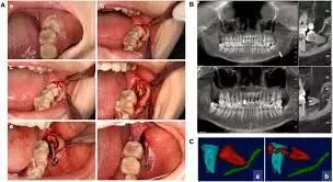
Compared to surgical removal of mandibular third molars, Coronectomy significantly impacted postoperative oral health-related QoL suggests a study published in the Journal of Oral and Maxillofacial Surgery.

Coronectomy is an alternative procedure for removing mandibular third molars near the inferior alveolar nerve. Limited research exists on the effect of coronectomy on the postoperative quality of life (QoL). This study compared postoperative QoL after coronectomy and complete surgical removal of mandibular third molars during the first postoperative week. This prospective cross-over study was conducted in the Oral and Maxillofacial Department of Amstelland Hospital, Amstelveen, The Netherlands. The study sample consisted of patients with indications for removal of both mandibular third molars, with one at increased risk of nerve injury undergoing coronectomy, while the other molar was extracted. Exclusion criteria were ibuprofen allergy, anticoagulant therapy, systemic disease, local pathology, or failure to complete the Oral Health Impact Profile-14 (OHIP-14) questionnaire. Results: The sample included 55 patients (18 male and 37 female) with a mean age of 24.6 ± 4.7 years. Mean OHIP-14 scores during the first 6 postoperative days were significantly higher after coronectomy compared to after surgical removal (day 1: 24.93 ± 9.82 vs 22.7 ± 9.5; day 6: 11.27 ± 10.36 vs 8.49 ± 10.93) (P < .05). Pain was significantly higher on the second to sixth postoperative days after coronectomy (day 2: 6.02 ± 1.92 vs 5.78 ± 1.73; day 6: 4.11 ± 2.49 vs 3.09 ± 2.41) (P < .05). Patients used more analgesics after coronectomy (day 2: 4.09 ± 2.53 vs 3.27 ± 1.9; day 6: 2.76 ± 2.62 vs 2.13 ± 2.49) (P < .05). They found no differences in outcomes for sex or molar impaction (P > .05). Coronectomy significantly impacted postoperative oral health–related QoL compared to complete surgical removal of mandibular third molars.

Reference:

Postoperative Outcome After Coronectomy Versus Surgical Removal of Impacted Mandibular Third Molars – Clinical and Oral Health-Related Quality of Life Follow-Up

Simons, Rashida N. et al. Journal of Oral and Maxillofacial Surgery, Volume 82, Issue 9, 1109 – 1120

Keywords:

Compared, surgical, removal, mandibular, third molars, Coronectomy, significantly, impacted, postoperative, oral health related QoL, Study, Journal of Oral and Maxillofacial Surgery

Powered by WPeMatico

New KECORT Study Offers Guidelines for Consistent Keloid Treatment Using Intralesional Corticosteroids

Netherlands: In a recent development in dermatology, the KECORT Study has produced essential guidelines on keloid treatment using intralesional corticosteroids. The international e-Delphi study brings together experts from various fields to address the complexities of keloid management, a condition known for its challenging nature and high recurrence rates.

This e-Delphi study offers significant clinical treatment recommendations regarding key aspects of intralesional corticosteroid administration (ICA) in keloids. The researchers note that implementing these guidelines is expected to enhance consistency in keloid treatment and improve overall outcomes. The findings were published online in the American Journal of Clinical Dermatology. 

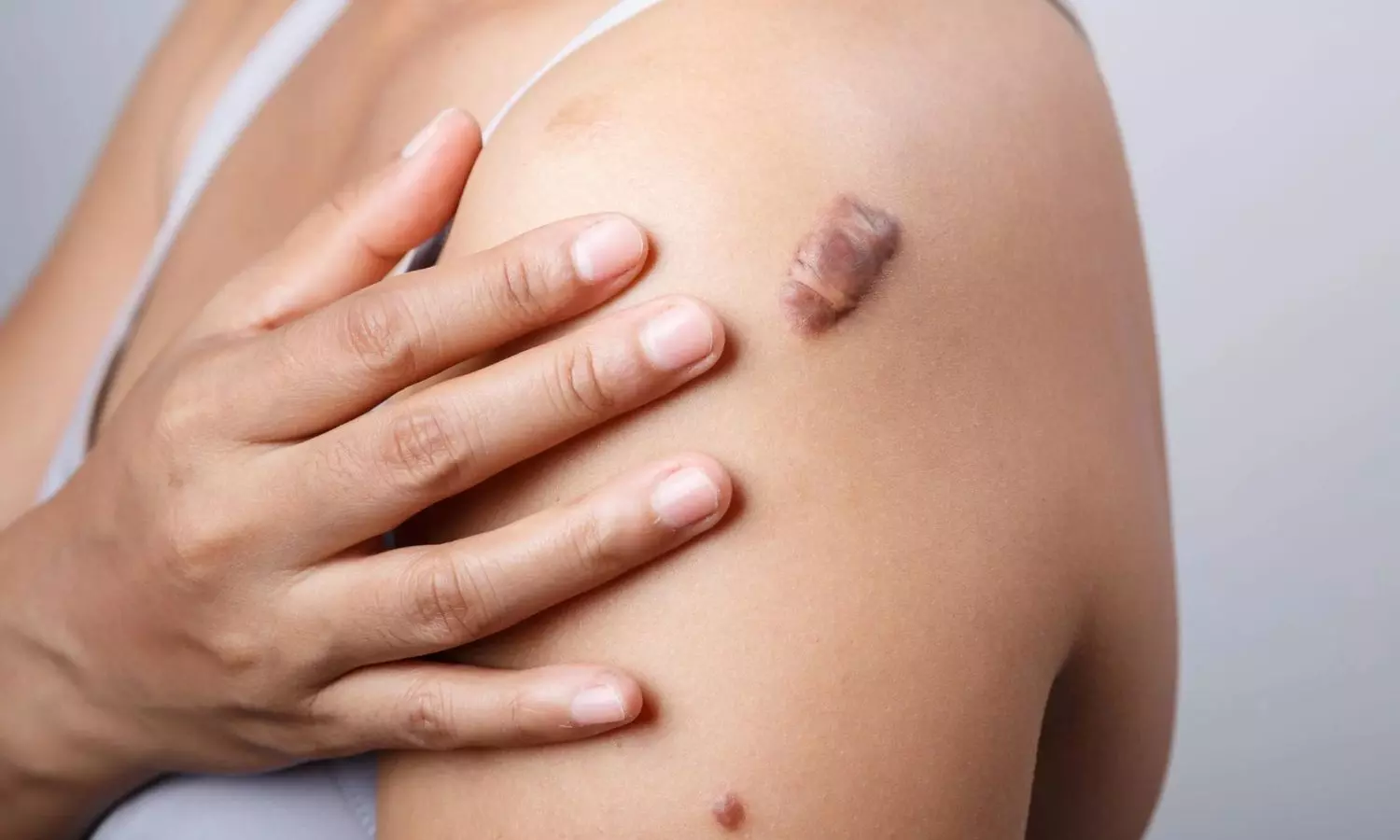
Keloids are raised scars that result from an overproduction of collagen during the healing process. They can cause discomfort and distress for patients, making effective treatment crucial. ICA is the primary treatment for keloids. However, there is considerable variability in its application across clinical and scientific practices, making it difficult to compare treatment outcomes effectively.

To address this issue, Qi Yin, Department of Dermatology, Amsterdam UMC location University of Amsterdam, Amsterdam, The Netherlands, and colleagues sought to reach a consensus on key aspects of intralesional corticosteroid administration using hypodermic needles for keloid treatment. They convened a global panel of dermatologists and plastic surgeons who specialize in this field, to develop consensus-based clinical recommendations for all physicians involved in managing keloids.

For this purpose, a panel of 12 dermatologists and 11 plastic surgeons specializing in keloids rated 30 statements over two online e-Delphi rounds, achieving a 100% response rate in both sessions. A total of 15 keloid experts took part in the final consensus meetings. Consensus was established when at least 75% of participants selected “agree” or “strongly agree” on a 7-point Likert scale.

The following were the key findings:

  • Consensus was achieved on several key treatment aspects, including treatment goals, the indication for intralesional corticosteroid administration, and the preference for triamcinolone acetonide (TAC) at a concentration of 40 mg/mL, with a maximum dosage of 80 mg per month administered at 4-week intervals.
  • Strategies to minimize pain during the procedure, the use of 1 mL syringes with 25 or 27-gauge needles, and blanching as an endpoint for successful infiltration were agreed upon. Participants also noted the importance of avoiding subcutaneous injections and the possibility of making multiple passes in very firm keloids before infiltration.
  • Consensus was not reached on TAC dosing, methods of prior local anesthesia, or the specific location of injection.

To conclude, this e-Delphi consensus study offers evidence-based clinical treatment recommendations for intralesional corticosteroid administration in keloids, targeting all physicians involved in keloid management. Key aspects covered include treatment goals, indications for ICA, choice of corticosteroid, dosing guidelines, treatment intervals, syringe and needle specifications, local anesthesia use, and manual injection techniques.

“Implementing these recommendations is expected to enhance consistency in ICA practices for keloid treatment, making outcomes more comparable and potentially improving overall treatment results,” the researchers concluded.

Reference:

Yin, Q., Wolkerstorfer, A., Lapid, O. et al. KECORT Study: An International e-Delphi Study on the Treatment of KEloids Using Intralesional CORTicosteroids in Clinical Practice. Am J Clin Dermatol (2024). https://doi.org/10.1007/s40257-024-00888-7

Powered by WPeMatico

Study Links Severe Atopic Dermatitis to Increased Risk of Glaucoma Surgery

Taiwan: A recent global population-based study has highlighted a significant association between severe atopic dermatitis (AD) and an increased risk of requiring glaucoma surgery. The findings, published in the Journal of Glaucoma, suggest that individuals with both conditions, particularly those with more severe forms of AD, may face a higher likelihood of needing surgical intervention to manage glaucoma.

Atopic dermatitis is a chronic inflammatory skin condition that causes itching, redness, and skin lesions. While it primarily affects the skin, recent research has explored its broader impacts on other health conditions, including its potential relationship with ocular diseases like glaucoma. Glaucoma, a leading cause of blindness worldwide, is characterized by damage to the optic nerve, often caused by increased intraocular pressure. It requires long-term management, including medical and sometimes surgical interventions.

The effect of atopic dermatitis on the prognosis of glaucoma patients has been rarely explored. To address this gap, Shu-Chun Kuo, MD, from the Department of Ophthalmology at Chi-Mei Medical Center in Tainan, Taiwan, and colleagues aimed to evaluate the risk of requiring glaucoma surgery in patients with glaucoma, comparing those with and without AD.

For this purpose, the researchers conducted a retrospective cohort analysis using the TriNetX database, assessing patients with glaucoma who were initially diagnosed between December 5, 2003, and December 3, 2018. The patients were divided into two groups: those with atopic dermatitis and those without. To ensure balanced baseline characteristics and comorbidities, the researchers performed 1:1 propensity score matching.

The study compared the risk and cumulative incidence of requiring glaucoma surgery—such as minimally invasive surgery, trabeculectomy, aqueous shunt, or transscleral cyclophotocoagulation—between cohorts. A subgroup analysis was also conducted for patients with severe AD.

The study revealed the following findings:

  • Out of 528,469 patients with glaucoma, 2,624 were in the atopic dermatitis group.
  • Among the AD group, 584 patients had severe AD.
  • The AD group showed a comparable risk of requiring glaucoma surgery to the non-AD group, with a hazard ratio of 1.03.
  • In contrast, the severe AD group demonstrated a significantly higher risk and cumulative incidence of requiring surgery, with a hazard ratio of 2.80 compared to the non-AD group.

“The findings showed that patients with glaucoma and severe atopic dermatitis are significantly more likely to require surgical intervention, with the severity of AD serving as a key factor contributing to the increased risk,” the researchers concluded.

Reference:

Chu, Yung-Yu MD*; Lee, Chia-Yi MD*; Huang, Wei-Yu MD*; Lin, Ju-Kuo MD*; Liu, Ching-Chih MD*; Lin, Hsing-Ying MD*; Ho, Chung-Han PhD†,‡; Chen, Yi-Chen MS†; Kuo, Shu-Chun MD*,§. Association of Atopic Dermatitis and Risk of Glaucoma Surgery: A Global Population-Based Study. Journal of Glaucoma 33(10):p 735-741, October 2024. | DOI: 10.1097/IJG.0000000000002464

Powered by WPeMatico

Sibeprenlimab Reduces Protein Levels in Adults with IgA Nephropathy shows Phase 3 VISIONARY study

USA: An interim analysis of the Phase 3 VISIONARY study revealed that sibeprenlimab achieved a significant and clinically meaningful reduction in the 24-hour urine protein-to-creatine ratio (uPCR) in adults with immunoglobulin A (IgA) nephropathy, successfully meeting its primary endpoint. 

The Otsuka Pharmaceutical Development & Commercialization, Inc. and Otsuka Pharmaceutical Co., Ltd. announced promising interim results from their ongoing Phase 3 trial of sibeprenlimab, an investigational treatment for immunoglobulin A nephropathy (IgA nephropathy) in adults. This announcement comes as a significant milestone in the search for effective therapies for this progressive kidney disease, which can lead to end-stage kidney disease (ESKD).

Sibeprenlimab is a monoclonal antibody that targets APRIL (A PRoliferation-Inducing Ligand), playing a crucial role in regulating the immune response that contributes to IgA nephropathy. By inhibiting the production of Gd-IgA1 and the formation of harmful immune complexes, sibeprenlimab aims to mitigate the disease’s progression. The treatment has already gained Breakthrough Therapy designation following encouraging results from the Phase 2 ENVISION clinical trial.

The interim analysis, conducted by an independent data monitoring committee, revealed that the Phase 3 VISIONARY study met its primary endpoint. Results showed that sibeprenlimab led to a statistically significant reduction in the 24-hour urine protein-to-creatine ratio compared to placebo after nine months of treatment. This multicenter, randomized, double-blind, placebo-controlled trial involved approximately 530 adult patients receiving standard care, making it the largest study in this patient population.

Dr. John Kraus, executive vice president and chief medical officer of Otsuka Pharmaceutical Development & Commercialization, remarked on the findings in a press release: “The positive interim data from this trial suggest that by targeting APRIL, we could provide a new therapeutic strategy for people living with this progressive kidney disease.” He expressed gratitude to the participants, caregivers, and investigators involved in the study.

Brian Pereira, CEO of Visterra, Inc., an Otsuka U.S. affiliate, also commented on the significance of the results, noting sibeprenlimab’s potential to offer a much-needed, disease-modifying treatment option for IgA nephropathy patients.

The ongoing Phase 3 study continues to assess kidney function changes over 24 months, using an estimated glomerular filtration rate (eGFR). The study is expected to conclude in early 2026, with further analyses planned to fully understand sibeprenlimab’s therapeutic potential. Otsuka aims to review these interim results with the FDA to facilitate a possible regulatory submission for accelerated approval.

About Sibeprenlimab

Sibeprenlimab (formerly VIS649) is an investigational monoclonal antibody developed by Visterra, Inc., a subsidiary of Otsuka. It targets APRIL (A PRoliferation-Inducing Ligand), a key player in the pathogenic cascade of IgA nephropathy. By blocking APRIL, sibeprenlimab aims to reduce levels of immunoglobulin A (IgA) and Gd-IgA1, potentially lowering auto-antibody production and immune complexes. This reduction may lead to fewer immune complex deposits in the kidneys, decreasing proteinuria and inflammation.

The drug’s mechanism could slow kidney damage and progression toward ESKD by addressing a specific driver of nephron loss in IgA nephropathy. Early-stage trials have shown promise, highlighting sibeprenlimab’s potential to provide a new therapeutic option for patients with this challenging condition.

Powered by WPeMatico

PG Medical Seat Scam: ED Issues Notice to Former Minister Malla Reddy

Hyderabad: While probing the allegations of illegal sale of postgraduate medical seats, the Enforcement Directorate (ED) has now issued notices to former Minister Malla Reddy.

Recently, the ED also questioned an administrative officer at Malla Reddy Institute of Medical Sciences, Surender Reddy, and recorded his statement as part of an ongoing investigation.

Medical Dialogues had earlier reported that investigating alleged seat-blocking fraud and irregularities in the financial transactions, ED officials had conducted raids on 12 medical colleges in Telangana.

Allegedly, some medical colleges, which are affiliated with Kaloji Narayana Rao University of Health Sciences (KNRUHS), sold the seats for higher prices by blocking them in a dubious manner last year.

Also Read: Seat Blocking Scam in Telangana: ED Raids 12 Medical Colleges to Investigate Financial Irregularities

The ED officials began simultaneous searches in the morning at multiple locations in Hyderabad, Karimnagar, Warangal, Mahabunagar and other places. Searches were on at Malla Reddy Medical College at Suraram on the outskirts of Hyderabad. The college is run by Malla Reddy group of institutions owned by Telangana labour minister Malla Reddy.

As per the latest media report by The Hans India, these 12 private medical colleges were linked to Minister Reddy and back then, ED had seized crucial documents, pen drives, hard disks, and other electronic evidence. Now, the Directorate has issued notices to the former Minister in connection with these alleged PG medical seat scam.

Hans India has reported that ED suspects that Minister Reddy, along with others, was involved in the illegal sale of PG medical seats and manipulation of seat allotments, potentially violating various norms. Reportedly, the raids were carried out in June following a tip-off and the authorities have claimed that significant evidence has been gathered to support these allegations. Malla Reddy served as Telangana Minister for Labour and Employment before his term ended.

Meanwhile, ED on Thursday questioned Surender Reddy, an administrative officer at Malla Reddy Institute of Medical Sciences. Reddy appeared after ED summoned him in connection to the probe regarding the alleged financial irregularities linked to former Minister Ch Malla Reddy’s institute.

The investigation primarily focuses on potential irregularities related to PG medical admissions, including the alleged misuse of student credentials in a seat-blocking scheme.

The Times of India has reported that sources indicated that ED is examining records and testimonies to trace the flow of funds and to unearth further details. In June 2023, ED had seized Rs 1.4 crore in cash from Malla Reddy Institute and froze assets worth 3 crore as part of its investigation under the Prevention of Money Laundering Act (PMLA). So far, the investigation by ED has uncovered evidence, including documents and digital records and referred to cash transactions and collection of premiums from students.

Also Read: ED officials conduct searches on 12 medical colleges in Telangana

Powered by WPeMatico

Bihar: Pregnant woman dies after quack performs C-section watching YouTube video

Begusarai: In a shocking case of medical fraud, a 30-year-old pregnant woman recently lost her life due to excessive bleeding after a quack allegedly performed C-section on her while taking guidance from a YouTube video at his clinic in the Begusarai district of Bihar. 

According to the sister of the deceased, the patient was kept in the operating room for 24 hours, during which the unqualified practitioner used a video tutorial to perform the surgery. She alleged that the patient’s condition worsened severely after the procedure, ultimately leading to her death.

Also read- Quack held for duping elderly woman of Rs 7.20 lakh with Fake Knee Surgery

It was later revealed that the doctor performing the surgery was a quack with no medical qualifications or experience who was running the clinic in the area. He allegedly targeted poor families by claiming to be a qualified physician.

The incident occurred after the patient from Arjunatol village in Cheria Bariarpur block was admitted to the clinic on November 2 with labour pain. As per a Hindustan news report, she underwent the surgery on the recommendation of the quack and gave birth to a girl. However, she died on the evening of November 5 due to excessive bleeding.

After the patient’s death, the family members created a ruckus in the clinic accusing the quack of negligence in treatment. Following this, the fake doctor, nurse and clinic staff reportedly fled from the spot.

The patient’s sister told Lokmat Times, “The doctor was performing the surgery after watching a video on YouTube. The operation had been going on for the past 24 hours. When my sister’s condition deteriorated and she died, the doctor fled on a bike.”

A police action regarding this incident remains pending. 

Also read- 16-year-old dies after quack administers injections for fever, Telangana Medical Council takes suo moto action

Powered by WPeMatico

Fluorescence-guided imaging technique could improve head and neck cancer surgery

Head and neck squamous cell carcinoma (HNSCC) ranks as one of the most common cancers globally, with over 650,000 new cases reported each year. Surgical intervention is often the primary treatment, but surgeons face a difficult challenge: they must completely remove the cancer while preserving as much surrounding healthy tissue as possible. This balance is crucial, as damage to nearby nerves can lead to significant post-surgical complications, affecting patients’ quality of life.

Powered by WPeMatico

Metabolic surgery may help reduce heart failure-related risk factors

Pennington Biomedical Research Center researchers at the Metamor Institute, along with colleagues from Our Lady of the Lake and LSU Health-New Orleans, have recently determined that metabolic surgery on patients with heart failure can result in a reduction in the need for oral diuretics, which are used to manage symptoms such as venous and vascular pressures.

Powered by WPeMatico

Biomarker identified for predicting inflammatory bowel disease treatment success

Not everyone responds equally well to treatments for inflammatory bowel disease (IBD). What will work for individual patients involves trial and error during the treatment process. Now, a team of researchers led by Charité—Universitätsmedizin, in collaboration with colleagues in Berlin and Bonn, has succeeded in identifying a biomarker that indicates whether or not treatment with a certain medication called an immunomodulator will be successful.

Powered by WPeMatico