Using Diaphragm Position on Chest Radiograph for Assessing Lung Volume in Infants not accurate and precise: JAMA
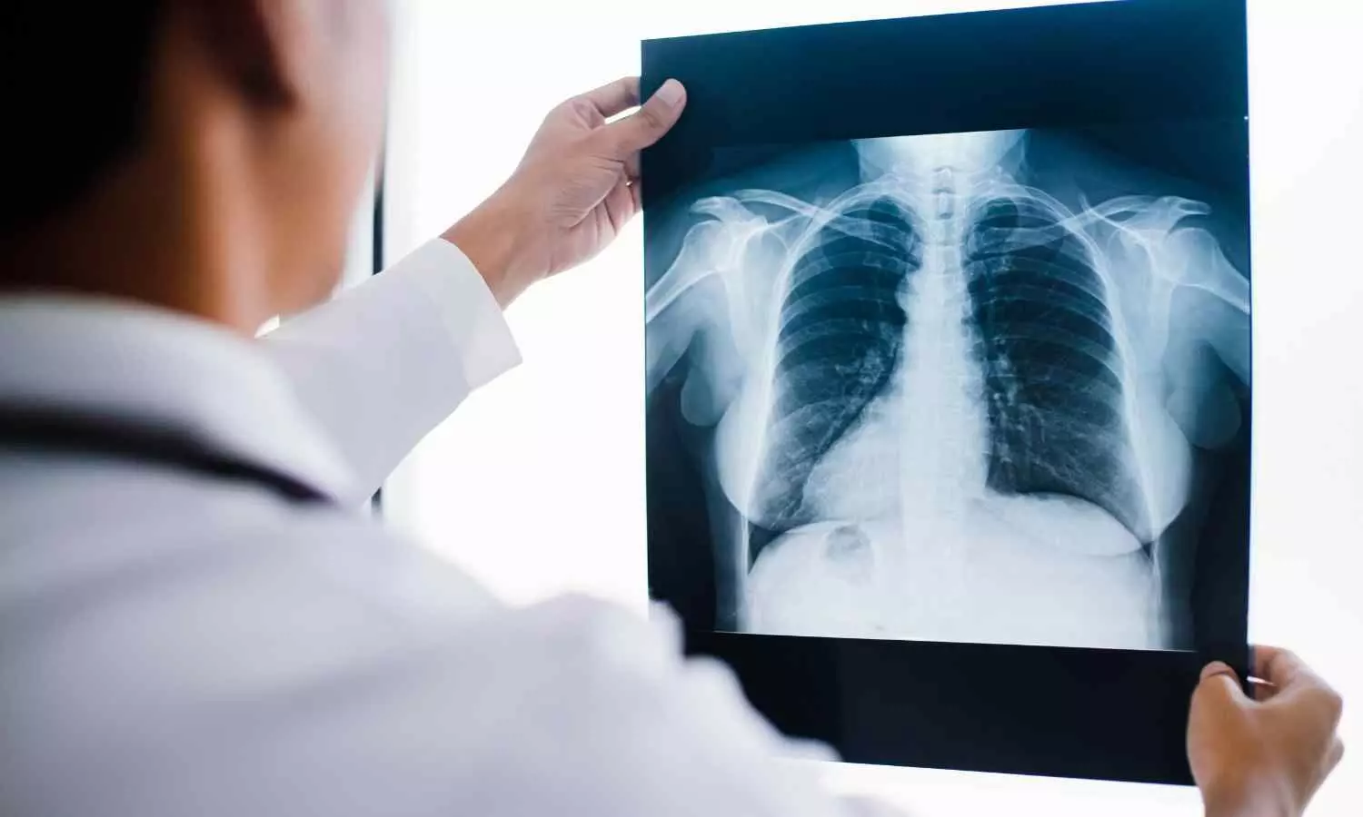
A new study published in the Journal of American Medical Association revealed that the use of diaphragm position on chest radiographs lacked the precision needed to accurately assess aerated lung volume and guide clinical decisions in infants in neonatal intensive care unit (NICU). Therefore it will be worthwhile to use a combination of clinical and diagnostic radiographic features to assess lung volume rather than diaphragm position alone.

Although it is standard procedure and advised by NICU guidelines, using chest radiographs to direct lung aeration during respiratory support in neonates has never been proven to be effective. Thus, this study was set to characterize the relationship between the infant’s diaphragm position on a chest radiograph and the computed tomography (CT) measurement of aerated lung capacity.

The Royal Children’s Hospital in Melbourne, Australia, was the site of this retrospective cross-sectional study. Infants without congenital lung pathology who had a chest CT scan within 30 days of delivery, from July 9, 2012, to December 31, 2022, were included. Analysis of the study’s data took place between December 2022 and September 2023.

CT semiautomated tissue segmentation was used to quantify lung volume, and a consistent definition was used to establish diaphragm position. The measures from the chest radiograph were unknown to all of the investigators who were examining the CT scans, and vice versa. The distribution and accuracy of the total lung volume at each of the diaphragm positions (6th-11th posterior rib) were the main results.

A total of 218 newborns (mean [SD] age, 37.9 [1.9] weeks gestation at birth; 119 male [55%]; median [IQR] age, 11 [3-20] days) had their imaging data examined. A primary cardiac diagnosis was made for 132 (61%) of the infants, whose mean (SD) weight at scan was 3055 (584) g. The diaphragm location was represented by 6 to 11 posterior ribs. Lung volume and diaphragm position were only weakly correlated (Kendall τ = 0.23; 95% CI, 0.16–0.31).

The degree of consolidation (Kendall τ = 0.30; 95% CI, 0.21-0.38), apex-diaphragm distance (Kendall τ = 0.40; 95% CI, 0.28-0.51), hemithorax (left, Kendall τ = 0.25; 95% CI, 0.15-0.34; right, Kendall τ = 0.21; 95% CI, 0.10-0.31), and Hounsfield unit values (Kendall τ = −0.05; 95% CI, −0.15 to −0.06) all showed a similar weak association. Overall, in response to these findings, the diaphragm position as determined by the number of posterior ribs on a chest radiograph may not be accurate enough for practical use as a surrogate of lung volume, even though it has long been clinically accepted.

Source:

Dahm, S. I., Sett, A., Gunn, E. F., Ramanauskas, F., Hall, R., Stewart, D., Koeppenkastrop, S., McKenna, K., Gardiner, R. E., Rao, P., & Tingay, D. G. (2025). Diaphragm position on chest radiograph to estimate lung volume in neonates. JAMA Pediatrics. https://doi.org/10.1001/jamapediatrics.2025.2108

Powered by WPeMatico

Mayo Clinic’s AI tool identifies 9 dementia types, including Alzheimer’s, with one scan

Mayo Clinic researchers have developed a new artificial intelligence (AI) tool that helps clinicians identify brain activity patterns linked to nine types of dementia, including Alzheimer’s disease, using a single, widely available scan-a transformative advance in early, accurate diagnosis.

The tool, StateViewer, helped researchers identify the dementia type in 88% of cases, according to research published online on June 27, 2025, in Neurology, the medical journal of the American Academy of Neurology. It also enabled clinicians to interpret brain scans nearly twice as fast and with up to three times greater accuracy than standard workflows. Researchers trained and tested the AI on more than 3,600 scans, including images from patients with dementia and people without cognitive impairment.

This innovation addresses a core challenge in dementia care: identifying the disease early and precisely, even when multiple conditions are present. As new treatments emerge, timely diagnosis helps match patients with the most appropriate care when it can have the greatest impact. The tool could bring advanced diagnostic support to clinics that lack neurology expertise.

The rising toll of dementia

Dementia affects more than 55 million people worldwide, with nearly 10 million new cases each year. Alzheimer’s disease, the most common form, is now the fifth-leading cause of death globally. Diagnosing dementia typically requires cognitive tests, blood draws, imaging, clinical interviews and specialist referrals. Even with extensive testing, distinguishing conditions such as Alzheimer’s, Lewy body dementia and frontotemporal dementia remains challenging, including for highly experienced specialists.

StateViewer was developed under the direction of David Jones, M.D., a Mayo Clinic neurologist and director of the Mayo Clinic Neurology Artificial Intelligence Program.

“Every patient who walks into my clinic carries a unique story shaped by the brain’s complexity,” Dr. Jones says. “That complexity drew me to neurology and continues to drive my commitment to clearer answers. StateViewer reflects that commitment — a step toward earlier understanding, more precise treatment and, one day, changing the course of these diseases.”

To bring that vision to life, Dr. Jones worked alongside Leland Barnard, Ph.D., a data scientist who leads the AI engineering behind StateViewer.

“As we were designing StateViewer, we never lost sight of the fact that behind every data point and brain scan was a person facing a difficult diagnosis and urgent questions,” Dr. Barnard says. “Seeing how this tool could assist physicians with real-time, precise insights and guidance highlights the potential of machine learning for clinical medicine.”

Turning brain patterns into clinical insight

The tool analyzes a fluorodeoxyglucose positron emission tomography (FDG-PET) scan, which shows how the brain uses glucose for energy. It then compares the scan to a large database of scans from people with confirmed dementia diagnoses and identifies patterns that match specific types, or combinations, of dementia.

Alzheimer’s typically affects memory and processing regions, Lewy body dementia involves areas tied to attention and movement, and frontotemporal dementia alters regions responsible for language and behavior. StateViewer displays these patterns through color-coded brain maps that highlight key areas of brain activity, giving all clinicians, even those without neurology training, a visual explanation of what the AI sees and how it supports the diagnosis.

Mayo Clinic researchers plan to expand the tool’s use and will continue evaluating its performance in a variety of clinical settings. 

Reference:

Leland Barnard, Hugo Botha, Nick Corriveau-Lecavalier, An FDG-PET–Based Machine Learning Framework to Support Neurologic Decision-Making in Alzheimer Disease and Related Disorders, Neurology, https://doi.org/10.1212/WNL.0000000000213831.

Powered by WPeMatico

Air pollution linked to adverse birth outcomes, reveals Indian study

Prenatal exposure to ambient fine particulate matter and climatic factors, such as temperature and rainfall, are associated with adverse birth outcomes in India, according to a study published July 2nd, 2025, in the open-access journal PLOS Global Public Health by Mary Abed Al Ahad from the University of St Andrews, U.K.

Ambient air pollution poses a global threat to human health, with a disproportionate burden of its detrimental effects falling on those residing in low and middle-income countries. Referred to as the silent killer, ambient air pollution is among the top five risk factors for mortality in both males and females. With a diameter of less than 2.5 microns, ambient fine particulate matter 2.5 (PM2.5), which primarily originates from the burning of fossil fuels and biomass, is considered the most harmful air pollutant. In the 2023 World Air Quality Report, India was ranked as the third most polluted country out of 134 nations based on its average yearly PM2.5 levels.

Ambient air pollution has been associated with a range of pediatric morbidities, including adverse birth outcomes, asthma, cancer, and an increased risk of chronic diseases. Most studies investigating the association between ambient air pollution and adverse birth outcomes have primarily been conducted in high-income countries. Despite the alarming rise in air pollution levels in India, there has been a paucity of research exploring its impact on adverse birth outcomes.

To address this knowledge gap, the researchers investigated the impact of ambient air pollution on adverse birth outcomes at the national level, focusing on low birth weight and preterm birth, and used different geospatial models to highlight vulnerable areas. The analysis provided evidence of the association between in-utero exposure to PM2.5 and adverse birth outcomes by leveraging satellite data and large-scale survey data. The individual-level analysis revealed that an increase in ambient PM2.5 is associated with a greater likelihood of low birth weight and preterm birth. Climatic factors such as rainfall and temperature were also linked to adverse birth outcomes. Children residing in the Northern districts of India appeared to be more susceptible to the adverse effects of ambient air pollution.

According to the authors, the geostatistical analysis underscores the need for targeted interventions, particularly in Northern districts. In addition, the National Clean Air Program should be intensified, with stricter emission standards and enhanced air quality monitoring. Climate adaptation strategies, such as developing heat action plans and improving water management, should be incorporated into public health planning to mitigate the effects of extreme temperatures and irregular rainfall. Public health initiatives should be implemented to raise awareness of the risks of air pollution and climate change, particularly among pregnant women.  

Reference:

Arup Jana, Malay Pramanik, In-utero exposure to PM2.5 and adverse birth outcomes in India: Geostatistical modelling using remote sensing and demographic health survey data 2019–21, PLOS Global Public Health, https://doi.org/10.1371/journal.pgph.0003798.

Powered by WPeMatico

Patients continue to lose weight in the years after ‘tummy tuck’: Study

Most patients undergoing “tummy tuck” surgery (abdominoplasty) to remove excess skin and tissue after weight loss continue to lose weight in the months and years after surgery, suggests a follow-up study in the July issue of Plastic and Reconstructive Surgery®, the official medical journal of the American Society of Plastic Surgeons (ASPS). The journal is published in the Lippincott portfolio by Wolters Kluwer.

“We found that patients not only maintained their weight loss after abdominoplasty, but also continued to lose weight over time – up to ten pounds, on average,” comments senior author John Y.S. Kim of Northwestern University Feinberg School of Medicine, Chicago. “This postoperative weight loss appears greater, and increases at later follow-up times, in patients with initially higher body mass index [BMI].”

Continued weight loss up to five years after tummy tuck

Abdominoplasty is a cosmetic surgical procedure to improve the appearance of the abdomen. In 2023, ASPS Member Surgeons performed more than 170,000 abdominoplasties, according to ASPS statistics. Many of these procedures are performed in patients with massive weight loss that leaves them with excess, sagging skin.

Plastic surgeons have observed that patients may continue to lose weight after abdominoplasty. However, there is little research evidence on this issue, including whether the abdominoplasty procedure itself contributes to long-term weight loss.

Dr. Kim and colleagues performed a study to assess changes in body weight in 188 patients who underwent abdominoplasty between 2018 and 2022. Ninety-seven percent of patients were women. The average preoperative weight was about 168 pounds with a BMI of 27.7. Most patients underwent liposuction or a further procedure to remove excess fat (lipectomy) at the same time as abdominoplasty. Trends in body weight were assessed through up to five years after surgery.

The results showed continued weight loss after abdominoplasty. At three to six months, average weight loss was between five and six pounds, with about a three percent decrease in BMI. From one to four years, weight loss was about five pounds, for a BMI reduction of about two percent. By five years (in a limited number of patients), average weight loss was nearly ten pounds, with more than a five percent decrease in BMI.

‘Near-constant negative change in body weight’ after abdominoplasty

Overall, about 60% of patients lost weight during follow-up. Further analysis showed a “near constant negative change in body weight that did not significantly change over time,” the researchers write.

After adjustment for other factors, continued weight loss was more likely for older patients, for those who underwent liposuction/lipectomy, and those who had never smoked. Weight loss was greater for patients who had higher body weight and BMI before surgery, and for a small number of patients who used the newer weight loss medication semaglutide.

The study adds new evidence that “post-abdominoplasty weight reduction is a quantifiable phenomenon and that patients undergoing abdominoplasty continue to lose a significant amount of weight for up to five years after surgery,” the researchers write. They note some key limitations of their study, including varying follow-up times and potential confounding factors.

The study cannot definitively explain why patients continue to lose weight after surgery. However, Dr. Kim and coauthors write, “We have found that patients who were able to achieve weight loss after their abdominoplasty succeeded in developing healthy habits that centered around nutrition and exercise.” They highlight the need for an “evidence-based platform” to assess weight changes after abdominoplasty and to identify factors associated with long-term weight loss.

Reference:

Bricker, Jonathan MD; Ferenz, Sarah BA; Moradian, Simon MD; Termanini, Kareem MD; Jackson, Brandon MD; Kim, John Y. S. MD. What Happens to Weight following Abdominoplasty: An Analysis of 188 Consecutive Cases. Plastic and Reconstructive Surgery 156(1):p 51e-58e, July 2025. | DOI: 10.1097/PRS.0000000000011959

Powered by WPeMatico

Psychedelics and cannabis offer treatment hope for people with eating disorders: JAMA

A pioneering international survey of people living with eating disorders has found that cannabis and psychedelics, such as ‘magic mushrooms’ or LSD, were best rated as alleviating symptoms by respondents who self-medicated with the non-prescribed drugs.

The worst-rated drugs were alcohol, tobacco, nicotine and cocaine.

Prescribed drugs, such as antidepressants, were generally not well rated for treating eating-disorder symptoms but were positively rated for effects on general mental health.

The research, led by PhD student Sarah-Catherine Rodan at the University of Sydney’s Lambert Initiative for Cannabinoid Therapeutics, is published today in JAMA Network Open.

Ms Rodan said: “Our results provide important insights into the lived experiences of people with eating disorders and their drug use, highlighting promising avenues for future research into treatments.

“The findings suggest more research, including large clinical trials, should be undertaken around the beneficial effects of cannabis and psychedelics for people with eating disorders.”

The Lambert Initiative researchers will shortly launch a clinical trial of psilocybin in treating anorexia nervosa in collaboration with the Inside Out Institute at the University of Sydney.

Scope and response of survey

The study analysed responses from over 7600 self-allocated participants in 83 countries, making it the most comprehensive survey ever conducted on this topic.

The research focused on how people with different types of eating disorders use prescription and non-prescription drugs, and how they perceive these substances’ effects on their mental health and eating disorder symptoms.

The major categories of eating disorders were well-represented in the survey: anorexia nervosa (40%); bulimia nervosa (19%); binge-eating disorder (11%); and avoidant/restrictive food intake disorder (ARFID) (9%). About one third of respondents were not formally diagnosed with an eating disorder but self-reported an eating disorder that caused distress.

Co-morbid mental health conditions, which are often found in these populations, were frequently reported including depression (65%), generalised anxiety disorder (55%), ADHD (33%), drug dependence (15%) and alcohol dependence (9%).

Respondents were predominantly female (94%), with most from English-speaking places, like Australia (30%), the UK (21.3%) and the US (18%).

The results revealed patients with eating disorders have high rates of cannabis and psychedelic use relative to the general populations and rate their effects positively in terms of managing symptoms. Notably, cannabis was highly rated by respondents with restrictive eating disorders such as anorexia and ARFID, most likely because it enhances the rewarding value of food, addressing a core issue in these eating disorders.

In contrast, prescription stimulants such as lisdexamfetamine, which have strong appetite suppressing effects and are sometimes prescribed for binge eating disorder (BED), were positively rated by people with BED but poorly rated by those with restrictive type disorders.

Psychedelics, typically only taken once or twice a year by respondents, were reported to have remarkable long-lasting benefits, supporting recent research showing their therapeutic potential in treating conditions such as depression and anxiety. Conversely, commonly prescribed medications – such as antidepressants – which are typically taken daily, were generally rated as relatively ineffective for reducing ED symptoms but were widely acknowledged to help with overall mental health.

The survey also found that drugs like alcohol, nicotine, and cocaine, although quite widely used, led to negative outcomes on eating disorder symptoms and general mental health.

Ms Rodan said: “These findings highlight an important pattern: with traditional medications often falling short in treating eating disorders directly, while many individuals are self-medicating with substances they perceive as helpful. This underlines the urgent need to better investigate these substances in rigorously controlled clinical trials.”

Next steps: clinical trials

The insights gained by this study have already prompted further research initiatives. The Lambert Initiative, in collaboration with the Inside Out Institute at the University of Sydney, is preparing to launch a clinical trial of psilocybin in treating anorexia nervosa. Additionally, a pilot study examining the therapeutic potential of the non-intoxicating cannabis component, cannabidiol (CBD), in treating severe anorexia in young people, is nearing completion.

Professor Iain McGregor, the senior author on this paper and Academic Director of the Lambert Initiative, said: “This research suggests that cannabis and psychedelics hold significant promise for improving quality of life in individuals suffering eating disorders. This is particularly salient since current pharmacological options for these patients are severely limited and current treatment outcomes so disappointing.

“Of course, rigorous clinical trials are needed to confirm these benefits and better determine safety profiles.”

Ms Rodan said: “I hope this study gives a voice to people living with eating disorders, revealing that their often-stigmatised experiences with drugs might in fact have therapeutic potential. We are extremely grateful to the many thousands of respondents who took the time to provide such detailed responses around their lived experiences. This should spur further research and open new treatment pathways for these challenging conditions.”

Reference:

Rodan S, Maguire S, Meez N, et al. Prescription and Nonprescription Drug Use Among People With Eating Disorders. JAMA Netw Open. 2025;8(7):e2522406. doi:10.1001/jamanetworkopen.2025.22406.

Powered by WPeMatico

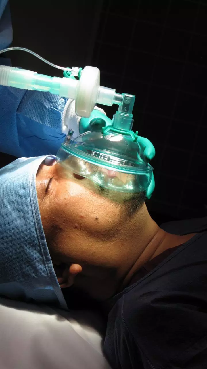
Cuddling the Circuit: How Circuit Insulation Enhances Gas Delivery in Anesthesia, study finds

Recent study explores the effect of breathing circuit insulation on the conditioning of inspired gases and the condensation of water vapor during general anesthesia, utilizing an in vitro mechanical lung model. Insufficient temperature and humidity of inspired gases may lead to mucus membrane dehydration, ciliary dysfunction, retention of secretions, and potential atelectasis, compromising postoperative pulmonary function. The methodology involved comparing three types of insulation (foam, cotton, and polyester) against non-insulated tubing (control), with measurements taken for temperature, absolute humidity (AH), and water vapor condensation after 120 minutes. The analysis employed Bonferroni-Holm adjustments to account for multiple testing.

Results of Insulation Performance

Results indicated enhanced performance of foam insulation, which resulted in higher temperatures and AH compared to the control and the other insulation types. Specifically, foam insulation elevated the mean temperature significantly (P < 0.001), achieving a mean difference of 1.07 °C from the control, while the cotton and polyester insulations also provided temperature elevations but fell short of the target range of 28-32 °C optimal for perioperative ventilation. Despite all insulation types improving AH levels significantly over the control, none reached the target levels, although they effectively exceeded 20 g•m-3 H2O.

Water Vapor Condensation Analysis

Water vapor condensation was markedly reduced in insulated circuits, with foam insulation demonstrating the lowest condensation rate at 1.59 mL•h-1, compared to 2.95 mL•h-1 in the control and 2.26 and 2.32 mL•h-1 in cotton and polyester, respectively. A strong negative correlation was observed between the amount of water vapor condensation and temperature increases.

Implications and Future Research

The study emphasizes the necessity of achieving adequate conditioning of inspired gases to mitigate complications arising from dry and cold gases during anesthesia. Limitations included the technical constraints of the mechanical lung model, such as a lack of physiological conditions, which may affect the external validity of the findings. Future research, ideally in vivo, is warranted to further investigate the clinical implications of insulation types concerning inspired gas conditioning in real-world anesthesia contexts.

Conclusions and Recommendations

Overall, the findings support the potential application of insulated breathing circuits as a method to enhance the conditioning of inspired gases under low-flow anesthesia, although achieving optimal clinical standards remains a challenge. The results advocate for foam insulation as the most effective option, encouraging further investigation into its practical applications in anesthetic practice.

Key Points

– The study evaluates the role of different insulation materials (foam, cotton, polyester, and control) on the conditioning of inspired gases during general anesthesia using an in vitro mechanical lung model, focusing on temperature and absolute humidity levels to prevent complications such as mucus membrane dehydration and postoperative pulmonary dysfunction.

– Foam insulation significantly outperformed the control and other materials, achieving a mean temperature increase of 1.07 °C (P < 0.001) but still failed to reach the optimal perioperative range of 28-32 °C; all insulation types improved absolute humidity but did not attain the ideal threshold, remaining above 20 g•m-3 H2O.

– Water vapor condensation rates were significantly lower in insulated circuits, with foam insulation recording the most effective performance at 1.59 mL•h-1, followed by cotton (2.26 mL•h-1) and polyester (2.32 mL•h-1), while the control exhibited the highest condensation rate at 2.95 mL•h-1; a negative correlation was noted between condensation rates and temperature increases.

– The investigation highlights the importance of proper conditioning of inspired gases to reduce the risk of adverse effects associated with poor thermal and humidity management in anesthetic practices, indicating a potential clinical significance for insulated breathing circuits.

– Limitations of the study were acknowledged, particularly the artificiality of the mechanical lung model, which may not fully replicate physiological conditions, suggesting that the external validity of the findings could be constrained.

– Future studies, ideally conducted in vivo, are proposed to further explore the clinical relevance of different insulation types in achieving optimal inspired gas conditioning in anesthesia, particularly focusing on the practical implementation of foam insulation in clinical settings.

Reference –

Nguyen-Minh, T., Hönemann, C., Zarbock, A. et al. Effects of breathing circuit insulation on inspired gas conditioning and water vapour condensation: an in vitro study. Can J Anesth/J Can Anesth 72, 780–790 (2025). https://doi.org/10.1007/s12630-025-02959-7

Powered by WPeMatico

Young Breast Cancer Patients Have Low Risk of Isolated Locoregional Recurrence After Surgery: JAMA

USA: A large retrospective study found that young women diagnosed with breast cancer have a relatively low risk of isolated locoregional recurrence (LRR) after initial treatment. Researchers concluded that long-term LRR risk should not influence surgical decision-making in this population.

Published in JAMA Surgery, the study was led by Dr. Laura S. Dominici from the Division of Breast Surgery at Brigham and Women’s Hospital, Boston, along with colleagues from the multicenter Young Women’s Breast Cancer Study. It aimed to evaluate the long-term rates of isolated LRR based on molecular subtypes in women aged 40 or younger at the time of diagnosis.

The cohort included 1,135 women with stage I to III breast cancer, diagnosed between 2006 and 2016. The median follow-up was 10.1 years, making it one of the most comprehensive evaluations of long-term outcomes in this demographic. Over the follow-up period, 59 patients experienced isolated local recurrence (5.2%), and four had isolated regional recurrence (0.4%), leading to an overall LRR rate of 5.6%.

Participants were stratified by age at diagnosis: 12.8% were under 30 years, 28% were aged 31 to 35, and the remaining 59.2% were between 36 and 40. The study also accounted for molecular subtypes and types of local therapy, such as breast-conserving treatment (BCT), unilateral mastectomy, and bilateral mastectomy.

Based on the study, the researchers reported the following findings:

  • Among the participants, 32% had luminal A–like tumors, 21% had luminal B–like tumors, 20% had luminal ERBB2-positive (formerly HER2-positive) tumors, 8% were ERBB2-positive, and 18% had triple-negative breast cancer.
  • Despite the variation in tumor subtypes, the cumulative incidence of isolated locoregional recurrence (LRR) remained low across all groups.
  • The LRR rates ranged from 2.2% in ERBB2-positive cases to 6.5% in patients with triple-negative breast cancer.
  • At the 10-year mark, LRR occurred in 6.7% of women who received breast-conserving therapy (BCT).
  • The LRR rate was 6.5% in women who underwent mastectomy without radiation.
  • The lowest LRR rate of 2.4% was observed in those who had mastectomy with radiation.
  • Although mastectomy with radiation showed the lowest recurrence rate in multivariable analysis, no significant differences in LRR were found when treatment outcomes were evaluated within individual molecular subtypes.

The findings challenge the long-held belief that young women with breast cancer are at a markedly higher risk of recurrence regardless of treatment strategy. Instead, this study suggests that surgical choices should not be dictated by concerns over long-term recurrence alone.

Researchers emphasized that these results underscore the importance of personalized treatment planning, rather than defaulting to more aggressive surgical approaches based solely on age or tumor subtype. The relatively low incidence of recurrence over a decade post-diagnosis supports shared decision-making that aligns with patient preferences and overall health goals.

Reference:

Dominici LS, Zheng Y, King TA, et al. Long-Term Locoregional Outcomes in a Contemporary Cohort of Young Women With Breast Cancer. JAMA Surg. Published online July 23, 2025. doi:10.1001/jamasurg.2025.2324

Powered by WPeMatico

Low-dose three-phase brain perfusion imaging and AI-based parameter map generation:Study

Computed Tomography Perfusion (CTP) is a critical tool for rapidly evaluating brain blood flow in suspected stroke patients, guiding time-sensitive treatment decisions. However, standard CTP requires continuously scanning the brain over 40-60 seconds, capturing numerous time points. This results in high cumulative radiation doses, poses risks for patients with kidney problems due to the contrast agent load, and is sensitive to patient movement, leading to complex processing and potential failure. While reducing the number of scans seems logical, randomly skipping time points often misses crucial peaks in blood flow, severely underestimating key parameters. Thus, how to optimize scanning protocols to reduce CTP radiation dose and address the complexity of current functional imaging processes are urgent issues to address.

Now, team from the First Affiliated Hospital of Jinan University and Southern Medical University, have developed an innovative CTP scanning protocol and a deep learning model that can generate the vital blood flow maps needed to assess stroke patients. This work has proofed the proposed low radiation dose imaging program can slash radiation exposure by over 80% compared to current methods. This innovation promised to make stroke diagnosis safer and more accessible, particularly for vulnerable patients.

Addressing Limitations in Conventional CTP

Despite its clinical value, traditional CTP is associated with significant drawbacks. These include:

  1. High Radiation Dose: Conventional protocols can reach cumulative doses around 5260 mGy·cm, notably higher than CTA (~3222 mGy·cm).
  2. Motion Sensitivity: Repeated scans across timepoints make the technique vulnerable to patient motion, requiring sophisticated registration algorithms.
  3. Workflow Complexity: The image processing burden and risk of failure hinder routine use in clinical settings.

Previous strategies for reducing radiation via temporal subsampling risk omitting critical arterial enhancement peaks, underestimating hemodynamic parameters. While multiphase CTA (mCTA) has shown promise in capturing arterial and venous phases, it requires large contrast volumes (~80 mL), posing risks for patients with impaired renal function, and lacks quantitative perfusion data.

A Three-Phase CTP with Deep Learning Enhancement

Inspired by the temporal structure of mCTA, the team introduced a three-phase CTP protocol that drastically reduces temporal sampling while preserving essential perfusion information. A generative adversarial network (GAN)-based model was developed to directly synthesize perfusion parameter maps from only three timepoints.

In internal validation datasets, the model-produced maps showed high structural and perceptual fidelity compared to ground truth, demonstrating its capability to reconstruct key perfusion features. Further experiments explored how variations in the selected three-phase combinations affected performance. Even with ±2-second deviations from the ideal timepoints, the model maintained high predictive accuracy, although performance dropped with deviations beyond 4 seconds. These findings support both the practical feasibility of the protocol and the robustness of the model.

Reference:

Cuidie Zeng, Xiaoling Wu, Fusheng Ouyang, Baoliang Guo, Xiao Zhang, Jianghua Ma, Dong Zeng, Bin Zhang. Perfusion Parameter Map Generation from 3 Phases of Computed Tomography Perfusion in Stroke Using Generative Adversarial Networks. Research. 2025;8:0689.DOI:10.34133/research.0689.

Powered by WPeMatico

A media–public disconnect exists on wild meat narratives in central Africa during COVID-19

A new study published by researchers from the University of Oxford, the Wildlife Conservation Society (WCS), CIFOR-ICRAF, and institutional partners reveals a disconnect between media and public perceptions of the risks of consuming wild meat in Central Africa during COVID-19, and sheds light on the complex relationship between media reporting, community beliefs, and behavior change—offering important lessons for wildlife management and public health strategies.

Powered by WPeMatico

Intense, persistent grief linked to nearly double mortality risk over 10 years

Grief after the loss of a loved one is a natural response—an inevitable part of living and loving. But in a minority of the bereaved, grief is so overwhelming that it can lead to physical and mental illness, even if they don’t necessarily qualify for a diagnosis of the mental health condition of prolonged grief disorder. For example, studies have shown that people who recently lost a loved one use health care services more often, and have an increased mortality rate over the short term.

Powered by WPeMatico