18F-FDG PET/CT effective in diagnosing refractory fracture-related infections, suggests study

A new study published in The Bone and Joint Journal showed that for the diagnosis of infections in the lower limbs associated with refractory fractures, positron emission tomography (PET)/CT is a useful diagnostic technique.

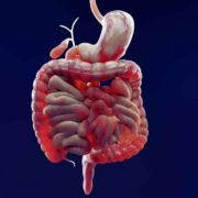
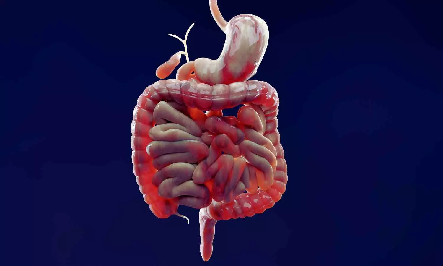
A significant clinical problem is fracture-related infection (FRI), which frequently arises following fractures or surgical operations that specifically impact the lower limbs. FRI is still difficult to diagnose, especially in chronic patients that frequently exhibit a wide range of unusual clinical symptoms. Even if a consensus has been presented by the AO international expert group, there are still no reliable and consistent diagnostic techniques, particularly when contrasted with the recognized standards for periprosthetic joint infections (PJIs).

By determining ideal maximum standardized uptake value (SUVmax) thresholds and examining sub-group-specific diagnostic performance, this study seeks to assess the diagnostic accuracy of 18F-FDG PET/CT for refractory FRIs in the lower limbs.

A total of 429 PET/CT images from a tertiary orthopaedic center conducted between November 2016 and October 2021 were included in this retrospective analysis. Included were patients with probable refractory FRI, which is defined as an infection that has persisted after at least 2 previous therapies.

Microbiological cultures, histological examination, intraoperative results, and follow-up data were all included in the reference standard. Receiver operating characteristic curve analysis was used to compute diagnostic performance parameters, such as sensitivity, specificity, and area under the curve (AUC). The patients were classified in sub-group analyses according to the time since prior surgery and the clinical presentation.

AUC for PET/CT was 0.84, indicating strong diagnostic performance. With a sensitivity of 70.7% and specificity of 85.6%, the ideal SUVmax threshold was 4.75. According to subgroup studies, customized thresholds increased diagnosis accuracy; at a threshold of 5.05., the “No signs + Early phase” group had the best accuracy (87.5%) and specificity (89.4%).

By separating infection from leftover metabolic activity, a more stringent cut-off of 3.95 in the “No signs + Over phase” group, on the other hand, reduced overdiagnosis. Given the difficulties in identifying chronic infections, the “Over phase” group had the highest false-positive rate (42.4%) and the best specificity (90.1%) at a cut-off of 4.65. These results highlight how SUVmax thresholds vary depending on the clinical context.

Overall, 18F-FDG With stratified SUVmax criteria that improve diagnosis accuracy dependent on the period since prior surgery and symptom manifestation, PET/CT is a dependable diagnostic tool for refractory FRI. By enhancing infection control and lowering recurrence rates, PET/CT proves to be cost-effective over the long run, even with its high initial cost.

Source:

Cai, W., Lu, Y., Ren, Z., Zhang, Y., Cheng, P., Chen, X., Han, P., & Xu, Z. (2025). Real-world experience of the role of 18F-fluorodeoxyglucose positron emission tomography/CT refractory fracture-related infection on lower limbs : a retrospective diagnostic study: A retrospective diagnostic study. The Bone & Joint Journal, 107-B(8), 846–856. https://doi.org/10.1302/0301-620X.107B8.BJJ-2024-1158.R2

Powered by WPeMatico

Neuroserpin’s Dual Role: A Potential Biomarker and Protector in Preeclampsia, finds study

Recent study evaluated maternal serum neuroserpin concentrations in pregnancies affected by preeclampsia and their relationship with disease severity, considering neuroserpin’s roles in anti-inflammation and neuroprotection. This prospective case-control investigation was conducted from September 2024 to January 2025 at a tertiary referral hospital in Ankara, Türkiye, with a total of 88 participants: 44 diagnosed with preeclampsia and 44 healthy gestational-age-matched controls. ### Methodology Participants comprised pregnant women aged 21 to 45 with singleton pregnancies, diagnosed with preeclampsia based on the American College of Obstetricians and Gynecologists (ACOG) criteria. Exclusion criteria included multiple gestations, existing maternal comorbidities, substance abuse, prior corticosteroid treatment, and fetal anomalies. Serum samples (≥ 3 mL) were collected post-fasting and preserved at -80 °C until analysis. Neuroserpin levels were quantified using a Human NSP ELISA Kit via the sandwich ELISA method, with statistical significance evaluated through various tests (e.g., Student’s t-test, Mann-Whitney U test) and assessed using receiver operating characteristic (ROC) curve analysis for diagnostic efficacy. ### Key Results

The results demonstrated significantly reduced serum neuroserpin levels in women with preeclampsia compared to the control group (p = 0.018). Furthermore, women with severe preeclampsia (Preeclampsia-SF) exhibited even lower neuroserpin levels, indicating a potential link between neuroserpin reduction and disease severity. Notably, these variations in neuroserpin levels occurred independently of maternal demographic factors, such as age and BMI, enhancing its potential as a biomarker for preeclampsia identification.

Diagnostic Analysis

ROC analysis established that a neuroserpin level threshold of ≤ 22.95 ng/mL could identify preeclampsia with a sensitivity of 86.4% and specificity of 43.2%. For severe cases, a threshold of ≤ 14.7 ng/mL produced a sensitivity of 50.0% and a specificity of 96.7%. These outcomes indicate that neuroserpin could function as a valuable diagnostic instrument for evaluating both the presence and severity of preeclampsia.

Neuroserpin Properties –

Neuroserpin, a serine protease inhibitor secreted mainly by neurons and monocytes, possesses known anti-inflammatory properties that could alleviate the endothelial and inflammatory dysfunctions associated with preeclampsia. Under normal physiological conditions, it is thought to mitigate extracellular damage, particularly in ischemic situations, which may be vital for preserving vascular integrity during pregnancy. These findings suggest that decreased neuroserpin levels in preeclamptic pregnancies may exacerbate unregulated vascular inflammation, thereby worsening endothelial dysfunction.

Study Limitations

Limitations of this study include its relatively small sample size and its focus on a single center, which may limit the generalizability of the findings. Additionally, the cross-sectional design does not permit longitudinal tracking of neuroserpin levels throughout pregnancy or during postpartum recovery. ### Conclusion

Implications

In conclusion, lower maternal serum neuroserpin levels are significantly linked to both the presence and severity of preeclampsia, indicating its potential as a biomarker for assessing disease burden and inflammatory activity. Future multi-center, longitudinal studies could further confirm these results and investigate the clinical implications of neuroserpin in predicting and managing preeclampsia.

The key points from the research paper are: 1. Maternal serum neuroserpin levels were significantly lower in women diagnosed with preeclampsia compared to healthy pregnant controls. Moreover, neuroserpin levels were further decreased in those with severe preeclampsia, suggesting a potential role of neuroserpin not only in the presence but also in the progression of the disease. 2. The observed suppression of neuroserpin in preeclamptic pregnancies may reflect impaired physiological upregulation in response to inflammatory vascular stress. In healthy pregnancies, neuroserpin expression may serve as a compensatory mechanism to maintain endothelial integrity, and its reduction in preeclampsia could facilitate unchecked vascular inflammation and endothelial dysfunction. 3. Receiver operating characteristic (ROC) curve analysis showed that a serum neuroserpin cutoff value of ≤ 22.95 ng/mL had a sensitivity of 86.4% and a specificity of 43.2% for identifying preeclampsia, while a cutoff of ≤ 14.7 ng/mL had a sensitivity of 50.0% and a specificity of 96.7% for identifying severe preeclampsia. These findings suggest that neuroserpin may serve as a candidate biomarker reflecting disease burden and inflammatory activity in preeclampsia.

Reference –

Belgin Savran Ucok et al. (2025). Investigation Of Serum Neuroserpin Levels In Pregnant Women Diagnosed With Pre-Eclampsia: A Prospective Case-Control Study. *BMC Pregnancy And Childbirth*, 25. https://doi.org/10.1186/s12884-025-07673-2.

Powered by WPeMatico

FDA Approves Ajovy for Preventive Treatment of Episodic Migraine in Children Aged 6–17

The FDA has approved Ajovy (fremanezumab-vfrm) for preventive treatment of episodic migraine in pediatric patients aged 6 to 17 years who weigh at least 45 kg.

With this approval, AJOVY becomes the first and only calcitonin gene-related peptide (CGRP) antagonist indicated for the preventive treatment of episodic migraine in pediatric patients and migraine in adults, marking a meaningful advancement in expanding preventive treatment options for those living with migraine.

Preventative treatment can help reduce the frequency of migraine attacks, helping children and adolescents to better manage the condition day-to-day. AJOVY is administered once a month and available for in-office or at home use, offering a treatment option that is intended to support adherence and reduce treatment burden for families.

“Migraines are a common yet invisible condition that can severely disrupt daily life for children and adolescents, often leaving them overlooked and misunderstood,” said Chris Fox, Executive Vice President, U.S. Commercial and Innovative Franchise Lead and Head of Global Marketing Business at Teva. “With this FDA approval, AJOVY now offers younger patients a new treatment option, addressing a long-standing gap in care and offering families added support as they navigate the challenges of this condition.”

1 in 10 children and adolescents in the U.S. suffer from migraine, one of the most common and disabling neurological conditions. Despite its widespread prevalence, pediatric migraine is often underrecognized and undertreated, contributing to missed school days, difficulties with schoolwork, and disrupted social activities.

“Pediatric migraine is a complex condition that can significantly impact a child’s daily life, from school performance to emotional well-being,” said Dr. Jennifer McVige, MD, MA, Pediatric Neurologist at the DENT Neurologic Institute. “Having an FDA-approved treatment like AJOVY offers an important option, providing a targeted approach to preventive treatment for episodic migraine that can help reduce the frequency of attacks in younger patients and help clinicians manage this often-overlooked condition.”

Building on its established success in adult patients since its U.S. approval in 2018, this expanded indication strengthens Teva’s commitment to broadening access to neuroscience therapies across age groups.2 AJOVY continues to demonstrate efficacy in addressing the underlying biology of migraine and now provides a treatment pathway for a patient population with historically limited preventive options.

About Migraine

Migraine attacks cause disabling pain, nausea, vomiting and sensitivities to light and sound, resulting in serious effects on the ability to complete daily tasks.5 Migraine can cause significant disability in children and adolescents, leading to absence from school, impaired educational performance and missed social activities.3

About AJOVY

AJOVY is indicated for preventive treatment of migraine in adults and episodic migraine in children and adolescent patients aged 6-17 years who weigh 45 kilograms (99 pounds) or more. AJOVY is available as a 225 mg/1.5 mL single dose injection in a pre-filled autoinjector or in a pre-filled syringe. AJOVY can be administered either by a healthcare professional or at home by a patient or caregiver. No starting dose is required to begin treatment.

Powered by WPeMatico

Guselkumab demonstrates superior efficacy in landmark clinical trials and offers new hope to Crohn’s disease patients

In a major advance for patients with Crohn’s disease, a new study led by researchers at Mount Sinai Health System found that guselkumab, a medication with a mechanism of action that is new to inflammatory bowel disease (IBD) treatment, outperformed an established standard of care in promoting intestinal healing and symptom relief.

These findings from two pivotal phase 3 trials known as GALAXI 2 and 3, published today in The Lancet, provided the basis for the recent Food and Drug Administration approval of guselkumab (brand name Tremfya) for the treatment of moderately to severely active Crohn’s disease.

Crohn’s disease affects roughly 780,000 people in the United States and often requires a lifetime of management. Despite numerous available biologic medications, many patients fail to achieve sustained remission. Guselkumab blocks the interleukin-23 (IL-23) pathway, a key driver of chronic intestinal inflammation.

“Suboptimal disease control despite the availability of biologic therapies remains a prevalent problem among patients with Crohn’s disease,” said Bruce E. Sands, MD, MS, the Dr. Burrill B. Crohn Professor of Medicine at the Icahn School of Medicine at Mount Sinai and senior author on this paper. “The GALAXI trials were especially impactful as they compared two dosing regimens of guselkumab to both placebo and ustekinumab over a 48-week period. Patients receiving guselkumab showed significantly higher rates of endoscopic healing and deep remission, critical indicators linked to fewer disease flares, hospitalizations, and long-term complications.”

Guselkumab is the first biologic shown in identically designed, 48-week, double-blind trials to outperform another leading biologic—ustekinumab, a monoclonal antibody that blocks both IL-12 and IL-23—for key markers of disease remission and gut healing in Crohn’s disease. The design allowed for head-to-head comparisons to both placebo and ustekinumab. The studies enrolled 1,048 patients worldwide and randomly assigned participants to one of four groups:

• guselkumab 200 mg intravenously (IV) at weeks 0/4/8, then guselkumab 100 mg subcutaneously (SC) every eight weeks beginning at week 16

• guselkumab IV at weeks 0/4/8, then guselkumab 200 mg every four weeks beginning at week 12

• ustekinumab ~6 mg/kg IV at week 0, then ustekinumab 90 mg SC every eight weeks beginning at week 8

• placebo

Guselkumab demonstrated statistically significant improvements across multiple endpoints, including endoscopic response and deep remission. Safety outcomes, based on adverse events and laboratory findings, were favorable and consistent with the known profile of guselkumab in approved indications.

Beyond symptom control, the therapy’s corticosteroid-sparing effect further underscores its clinical value, especially for patients seeking alternatives to long-term steroid use.

The GALAXI 2 and 3 trials were sponsored by Johnson & Johnson and enrolled patients with prior biologic treatment failures.

Dr. Sands, the Chief of Mount Sinai’s Dr. Henry D. Janowitz Division of Gastroenterology, is a renowned expert in inflammatory bowel disease. He previously co-authored the pivotal UNITI trial, and was both the lead author and corresponding author on the UNIFI trials. Both of these trials helped establish ustekinumab’s place in IBD care. His latest work builds on that legacy, demonstrating not only the effectiveness of IL-23 inhibition but its potential to redefine front-line treatment.

Powered by WPeMatico

Tumor diagnostics: AI model detects more than 170 types of cancer

The MRI image shows a brain tumor in an inauspicious location, – and a brain biopsy will entail high risks for the patient, who had consulted us due to double vision. Situations such as this case discussion, cited by way of example, in a multidisciplinary team of cancer medicine experts prompted researchers at Charité – Universitätsmedizin Berlin, together with cooperation partners, to look for new diagnostic procedures. The result is an AI model. The model makes use of specific characteristics in the genetic material of tumors – their epigenetic fingerprint, obtained for example from cerebrospinal fluid, among other things. As the team shows in the journal Nature Cancer*, the new model classifies tumors quickly and very reliably.

Today, far more types of tumors are known than the organs from which they arise. Each tumor has its own characteristics, certain tissue features, growth rates and metabolic peculiarities. Nevertheless, tumor types with similar molecular characteristics can be grouped together. The treatment of the individual disease depends decisively on the type of tumor. New, targeted therapies address certain structures of tumor cells or block their signaling pathways in order to halt the pathological tissue growth. Chemotherapies can be selected according to tumor type and their dosage adjusted accordingly. Particularly in the case of rare tumor types, it may be possible to pursue innovative therapies as part of studies.

“Against the backdrop of increasingly personalized, rapidly developing cancer medicine, precise diagnosis at a certified tumor center is the way forward for successful treatment,” as Prof. Martin E. Kreis, Chief Medical Officer at Charité, stated. While a comprehensive molecular, cellular and functional analysis of a tumor based on tissue samples provides the necessary information, physicians are also confronted with cases in which it is not possible or very risky to extract tissue samples from the tumor. What’s more, even a histological examination alone is not capable of providing as precise a diagnosis as the new AI model.

Looking into the genome instead of into the tissue

A method has been established for characterizing brain tumors that is not based on conventional microscopic diagnostics, but instead on modifications of the tumor‘s genetic material, the epigenetic characteristics. They are part of the memory of every cell and determine which parts of the genetic information are read and when. “Hundreds of thousands of epigenetic modifications act as on and off switches for individual gene sections. Their patterns form a unique, unmistakable fingerprint,” explains Dr. Philipp Euskirchen, scientist at the Berlin site of the German Cancer Consortium and at the Institute of Neuropathology at Charité, who heads the recently published study. “In tumor cells, the epigenetic information is altered in a characteristic way. Based on their profiles, we can differentiate between tumors and classify them.” In the case of brain tumors, even a sample of the cerebrospinal fluid is sufficient in some cases, and can be obtained relatively easily – dispensing with surgery altogether.

In order to compare an unknown fingerprint with thousands of known fingerprints of different cancers and assign it to a specific tumor type, machine learning methods, i.e., artificial intelligence, are required given that the data are very extensive and complex. What’s more, different DNA sequencing methods were applied in the past. In addition, epigenetic analyses are usually limited to defined patterns and gene segments that are typical for individual tumor types. “Consequently, our aim was to develop a model that accurately classifies tumors, even if they are only based on parts of the entire tumor epigenome or the profiles were collected by means of different techniques and varying degrees of accuracy,” as bioinformatician Dr. Sören Lukassen, head of the Medical Omics working group at the Berlin Institute of Health at Charité (BIH), stated.

Reliable and traceable

A newly developed AI model goes by the name of crossNN, whose architecture is based on a simple neural network. The model was trained with a large number of reference tumors and subsequently tested on more than 5,000 tumors. “Our model allows a very precise diagnosis of brain tumors in 99.1 percent of all cases and is more accurate than the AI solutions at work to date,” as Philipp Euskirchen related. “In addition, we were able to train an AI model in the same way that can differentiate between over 170 tumor types from all organs, while achieving accuracy of 97.8 percent. This means that it can be used for cancers of all organs, in addition to the relatively rare brain tumors.” The decisive factor for future approvals in clinical application is that the models are fully explainable, i.e., it must be possible to understand how the decisions are arrived at.

The molecular fingerprint that the AI model receives for determination can stem from a tissue sample or from body fluids. In the case of specific brain tumors, the Department of Neuropathology at Charité already offers non-invasive diagnostics based on cerebrospinal fluid, known as liquid biopsy. This allows a diagnosis to be made without a stressful operation, also in difficult situations. The patient who consulted us with double vision was one of the beneficiaries. “We examined the cerebrospinal fluid using nanopore sequencing, a novel, very fast and efficient form of genetic analysis. The classification by our models revealed that it was a lymphoma of the central nervous system, enabling us to promptly initiate appropriate chemotherapy,” Philipp Euskirchen explains.

crossNN in clinical trials

The accuracy of the methodology even took the researchers by surprise. “Although the architecture of our AI model is far more simple than previous approaches and therefore remains explainable, it delivers more precise predictions and therefore greater diagnostic certainty,” says Sören Lukassen. Together with the German Cancer Consortium (DKTK), the research team is therefore planning clinical trials with crossNN at all eight DKTK locations in Germany. In addition, intraoperative use is also to be tested. The aim is to transfer the precise and comparatively inexpensive tumor determination based on DNA samples to routine care.

Reference:

Yuan, D., Jugas, R., Pokorna, P. et al. crossNN is an explainable framework for cross-platform DNA methylation-based classification of tumors. Nat Cancer 6, 1283–1294 (2025). https://doi.org/10.1038/s43018-025-00976-5.

Powered by WPeMatico

Long-Term Passive Smoking Doubles COPD Risk in Women, Study Finds

China: A new community-based study from Jinan, China, has revealed that women with long-term exposure to secondhand smoke face a significantly higher risk of developing chronic obstructive pulmonary disease (COPD), alongside more severe symptoms and greater lung function decline. The research, led by Dr. Jiling Lv from the Department of Respiratory Medicine at Shandong Second Provincial General Hospital, was published in the International Journal of Chronic Obstructive Pulmonary Disease. 

COPD, a progressive lung condition often associated with smoking, is increasingly being recognized in non-smokers—particularly women exposed to secondhand smoke over extended periods. The study sought to explore the relationship between prolonged passive smoking and the onset of COPD in women, shedding light on a preventable but often overlooked risk factor.

Between October 2022 and April 2023, the research team examined 2,360 women aged 40 years and older. Participants underwent a battery of assessments, including detailed respiratory questionnaires, lung function measurements through spirometry, and blood tests to detect inflammation. Based on their history of secondhand smoke exposure, they were divided into two groups: long-term passive smokers (LPS, 610 participants) and non-passive smokers (NPS, 1,750 participants).

Key Findings:

  • Women in the long-term passive smoking (LPS) group showed significantly poorer respiratory health compared to those without passive smoke exposure.
  • The LPS group had a lower average forced expiratory volume in one second (FEV1: 2.97 vs 3.25 liters).
  • Predicted FEV1 percentages were reduced in the LPS group.
  • The LPS group had a lower FEV1/forced vital capacity ratio (83.3% vs 87.2%).
  • Small airway function was more impaired in the LPS group, with reduced MEF75% (77.6 vs 86.1 L/s).
  • MEF50% was lower in the LPS group (62.8 vs 89.4 L/s).
  • Maximal mid-expiratory flow rate was also reduced in the LPS group (80.9 vs 87.5 L/s).
  • Prevalence of preserved ratio impaired spirometry (PRISm) was almost twice as high in the LPS group (5.74% vs 2.91%).
  • Women in the LPS group reported more frequent COPD exacerbations.
  • The LPS group experienced more severe respiratory symptoms.
  • Systemic inflammatory markers were higher in the LPS group.

The authors conclude that long-term passive smoking should be considered an independent risk factor for COPD in women, capable of accelerating disease progression and increasing both symptom burden and frequency of acute exacerbations. They argue that these findings strengthen the case for stricter public health measures aimed at minimizing secondhand smoke exposure, particularly in environments where women may be regularly exposed, such as homes or workplaces.

While the study provides compelling evidence, the researchers caution that certain limitations—such as potential selection bias and unmeasured lifestyle factors—could influence the results. They recommend that future large-scale, longitudinal studies be conducted to confirm these associations and further refine public health strategies.

“In the meantime, the message from this study is clear: for women, avoiding long-term exposure to secondhand smoke could play a crucial role in preventing the onset and progression of COPD,” the authors concluded.

Reference:

Liu Z, Jiao M, Lv J, Han Q. Increased Incidence of Chronic Obstructive Pulmonary Disease in Women Due to Long-Term Passive Smoking. Int J Chron Obstruct Pulmon Dis. 2025;20:2745-2752. https://doi.org/10.2147/COPD.S534060

Powered by WPeMatico

Sunlight Exposure May Reduce Risk of Postoperative Hypoparathyroidism, suggests study

Researchers have discovered in a new study that patients in regions with higher sunlight exposure appear to have lower rates of postoperative hypoparathyroidism, suggesting sunlight may play a protective role in its prevention.

Postoperative hypoparathyroidism is a common complication of thyroid surgery. Sunlight is a natural source of ultraviolet B (UVB) radiation, which facilitates the synthesis of vitamin D3 in the skin. Inadequate sunlight exposure has been linked to vitamin D deficiency, potentially exacerbating the risk of hypocalcemia in patients undergoing thyroid surgery. The aim of the present study is to evaluate the effect of sunshine levels on postoperative hypoparathyroidism. Method: We retrospectively evaluated patients that underwent total thyroidectomies at two different centers (Thessaloniki and Rhodes) by the same surgical team from 2021 to 2023 in terms of postoperative hypoparathyroidism. We compared the sunshine levels at each center the year before surgery and correlated them with postoperative levels of parathyroid hormone, serum ionized calcium, and phosphorus. Results: One-hundred twenty patients (Group Thessaloniki = 60 patients, Group Rhodes = 60 patients) who were matched for demographic characteristics and type of thyroid disease and surgery were enrolled in our study. The sunshine levels were different between the two centers (Rhodes > Thessaloniki, p < 0.001). It was found that sunshine levels affect preoperative serum ionized calcium (p = 0.002) and postoperative parathyroid hormone levels (p = 0.025). Conclusions: Sunlight exposure levels may play a crucial role in preventing postoperative hypoparathyroidism. Patients living in locations with higher sunshine levels may have lower rates of postoperative hypoparathyroidism.

Reference:

Chorti A, Pliakos I, Moysidis M, Smprini A, Bakkar S, Papavramidis T. The Impact of Sunlight Exposure on Postoperative Hypoparathyroidism: A Retrospective Analysis from Two Greek Centers. Journal of Clinical Medicine. 2025; 14(13):4418. https://doi.org/10.3390/jcm14134418

Keywords:

Sunlight, Exposure, May, Reduce, Risk, Postoperative , Hypoparathyroidism, Chorti A, Pliakos I, Moysidis M, Smprini A, Bakkar S, Papavramidis T, sunshine; postoperative hypoparathyroidism; postoperative hypocalcemia; vitamin D

Powered by WPeMatico

DMFT Score associated with increased risk of Buccal Mucosa Cancer: Study

A new study published in JCO Global Oncology has revealed that higher DMFT (Decayed, Missing, and Filled Teeth) scores are significantly associated with an increased risk of buccal mucosa cancer (BMC), a common subtype of oral cancer. Conducted by researchers including Abhijit Das and colleagues, the study emphasizes the importance of oral hygiene markers—especially DMFT—as predictors of cancer risk. Drawing from a hospital-based case-control analysis involving 358 individuals in India, the researchers found that individuals with poor dental health, as measured by higher DMFT scores, had a markedly greater likelihood of developing buccal mucosa cancer. The findings suggest that DMFT scores should be prioritized in national oral health screening programs, not only as indicators of general dental health but also as part of preventive strategies against oral cancers (OCs). The study further reinforces the link between poor oral hygiene, chronic inflammation, and carcinogenesis in the oral cavity. This adds to the growing evidence that routine dental assessments may offer critical insight into broader health risks. The authors recommend that oral health programs integrate DMFT tracking with community-based cancer awareness and screening initiatives, particularly in populations with high tobacco and betel nut use. Incorporating dental assessments into public health policy may help with early detection and long-term cancer prevention, especially in low-resource settings.

Keywords: DMFT score, buccal mucosa cancer, oral cancer prevention, oral hygiene indicators, dental caries, JCO Global Oncology, Abhijit Das, oral cancer screening, decayed missing filled teeth, inflammation and cancer, national oral health programs

Citation (APA):
Das, A., Kumar, A., & Ghosh, S. (2024). Association of DMFT score with risk of buccal mucosa cancer: A case-control study from India. JCO Global Oncology. https://doi.org/10.1200/GO-25-00119


Powered by WPeMatico

OJEE Revises NEET 2025 Counselling Schedule for MBBS, BDS Admissions, choice filling till August 15

Odisha- The Odisha Joint Entrance Examination (OJEE) has revised the tentative schedule in line with the Medical Counselling Committee (MCC) Counselling extension for the state quota counselling and admission in the MBBS and BDS Courses for the academic year 2025-26.

In this regard, OJEE has released the revised tentative schedule for the National Eligibility and Entrance Test-Undergraduate (NEET UG) Round 1 Counselling 2025 on its official website. Therefore, candidates willing to participate in the said counselling should check the new dates

As per the revised tentative schedule, the last date for the Choice locking is now extended to August 15. Following this, the 1st Round Provisional Allotment will be declared on 16 August 2025 and Invitation for Query, if any, related to Provisional Allotment will be done on 17th August 2025. Below is the complete schedule-

REVISED TENTATIVE SCHEDULE

S.NO

ACTIVITY

DATE & TIME

1

Publication of Revised State Merit List.

03 August 2025, 5:00 PM

2

Choice Filling and Locking.

31 July 2025, 02:00 PM to 15 August 2025, 04:30 PM

3

1st Round Provisional Allotment.

16 August 2025, 11.00 AM

4

Invitation for Query, if any, related to Provisional Allotment.

17 August 2025 till 05.00 PM

5

Publication of 1 st Round Allotment and Downloading of Allotment Letter.

18 August 2025, 5.00 PM

6

Provisional Admission by Paying Admission Fees and Reporting at OJEE Cell, Gandamunda with original documents and signed bond.*

19 to 24 August 2025

7

Withdrawal (Resignation) from Admission Process.

19 August 2025, 11.00 AM to 25 August 2025, 11.55 PM

Meanwhile, it is to be noted that the candidates who have taken admission in 1st/ 2 nd or 3rd round of counselling (All India/ State) are not eligible to participate in Spot Counselling. Also, candidates who have taken admission at the end of All Indai 3rd round Counselling are not allowed to participate in state counselling. Candidates who have taken admission at the end of State 3 rd round Counselling are not allowed to resign.

To view the schedule, click the link below

https://medicaldialogues.in/pdf_upload/ojee-revises-neet-2025-counselling-schedule-for-mbbs-bds-admissions-choice-locking-deadline-extended-to-august-15-297203.pdf

Powered by WPeMatico

NEET 2025: 2675 MBBS seats in 19 Odisha Medical Colleges this year

New Delhi: The Medical Counselling Committee (MCC) recently released the MBBS seat matrix, and in Odisha for the academic year 2025-26, there are a total of 2,675 MBBS seats across 19 medical colleges.

This includes premier institutions like SCB Medical College, Cuttack, and MKCG Medical College, Berhampur with 250 seats each, and Veer Surendra Sai Institute, Burla, with 200 seats.

New and renamed government colleges like Bhima Bhai Medical College, Fakir Mohan Medical College, JKMC Jajpur, and Keonjhar Medical College have also been allotted seats. Private medical colleges such as Kalinga Institute of Medical Sciences, SUM Hospital, and Hi-Tech Medical Colleges contribute with up to 250 seats each.

AIIMS Bhubaneswar offers 125 seats under the central government allocation. This distribution strengthens Odisha’s medical education infrastructure and expands opportunities for NEET-UG 2025 aspirants.

Odisha Medical Colleges & MBBS Seats (2025)

S.No. Name of college No. of seats for
AY 2025-26
1 All India Institute
of Medical Sciences, Bhubaneswar
125
2 DRIEMS INSTITUTE OF
HEALTH SCIENCES AND HOSPITAL
50
3 Government Medical
College & Hospital (Renamed as Bhima Bhai Medical College & Hospital)
100
4 Government Medical
College & Hospital (Renamed as Fakir Mohan Medical College & hospital)
100
5 GOVERNMENT MEDICAL
COLLEGE AND HOSPITAL SUNDARGARH
100
6 Government Medical
college, Jajpur (JKMC), Odisha
50
7 Government Medical
College, Keonjhar
100
8 Hi-Tech Medical
College & Hospital, Rourkela
100
9 Hi-Tech Medical
College and Hospital Bhubaneswar
150
10 Institute of Medical
Sciences and SUM Hospital Campus II
150
11 Instt. of Medical
Sciences and SUM Hospital
250
12 Kalinga Institute of
Medical Sciences, Bhubaneswar
250
13 MKCG Medical College,
Berhampur
250
14 Pt. Raghunath Murmu
Medical College and Hospital, Baripada, Odisha
125
15 Saheed Laxman Nayak
Medical College & Hospital, Koraput
125
16 Saheed Rendo Majhi
Medical College and Hospital, Bhawanipatna,
Xalahandi, Odisha
100
17 SCB Medical College,
Cuttack
250
18 Sri Jagannath Medical
College & Hospital, Puri
100
19 Veer Surendra Sai
Institute of Medical Sciences and Research, Burla
200
Total Seats 2675

SCHEDULE FOR ONLINE COUNSELING (ALLOTMENT PROCESS) FOR NEET-UG (MBBS/ BDS/ B.SC (NURSING) – 2025

ALL INDIA QUOTA (15%)/DEEMED/CENTRAL UNIVERSITIES/ALL AIIMS INSTITUTES/JIPMER (PUDUCHERRY & KARAIKAL)

ROUND I

SL.

NO.

VERIFICATION OF TENTATIVE SEAT MATRIX BY THE PARTICIPATING INSTITUTES AND NMC

REGISTRATION/PAYMENT

CHOICE FILLING/LOCKING

PROCESSING OF SEAT ALLOTMENT

RESULT

REPORTING/JOINING

VERIFICATION OF JOINED CANDIDATE’S DATA BY INSTITUTES SHARING OF DATA TO MCC

1

18th to 19th July, 2025

21st July, 2025 to 6th August 2025 up to 03:00 PM ( as per Server Time) *

Payment facility will be available up to 06:00 PM on 6th August 2025 as per Server Time

22nd July, 2025 to 7th August 2025 (up to 08:00 A.M of 7th August 2025 (as per Server Time)

Choice Locking will be available from 08:00 PM of 6th Aug, 2025, upto 08:00 AM of 7th August 2025 (as per Server Time)

7th to 8th August 2025

9th August 2025

9th August 2025 to 18th August 2025

19th to 20th August 2025

DAYS

(02-Days)

(17-Days)

(17-Days)

(02-Days)

(01-Day)

(06-Days)

(02-Days)

ROUND II

SL.

NO.

VERIFICATION OF TENTATIVE SEAT MATRIX BY THE PARTICIPATING INSTITUTES AND NMC

REGISTRATION/PAYMENT

CHOICE FILLING/LOCKING

PROCESSING OF SEAT ALLOTMENT

RESULT

REPORTING/JOINING

VERIFICATION OF JOINED CANDIDATE’S DATA BY INSTITUTES SHARING OF DATA TO MCC

1

21st August 2025

21st to 26th August 2025 upto 12:00 NOON as per Server Time

Payment facility will be available up to 03:00 PM on 26th August 2025 as per Server Time

22nd to 26t August 2025 (till 11:55 PM) as per Server Time Choice

Locking will be available from 04:00 PM on 26th August 2025 upto 11:55 PM on 26th August 2025 as per Server Time

27th to 28th August 2025

29th August 2025

30th August 2025 to 5th September 2025

6th to 8th September 2025

DAYS

(01-Day)

(06-Days)

(05-Days)

(02-Days)

(01-Day)

(07-Days)

(03-Days)

ROUND III

SL.

NO.

VERIFICATION OF TENTATIVE SEAT MATRIX BY THE PARTICIPATING INSTITUTES AND NMC

REGISTRATION/PAYMENT

CHOICE FILLING/LOCKING

PROCESSING OF SEAT ALLOTMENT

RESULT

REPORTING/JOINING

VERIFICATION OF JOINED CANDIDATE’S DATA BY INSTITUTES SHARING OF DATA TO MCC

1

9th 2025

9th to 14th September 2025

(12:00 NOON as per Server Time)

Payment facility will be available upto

03:00 PM of 14th September 2025 as per Server Time

10th to 14th September 2025

(till 11:55 PM of 14th September 2025) as per Server Time

Choice Locking will be available from 04:00 PM on September 2025, upto to 11:55 PM on 14th September 2025 as per Server Time

15th to 16th September 2025

17th September 2025

18th to 25th September 2025

26th to 28th September 2025

DAYS

(01-Day)

(06-Days)

(05-Days)

(02-Days)

(01-Day)

(08-Days)

(03-Days)

ONLINE STRAY VACANCY ROUND

SL.

NO.

VERIFICATION OF TENTATIVE SEAT MATRIX BY THE PARTICIPATING INSTITUTES AND NMC

REGISTRATION/PAYMENT

CHOICE FILLING/LOCKING

PROCESSING OF SEAT ALLOTMENT

RESULT

REPORTING/JOINING

VERIFICATION OF JOINED CANDIDATE’S DATA BY INSTITUTES SHARING OF DATA TO MCC

1

29th September 2025 2nd October 2025

30th September to 2nd October (03:00 PM as per Server Time)

Payment facility will be available upto

06:00 PM of 2nd October 2025 as per Server Time

30th September to 3rd October 2025 (till 08:00 AM of 3rd October 2025) as per Server Time

Choice Locking will be available from 08:00 PM of 2nd October 2025, upto 08:00 AM of 3rd. October 2025 as per Server Time

3rd October to

4th October 2025

5th to 10th October 2025

DAYS

(01-Day)

(03-Days)

(04-Days)

(02-Days)

(01-Day)

(06-Days)

ALL INDIA QUOTA/DEEMED/CENTRAL STATE QUOTA

S.NO.

SCHEDULE FOR ADMISSION

ALL INDIA QUOTA/DEEMED/CENTRAL UNIVERSITIES

VERIFICATION OF JOINED CANDIDATES’ DATA BY MCC

STATE COUNSELLING

VERIFICATION OF JOINED CANDIDATES

1

1st Round of Counselling

21st July 2025 to 9th August 2025

19th to 20th August 2025

9th to 18th August 2025

25th to 26th August 2025

2

Last date of Joining

18th August 2025

24th August 2025

3

2nd round of Counselling

21st to 29th August 2025

6th to 8th September 2025

27th August 2025 to 5th September 2025

12th to 13th September 2025

4

Last date of joining

5th September 2025

11th Sept, 2025

5

Round-3

9th to 17th September 2025

26th 28th September 2025

15th to 25th September 2025

1st October 2025

6

Last date of joining

25th September 2025

30th September 2025

7.

Stray Vacancy

30th September 2025 to 4th October 2025

2nd to 5th October 2025

10th October 2025

8.

Last date of joining

10th October 2025

10th October 2025

9.

Commencement of Academic Session for UG Courses

5th September, 2025`

Powered by WPeMatico