Ultra-low dose CT aids pneumonia diagnosis in immunocompromised patients: Study

Denoised ultra-low dose CT can effectively diagnose pneumonia in immunocompromised patients using only 2% of the radiation dose of standard CT, according to a study published today in Radiology: Cardiothoracic Imaging, a journal of the Radiological Society of North America (RSNA).

“For patients with weakened immune systems, lung infections can be life threatening,” said lead study author Maximiliano Klug, M.D., a radiologist in the division of diagnostic imaging at the Sheba Medical Center in Ramat Gan, Israel. “CT scans are the gold standard for detecting pneumonia, but repeated scans can expose patients to significant radiation.”

While the early diagnosis of lung infections in immunocompromised patients is important, the risks of cumulative radiation dose exposure from frequent CT scans is a concern.

Ultra-low dose CT reduces radiation exposure but can result in poor image quality due to added “noise,” which manifests as a grainy texture throughout the image. This reduction in image quality can affect the accuracy of diagnosis. Therefore, Dr. Klug and colleagues sought to test the denoising capabilities of a deep learning algorithm on ultra-low dose CT scans.

From September 2020 to December 2022, 54 immunocompromised patients with fevers were referred to Dr. Klug’s division to undergo two chest CT scans: a normal dose scan and an ultra-low dose scan. A deep learning algorithm was applied to denoise all 54 of the ultra-low dose CT scans.

Radiologists individually assessed and documented their findings from the normal dose CT, ultra-low dose CT and denoised ultra-low dose CT scans. They were blinded to all patient clinical information.

The deep learning algorithm significantly improved the image quality and clarity of the ultra-low dose CT scans and reduced false positives. Nodules were also more easily identified on the denoised scans.

The average effective radiation dose for ultra-low dose scans was 2% of the average effective radiation dose of the standard CT scans.

“This study paves the way for safer, AI-driven imaging that reduces radiation exposure while preserving diagnostic accuracy,” Dr. Klug said.

The researchers note that deep learning-based denoising on ultra-low dose CT scans can be beneficial in other patient groups, such as young patients.

“This pilot study identified infection with a fraction of the radiation dose,” Dr. Klug said. “This approach could drive larger studies and ultimately reshape clinical guidelines, making denoised ultra-low dose CT the new standard for young immunocompromised patients.”

Reference:

Maximiliano Klug, Tamer Sobeh, Michael Green, Denoised Ultra-Low-Dose Chest CT to Assess Pneumonia in Individuals Who Are Immunocompromised, Radiology Cardiothoracic Imaging, https://doi.org/10.1148/ryct.240189

Powered by WPeMatico

Fremanezumab Shows Dual Efficacy in Treating Migraine and Comorbid Major Depressive Disorder: JAMA

Researchers have found in a new study that Fremanezumab proved effective in reducing symptoms of both migraine and comorbid major depressive disorder in patients suffering from both conditions. The study was conducted by Richard B. L. and fellow researchers published in JAMA Neurology. Although migraine and major depressive disorder (MDD) frequently co-occur, evidence for effective treatment approaches for both conditions at the same time has been sparse. The UNITE study bridges this gap by evaluating fremanezumab, a calcitonin gene–related peptide (CGRP) monoclonal antibody indicated for migraine prevention in a patient population with both migraine (episodic or chronic) and MDD with active symptoms.

The UNITE trial was a double-blind, placebo-controlled, parallel-group trial from July 9, 2020, to August 31, 2022, at 55 centers in 12 countries. The trial consisted of a 4-week screening period, a 12-week double-blind treatment period, and a 12-week open-label extension (OLE).

540 adults were screened, of whom 353 patients were enrolled and randomized: 175 to fremanezumab (225 mg per month), and 178 to placebo. Participants were eligible if they had episodic migraine (48%) or chronic migraine (52%), along with a DSM-5-diagnosis major depressive disorder for ≥12 months, and a score of active depressive symptoms (PHQ-9 ≥10) at screening. For the OLE phase, all patients received quarterly fremanezumab (675 mg).

Key Findings

  • The study population mean age was 42.9 years, and 88% (310/353) of the participants were female.

  • Fremanezumab led to a mean decrease of −5.1 monthly migraine days (SE 0.50; 95% CI: −6.09 to −4.13) versus −2.9 days with placebo (SE 0.49; 95% CI: −3.89 to −1.96), a statistically significant difference (P <.001).

  • At week 8, mean change from baseline in Hamilton Depression Rating Scale–17 Items score was −6.0 points (SE 0.55) with fremanezumab and −4.6 points (SE 0.54) with placebo. The between-group difference was −1.4 points (SE 0.61; 95% CI: −2.61 to −0.22; P = .02).

  • The therapeutic effects of fremanezumab were sustained during the open-label extension period.

  • Adverse events seen were as expected with prior fremanezumab trials, with no new safety issues identified in the study.

The UNITE study offers strong evidence that fremanezumab substantially decreases monthly migraine days and depressive symptoms in patients with both migraine and major depressive disorder, with no new safety issues. This dual effect represents a breakthrough in the treatment of comorbid neurological and psychiatric disorders, highlighting the promise of CGRP-targeting treatments such as fremanezumab in more integrated patient care approaches.

Reference:

Lipton RB, Ramirez Campos V, Roth-Ben Arie Z, et al. Fremanezumab for the Treatment of Patients With Migraine and Comorbid Major Depressive Disorder: The UNITE Randomized Clinical Trial. JAMA Neurol. Published online May 05, 2025. doi:10.1001/jamaneurol.2025.0806

Powered by WPeMatico

Adolescents with mental health conditions use social media differently than their peers, study suggests

Young people with a diagnosable mental health condition report differences in their experiences of social media compared to those without a condition, including greater dissatisfaction with online friend counts and more time spent on social media sites.

This is according to a new study led by the University of Cambridge, which suggests that adolescents with “internalising” conditions such as anxiety and depression report feeling particularly affected by social media.

Young people with these conditions are more likely to report comparing themselves to others on social media, feeling a lack of self-control over time spent on the platforms, as well as changes in mood due to the likes and comments received.

Researchers found that adolescents with any mental health condition report spending more time on social media than those without a mental health condition, amounting to an average of roughly 50 minutes extra on a typical day.

The study, led by Cambridge’s Medical Research Council Cognition and Brain Sciences Unit (MRC CBU), analysed data from a survey of 3,340 adolescents in the UK aged between 11 and 19 years old, conducted by NHS Digital in 2017.

It is one of the first studies on social media use among adolescents to utilise multi-informant clinical assessments of mental health. These were produced by professional clinical raters interviewing young people, along with their parents and teachers in some cases.

“The link between social media use and youth mental health is hotly debated, but hardly any studies look at young people already struggling with clinical-level mental health symptoms,” said Luisa Fassi, a researcher at Cambridge’s MRC CBU and lead author of the study, published in the journal Nature Human Behaviour.

“Our study doesn’t establish a causal link, but it does show that young people with mental health conditions use social media differently than young people without a condition.

“This could be because mental health conditions shape the way adolescents interact with online platforms, or perhaps social media use contributes to their symptoms. At this stage, we can’t say which comes first – only that these differences exist,” Fassi said.

The researchers developed high benchmarks for the study based on existing research into sleep, physical activity and mental health. Only findings with comparable levels of association to how sleep and exercise differ between people with and without mental health conditions were deemed significant.

While mental health was measured with clinical-level assessments, social media use came from questionnaires completed by study participants, who were not asked about specific platforms.

As well as time spent on social media, all mental health conditions were linked to greater dissatisfaction with the number of online friends. “Friendships are crucial during adolescence as they shape identity development,” said Fassi.

“Social media platforms assign a concrete number to friendships, making social comparisons more conspicuous. For young people struggling with mental health conditions, this may increase existing feelings of rejection or inadequacy.”

Researchers looked at differences in social media use between young people with internalising conditions, such as anxiety, depression and PTSD, and externalising conditions, such as ADHD or conduct disorders.

The majority of differences in social media use were reported by young people with internalising conditions. For example, “social comparison” – comparing themselves to others online – was twice as high in adolescents with internalising conditions (48%, around one in two) than for those without a mental health condition (24%, around one in four).

Adolescents with internalising conditions were also more likely to report mood changes in response to social media feedback (28%, around 1 in 4) compared to those without a mental health condition (13%, around 1 in 8). They also reported lower levels of self-control over time spent on social media and a reduced willingness to be honest about their emotional state when online.

“Some of the differences in how young people with anxiety and depression use social media reflect what we already know about their offline experiences. Social comparison is a well-documented part of everyday life for these young people, and our study shows that this pattern extends to their online world as well,” Fassi said.

By contrast, other than time spent on social media, researchers found few differences between young people with externalising conditions and those without a condition.

“Our findings provide important insights for clinical practice, and could help to inform future guidelines for early intervention,” said Cambridge’s Dr Amy Orben, senior author of the study.

“However, this study has only scratched the surface of the complex interplay between social media use and mental health. The fact that this is one of the first large-scale and high-quality studies of its kind shows the lack of systemic investment in this space.”

Added Fassi: “So many factors can be behind why someone develops a mental health condition, and it’s very hard to get at whether social media use is one of them.”

“A huge question like this needs lots of research that combines experimental designs with objective social media data on what young people are actually seeing and doing online.”

“We need to understand how different types of social media. content and activities affect young people with a range of mental health conditions such as those living with eating disorders, ADHD, or depression. Without including these understudied groups, we risk missing the full picture.”

Reference:

Fassi, L., Ferguson, A.M., Przybylski, A.K. et al. Social media use in adolescents with and without mental health conditions. Nat Hum Behav (2025). https://doi.org/10.1038/s41562-025-02134-4
Powered by WPeMatico

Maternal diabetes increases risk of neurodevelopmental disorders among children: Lancet

A new study published in the journal of The Lancet Diabetes & Endocrinology showed that children with maternal diabetes have a higher chance of developing neurodevelopmental problems and perform worse in these domains. Diabetes in the mother may change how the fetus develops its brain. Thus, to synthesize and assess the existing data about the impact of maternal diabetes on children’s neurodevelopmental outcomes, Wenrui Ye and colleagues carried out this investigation.

From the beginning until December 1, 2024, this study searched the PubMed, Web of Science, Embase, and EBSCO databases for research on the neurodevelopmental outcomes of children born to women with diabetes. According to the Diagnostic and Statistical Manual of Mental Disorders, Fifth Edition, and the International Classification of Diseases, Eleventh Revision, the main result was neurodevelopmental disorders. Information was taken from reports that were published. Random-effects models were used to pool the data, and the results were shown as standard mean differences with 95% confidence intervals or risk ratios.

The meta-analysis includes 202 studies with 56 082 462 mother-child pairings. Of them, 80 (40%) looked at pre-gestational diabetes, and 110 (54%) looked at gestational diabetes. 169 (84%) of the total examined studies were only concerned with children and adolescents up to the age of 18. Maternal diabetes was linked to poorer IQ and psychomotor scores, as well as higher odds of all forms of neurodevelopmental problems, in studies that controlled for at least one significant confounder.

Children exposed to maternal diabetes had a higher risk of any neurodevelopmental disorder, attention-deficit hyperactivity disorder, autism spectrum disorder, intellectual disability, communication disorder, specific developmental disorders, motor disorder, and learning disorder than children who were not exposed, according to studies controlling for multiple confounders (n=98, 49%). When compared to gestational diabetes, maternal pre-gestational diabetes was more significantly linked to the risk of the majority of neurodevelopmental problems in offspring.

Overall, children with diabetes had a higher chance of developing neurodevelopmental problems. Specific developmental problems, intellectual impairment, ADHD, and autism spectrum disorder were more prevalent. The risk of pre-gestational diabetes was higher than that of gestational diabetes. To prove causation and elucidate the relationships between certain forms of diabetes and the entire range of neurodevelopmental problems, more excellent research is required.

Source:

Ye, W., Luo, C., Zhou, J., Liang, X., Wen, J., Huang, J., Zeng, Y., Wu, Y., Gao, Y., Liu, Z., & Liu, F. (2025). Association between maternal diabetes and neurodevelopmental outcomes in children: a systematic review and meta-analysis of 202 observational studies comprising 56·1 million pregnancies. The Lancet. Diabetes & Endocrinology. https://doi.org/10.1016/S2213-8587(25)00036-1

Powered by WPeMatico

FDA Clears AI Tool for Lung Sound Detection in Clinics and Homes

The US food and Drug Administration (FDA) has cleared TytoCare’s AI-powered device, Tyto Insights for Lung Sounds, to detect rhonchi, adding to its existing capabilities for identifying wheeze and crackles. Designed for use in primary care settings and patient homes, the tool brings specialist-level lung sound detection to more accessible environments.

With this latest clearance, TytoCare’s platform now offers comprehensive, AI-driven analysis of the three most clinically significant abnormal lung sounds—wheezes, crackles, and rhonchi-directly within its Home Smart Clinic and Pro Smart Clinic solutions. The diagnosis of these abnormal lung sounds presents a recognized challenge for trained medical professionals, as noted in the FDA clearance, underscoring the significance of this advancement as a major milestone in the evolution of virtual care. 

This milestone marks a major leap forward in TytoCare’s mission to transform virtual respiratory care. By combining smart digital stethoscope technology with AI that identifies key lung sound abnormalities, TytoCare enables providers to deliver faster, more accurate diagnoses, while patients will get timely, high-quality care from the comfort of their homes, reducing unnecessary clinic visits and improving health outcomes through earlier detection and intervention.

“Completing our AI lung sound suite is a turning point for virtual care,” said Dedi Gilad, CEO and Co-Founder of TytoCare. “Wheezes, crackles, and rhonchi are critical markers in diagnosing respiratory conditions, and now our AI assists clinicians in detecting all three using TytoCare during a virtual exam. This enhances decision-making and ensures that patients receive the right care at the right time.”

Tyto InsightsTM for Lung Sounds is built on TytoCare’s proprietary database of 1.8 million lung sound recordings and leverages machine-learning algorithms trained and validated by pulmonologists. Each component of the suite-wheeze detection (FDA-cleared in 2023), crackle detection (FDA-cleared in 2024), and now rhonchi detection-analyzes lung acoustic signals in real-time and flags potential abnormalities for clinical review. The Rhonchi device was cleared as an Rx device, and for the end user as well.

Clinical validation demonstrated that the device performs equivalently to, or better than, non-pulmonologist clinicians-confirming that the algorithm is a powerful and reliable detector of rhonchi. The performance and accuracy of the Tyto Insights software for detecting wheeze, crackles, and rhonchi were evaluated using retrospective validation datasets sourced from real-world use of the FDA-cleared compatible Tyto Stethoscope.

The algorithm demonstrated strong discriminative ability across all three sound types, as measured by the area under the receiver operating characteristic curve (AUC). The algorithm’s AUC was 95.85%, compared to 79.45% for general practitioners and 83.03% for experts. While multiple performance indicators were assessed, the statistics reported above refer specifically to AUC values-an overall performance measure that evaluates both positive and negative detections and reflects the level of confidence clinicians have in the results.

Lung sound analysis is a core element of TytoCare’s Home and Pro Smart Clinics, which replicate the in-person clinical experience virtually and support exams of the heart, lungs, throat, ears, and skin. Respiratory conditions account for more than 40% of diagnoses made using TytoCare’s platform, underscoring the importance of intelligent respiratory diagnostics in home-based care, while Rhonchi is associated with bronchitis and COPD

Powered by WPeMatico

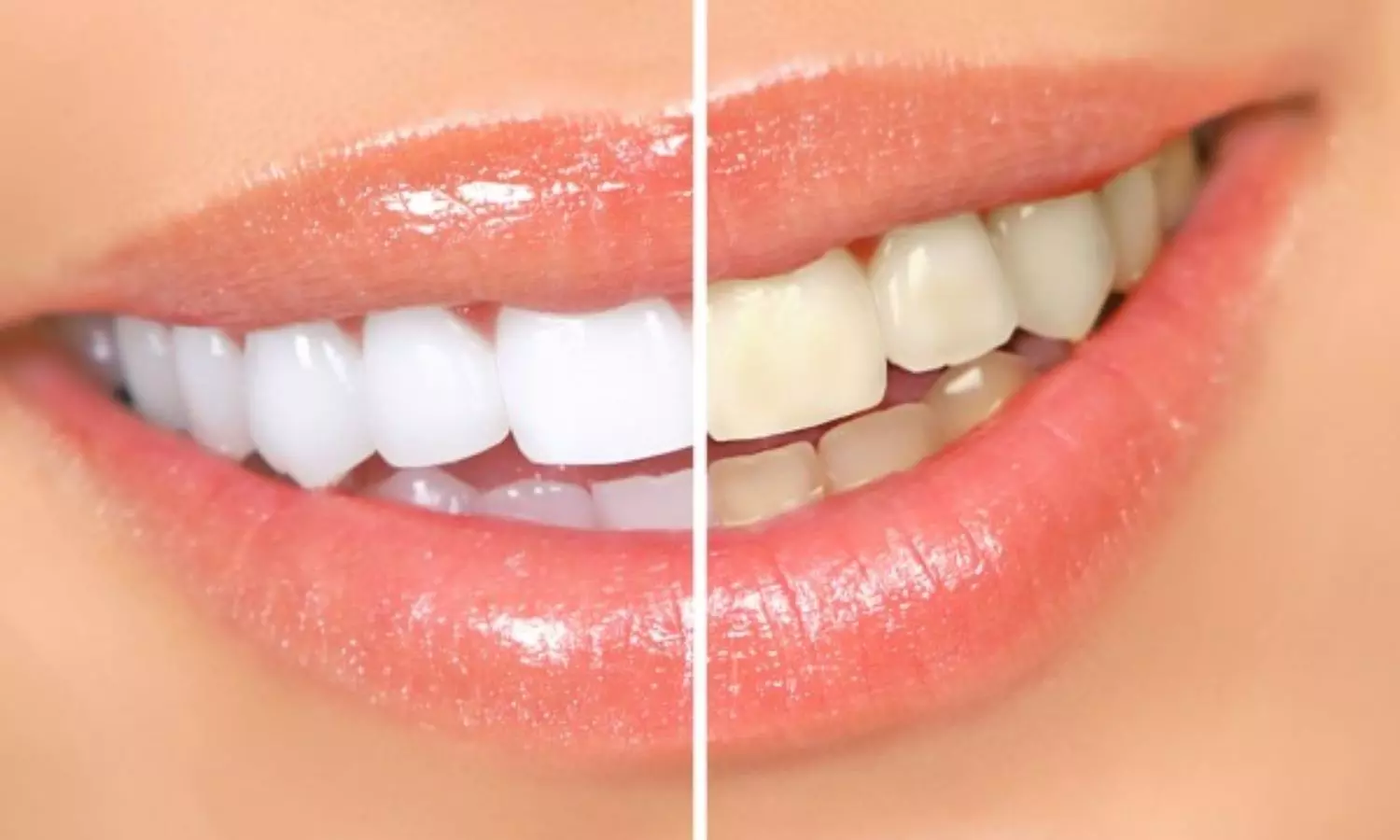
At-Home Bleaching equally effective for Upper and Lower Dental Arches, suggests study

A new study published in the Journal of Dentistry unveiled a curious phenomenon in cosmetic dentistry that teeth whitening treatments may be more effective on upper canines when compared to their lower counterparts. 

Teeth bleaching remains one of the most sought-after aesthetic dental procedures, designed to counter both external and internal tooth discoloration. Yet, dental professionals have long observed inconsistent results between upper and lower arches during treatment. This research assesses the role of sublingual salivary glands, which may increase saliva contamination of bleaching trays in the lower jaw which might accelerate the degradation of bleaching gel.

This prospective study included 32 participants with a standardized three-week at-home bleaching protocol using 16% carbamide peroxide gel. The research team monitored color changes weekly, employing objective color measurement systems: CIELAB, CIEDE2000, and the Whiteness Index for Dentistry (WID), through spectrophotometric analysis.

The results of this study found no statistically significant differences in whitening results between the upper and lower arches (p > 0.5) for incisors. However, disparities emerged when examining the canines, or eye teeth. By the second and third weeks of treatment, upper canines expressed significantly better bleaching results than their lower counterparts, with statistical significance confirmed using both the CIELAB and CIEDE2000 color difference formulas (p < 0.05).

Also, the WID formula for assessing tooth whiteness did not detect significant differences in any of the tooth groups studied, suggesting that while instrumental measurements show variations, these differences may not be easily noticeable in everyday interactions.

The average color difference (ΔE) values in upper canines increased more rapidly than those in lower canines during the second and third weeks, indicating more pronounced lightening. The largest gap was observed during week three, coinciding with the phase when bleaching trays tend to show diminished gel efficacy.

Despite the statistical variances, the study concludes that the slightly reduced bleaching effect in lower canines is not clinically visible, confirming that both arches benefit substantially from carbamide peroxide-based treatments. Overall, these findings may prompt dental professionals to tailor bleaching regimens more precisely and consider anatomical and physiological differences when planning treatments.

Source:

Pereira-Lores, P., Gancedo-Gancedo, T., Martín-Biedma, B., Varela-Aneiros, I., Dablanca-Blanco, A. B., Villasenín-Sánchez, C., Martín-González, J., Alonso de la Peña, V., & Castelo-Baz, P. (2025). Is at-home bleaching more effective on the upper arch than the lower arch? A prospective cohort study. Journal of Dentistry, 157(105729), 105729. https://doi.org/10.1016/j.jdent.2025.105729

Powered by WPeMatico

Children with liver disease face dramatically higher risk of early death: Study

Researchers from University of California San Diego have found that children diagnosed with metabolic dysfunction-associated steatotic liver disease (MASLD) are at significantly increased risk of premature death and serious long-term health complications. The findings, published April 22, 2025 in Hepatology, the scientific journal of the American Association for the Study of Liver Diseases, come from the Longitudinal InVestigation Evaluating Results of Steatosis (LIVERS) study, which followed 1,096 children over an average of 8.5 years. Nearly half of all deaths in the cohort were liver-related, and the overall mortality rate was 40 times higher than that of similar peers in the general U.S. population.

“Every child or young adult who died was a tragedy,” said Jeffrey Schwimmer, M.D., professor of pediatrics at UC San Diego School of Medicine and director of the Fatty Liver Clinic at Rady Children’s Hospital. “MASLD poses a real and measurable threat to pediatric health. To truly change outcomes, we need better tools to diagnose this disease, treatments that work for children, and systems that ensure every child has access to that care.”

MASLD, formerly known as nonalcoholic fatty liver disease (NAFLD), is the most common chronic liver disease in children, affecting nearly 10% of all youth and up to 25% of those with obesity. The reclassification to MASLD reflects growing recognition that fatty liver disease in children is closely linked to metabolic dysfunction, including obesity, hypertension and type 2 diabetes. Yet despite its prevalence, little has been known about the long-term risks it poses to children.

The LIVERS study is the most comprehensive assessment to date of clinical outcomes in pediatric MASLD. Conducted at Rady Children’s Hospital, the retrospective cohort study used medical records and National Death Index data to follow children ages 2 to 18 who were diagnosed with MASLD between 2000 and 2017. Over an average of 8.5 years of follow-up, 3.4% of children had died — a mortality rate more than 40 times higher than expected based on national averages and more than 50 times higher than expected based on state averages. These mortality rates are far higher than what has been reported in children with obesity or type 2 diabetes alone, suggesting that MASLD may confer independent risk beyond associated metabolic conditions.

The risk of death was higher in certain children, especially boys and those with low levels of high-density lipoprotein (HDL), often called the “good” cholesterol. Nearly half of the deaths were caused by liver disease itself, while others were due to heart-related conditions, accidents or suicide.

In addition to the risk of early death, many children in the study developed serious health problems while still in their teens or twenties. These included high blood pressure (14%), obstructive sleep apnea (9.5%) and type 2 diabetes (7.3%). Problems with blood fats, such as high triglycerides or low HDL, were even more common — making dyslipidemia, the presence of abnormal levels of fats (lipids) in the blood, the most frequent complication overall.

While many children showed improvement with clinical care, a substantial portion still experienced disease progression. The findings highlight how much we have yet to learn about the natural course of MASLD in children and reinforce the need for better tools to detect, monitor and manage this condition.

“We have shown that MASLD in children is a serious disease with life-threatening potential,” Schwimmer said. “Our hope is that these findings lead to greater awareness and stronger investment in pediatric-specific diagnostics, treatments and systems of care. With the right tools and commitment, we can make a meaningful difference in outcomes.”

Future research is needed to identify which children are at highest risk for progression to cirrhosis and early death, and to determine how lifestyle, medication or surgical interventions might change the disease course. For now, researchers emphasize that early recognition, consistent follow-up and greater public and provider awareness remain critical to protecting the health of children with MASLD.

Reference:

Schwimmer, Jeffrey B, Long-term mortality and extrahepatic outcomes in 1,096 children with MASLD: A retrospective cohort study, Hepatology, DOI: 10.1097/HEP.0000000000001357.

Powered by WPeMatico

Breakthrough approach for diagnosing TB could significantly improve detection

A new strategy for tuberculosis (TB) screening, proposed by a team of researchers led by Queen Mary University of London, provides a solution to problems with current TB screening, which does not always accurately detect disease. Simultaneously screening for both active and dormant TB infection could save lives, curtail infection rates, and rewrite the story of the continued spread of this disease. Researchers hope that this new approach will inform guidance from global health organisations and key decision-makers on the most effective way to screen for TB.

TB is the deadliest infectious diseases globally, with 10.8 million new cases and 1.25 million deaths in 2023. Effective screening for TB is an important intervention to quickly detect the disease, prevent its spread and ultimately eliminate the disease. TB can remain dormant in the body for some years without causing the symptoms of the disease but may cause active disease in the future especially among certain people at high risk. Accurate testing of both active and dormant infection is therefore of critical importance in screening programmes.

Testing for TB can take a range of forms, including chest x-rays, sputum cultures, molecular testing, screening for symptoms, and skin or blood tests to measure dormant TB infection (TBI). All types of tests aim to accurately detect TB with low numbers of false positive results to avoid unnecessary treatment.

A team led by Dr Dominik Zenner, Clinical Reader in Infectious Disease Epidemiology at Queen Mary, analysed the efficacy of combinations of commonly used tests for TB. Their study, published today in the European Respiratory Journal, examined data on 13 different TB tests as described in 437 original studies and published systematic reviews. These data were then used to estimate how good screening tests are at both correctly identifying TB and avoiding false positive results.

Unlike previous studies, the researchers found that including some immunological tests for dormant TB infection (TBI tests) added value to TB screening algorithms. TBI tests could also support earlier diagnosis of harder to detect TB, including extrapulmonary TB (disease that occurs outside the patient’s lungs) or TB in children.

This novel approach challenges existing protocols for TB testing, which reserve TBI tests for diagnosing dormant TB only. Importantly, combining diagnostic tests for both dormant and active TB would allow for both forms of the disease to be detected concurrently. This opportunity to improve detection of TB across large populations is of interest to policy makers and public health organisations, including WHO and the European Centre for Disease Prevention and Control.

Dr Zenner said: “Global TB control requires early identification and treatment of TB in risk groups. Our novel screening algorithms show that screening for active and dormant TB can be done simultaneously with high accuracy for migrants from countries where TB is common to improve individual and population benefits.”

Mario Raviglione, former Director of the Global Tuberculosis (TB) Programme at the World Health Organization and currently Professor of Global Health at the University of Milan, said: “This is a very sophisticated and well-thought study with major implications for clinical and public health practice, as well as policy change. By reviewing sound evidence and applying modern statistical methods, the authors have concluded that adopting an Interferon Gamma Release Assay (IGRA) test – a blood test that detects the response of white blood tests to TB antigens – on top of more traditional TB screening methods, the accuracy of screening migrants for active TB increases significantly.”

East London has the highest number of newly diagnosed TB cases in Western Europe with TB disproportionately affecting the most deprived populations. Researchers at Queen Mary are at the forefront of changing this picture, both locally and globally, and last month they partnered with Barts Health NHS Trust to form a new centre of excellence for TB research and treatment. 

Reference:

Dominik Zenner Hassan Haghparast-Bidgoli Tahreem Chaudhry, How to diagnose TB in migrants? A systematic review of reviews and decision tree analytical modelling exercise to evaluate properties for single and combined TB screening tests, European Respiratory Journal, https://doi.org/10.1183/13993003.02000-2024.

Powered by WPeMatico

DECT Leads to Faster Tophus Resolution Than Ultrasound in Gout Study

A new study published in the journal of Joint Bone Spine showed that dual-energy computed tomography (DECT) results in quicker complete tophus resolution compared to ultrasound in patients with gout.

Gout is the most prevalent cause of inflammatory arthritis globally and is brought on by the accumulation of monosodium urate (MSU) crystals following prolonged hyperuricemia. The inflammatory reaction to these MSU crystal deposits results in tophus development, gout flares, persistent arthritis, and ultimately erosive gout. The goal of managing gout is to use urate-lowering treatment (ULT) to completely dissolve MSU crystals, enabling flare-free gout remission.

Since ultrasound (US) may identify MSU deposits on the surface and tophi of cartilage, rheumatologists are increasingly using it as an addition to their clinical assessment. Dual-energy computed tomography offers direct assessment of substantial MSU crystal deposits and is a helpful technique for differentiating between urate, bone, and soft tissue. The study’s primary goal was to assess the tophus volume dynamics in gout patients receiving treat-to-target (T2T) urate-lowering medication throughout the first 24 months of treatment using dual-energy CT and ultrasound.

The patients with gout and US tophi who had never used ULT were included in this multicenter, prospective, 24-month longitudinal pilot research. At baseline, 6, 12, and 24 months, clinical visits were conducted, along with DECT and US scans of the knees and foot. The index tophus was determined to be the largest tophus recognized by the United States. The Spearman correlation coefficient (ρ) was used to evaluate the correlation between the tophus index volume measured with US and with DECT at all timepoints, as well as the change in absolute volume and relative change from baseline.

The study comprised 55 patients, most of whom were male, aged 63.1 (12.3), and whose baseline blood urate levels were 8.73 mg/dL [7.93; 9.52]. From 0.1 cm3 [0; 0.63] at baseline to 0 cm3 [0; 0] at month 24, the index tophus volume assessed with US changed from the median [inter-quartile range] 0.61 cm3 [0.30; 1.20] at baseline to 0.07 cm3 [0; 0.50] at month 24.

The index tophus volume changed by −56% [−90; 0] and −96% [−100; −34] at M6, −84% [−100; −13] and −100% [−100; −89] at M12, and −96% [−100; −72] and −100% [−100; −100] at M24 for the same measurements. Relative tophus volume change showed a slight association at month 6 and a strong correlation at months 12 and 24. Overall, there is more variation in volumetric measurements during follow-up since complete tophus resolution is achieved at 24 months of T2T in DECT but not in the US.

Reference:

Pascart, T., Richette, P., Bousson, V., Ottaviani, S., Ea, H.-K., Lioté, F., Latourte, A., Bardin, T., Ora, J., Pacaud, A., Vandecandelaere, M., Luraschi, H., Jauffret, C., Laurent, V., Boissel, M., Norberciak, L., Legrand, J., Lefevre, G., Ducoulombier, V., & Budzik, J.-F. (2025). Time-course of tophus resolution on Dual-energy CT and ultrasound after 24 months of a treat-to-target strategy: Results from GOUT-DECTUS study. Joint, Bone, Spine: Revue Du Rhumatisme, 92(4), 105892. https://doi.org/10.1016/j.jbspin.2025.105892

Powered by WPeMatico

Elevated Serum Uric Acid Increases Risk of Diabetic Retinopathy and Kidney Disease: Study

Researchers have discovered in a new study that high serum uric acid (SUA) levels are highly correlated with the risk of diabetic retinopathy (DR) and chronic kidney disease (CKD) in patients with long-term diabetes. Eye and kidney complications of diabetes are a serious problem in clinical settings. The cross-sectional study at Shanghai General Hospital found an evident correlation of increased SUA levels with the progression of the disease. Based on these observations, it seems that SUA may be an important risk factor for diabetic complications. The study was published in BMC Diabetology & Metabolic Syndrome by Hanying Wang and colleagues.

This research studied 1015 diabetic patients who were assessed from October 2018 to October 2021. Patients were screened with SUA measurement, renal function tests, and DR examination by fundus photography. Multivariable ordinal logistic models were applied to obtain odds ratios (ORs) and 95% confidence intervals (CIs) to evaluate DR and CKD progression. In addition, receiver operating characteristics (ROC) curves were employed to determine SUA thresholds, by which subjects could be divided into low and high SUA groups.

Key Findings

  • SUA Levels and CKD Progression: Increased SUA levels were observed in patients with later CKD stages versus stage 1 CKD (p < 0.001).

  • SUA and Vision-Threatening Diabetic Retinopathy (VTDR): The levels of SUA were higher in VTDR patients than in patients with no DR (p = 0.019).

  • Risk of DR: Higher SUA levels indicated a higher probability of DR (OR: 1.002; 95% CI: 1.001–1.004).

  • Risk of CKD: Increased SUA levels indicated a higher risk of CKD (OR: 1.008; 95% CI: 1.006–1.011).

  • Thresholds for Risk Rise: SUA values above 354.0 µmol/L (95% CI: 318.9–393.2) were linked to a 1.571-fold higher risk of DR (95% CI: 1.139–2.099, P = 0.006).

  • SUA and Risk of CKD: SUA values above 361.0 µmol/L (95% CI: 339.2–386.3) were associated with a 1.395-fold higher risk of CKD (95% CI: 1.033–1.885, P = 0.030).

  • Gender-Specific Analysis: Positive correlation of elevated SUA levels with DR and CKD in both male and female patients.

This research offers robust evidence for an independent association between high SUA levels and diabetic retinopathy and chronic kidney disease risks in long-term diabetic patients. The results highlight the probable predictive value of SUA in the prediction of diabetic complications, highlighting the importance of routine SUA monitoring in diabetes care.

Reference:

Wang, H., Gu, L., Ma, Y. et al. High levels of serum uric acid are associated with microvascular complications in patients with long-term diabetes. Diabetol Metab Syndr 17, 106 (2025). https://doi.org/10.1186/s13098-025-01656-1

Powered by WPeMatico