Real-World Study Confirms Baricitinib’s Effectiveness for Alopecia Areata

A recent real-world study conducted in China has confirmed that baricitinib, a Janus kinase (JAK) inhibitor, significantly improves hair regrowth in patients with various forms of alopecia areata, including totalis, universalis, and eyebrow/eyelash loss. The study observed a substantial reduction in the Severity of Alopecia Tool (SALT) scores, with an average improvement from 70.21 at baseline to 48.51 after treatment. A total of 47 patients met inclusion criteria. At weeks 12 and 24, 47% and 53% achieved SALT 20, respectively. By the end of treatment (24–104 months), 78.7% achieved SALT 20. The overall SALT score improvement before and after treatment was 48.51 (±30.36), with a hair regrowth rate of 68.10% ± 35.45%. Patients with disease duration ≤ 4 years showed significantly greater SALT score improvement and shorter time to hair regrowth onset compared to those with > 4 years. At week 24, 85.2% and 90.4% achieved ClinRO scores of 0/1 for eyebrow and eyelash alopecia, respectively. Negative correlations were observed between disease duration and hair regrowth rate, a positive correlation was found between treatment duration and hair regrowth rate. Baricitinib was well tolerated with no serious adverse events. These findings align with previous research supporting baricitinib’s efficacy and safety in treating severe alopecia areata. The study’s results suggest that baricitinib is a promising treatment option for patients with various forms of alopecia areata, offering significant improvements in hair regrowth without serious side effects.

Reference:

Wada-Irimada, M., Takahashi, T., Sekine, M., et al. (2025). Long-term real-world outcomes of baricitinib in severe alopecia areata: A 104-week retrospective analysis from a single institute. Journal of Dermatology. https://doi.org/10.1111/1346-8138.17829

Keywords: baricitinib, alopecia areata, hair regrowth, SALT score, real-world study, JAK inhibitor, totalis, universalis, eyebrow loss, eyelash loss.


Powered by WPeMatico

Pregnancy Physical Activity Questionnaire reliable tool for assessing Physical Activity in pregnant women: Study

Physical activity (PA) is associated with significant
positive impacts on both physical and mental health in non-pregnant populations.
Several health organisations have strongly suggested that PA is directly
correlated to a lower risk of heart disease, diabetes type 2, metabolic
syndrome, anxiety, and some types of cancer. Considering the physical changes
that occur during pregnancy, the maternal lifestyle can be a modifying factor
for both maternal and foetal health. For example, pregnant women are at risk of
pregnancy complications such as gestational diabetes and preeclampsia, which
are related to maternal obesity and inactivity.

Traditionally, pregnancy constitutes a barrier to PA due to
certain factors, beliefs, and lifestyle restraints. Moreover, pregnancy related
symptoms, including vomiting, abdominal discomfort, and easy fatigue, along
with safety concerns as well as limited scientific in formation and social
support, have steered pregnant women away from being active. However, over the
last few decades, scientific societies have issued guidelines recommending that
pregnant women without complications should engage in PA. For example, the
American College of Obstetricians and Gynecologists (ACOG) recommends at least
150 min of moderate-intensity PA per week. Despite these recommendations, only
a small percentage of pregnant women actually meet them. Few studies, with
limited numbers of cases, have examined PA levels during pregnancy in some
parts of the world. These studies used questionnaires with a small number of
participants and compared the data with the data collected using smartwatches
to estimate actual PA levels.

The pregnancy physical activity questionnaire (PPAQ) is
specially designed for pregnant women, which considers several hinderances that
women face during pregnancy. Several advantages, such as low cost, easy
implementation, self-administration, and ease of accessing a high volume of
individuals, make the questionnaire a suitable means to evaluate and promote PA
in a large population. The PPAQ has been translated into several languages and
adopted by various cultures, including the recently introduced Greek version.
This study aimed to record the degree of PA in pregnant Greek women during the
three trimesters of pregnancy and examine possible associations with
gestational age (GA), maternal age, education, and population of the
residential area. Authors tested the Greek version of the PPAQ and examined the
reliability of answers using a smartwatch in several cases.

This prospective study comprised two stages. The first stage
was the completion of the PPAQ Greek version twice, with a one-week interval
between the two rounds, to assess internal consistency and repro ductivity. The
validity was evaluated by comparing the PPAQ results with data from
smartwatches that recorded step counts and metabolic equivalent tasks (METs).
The second stage included pregnant women from various regions of Greece that
completed the PPAQ once during their pregnancy. The level of PA during
pregnancy and consistency with the international PA recommendations was
evaluated with the use of METs.

A total of 1058 pregnant women filled the PPAQ Greek
version, while eighteen women randomly participated in the reliability and
validity analyses of the PPAQ. Results from the PPAQ completed one week apart
showed strong agreement (Intraclass Correlation Coefficient [ICC]: 0.82–0.96).
A comparison between the PPAQ results and smartwatch data revealed no
significant differences in PA levels (correlation coefficient: 0.542; p =
0.022). The median total PA for all the participants was 142.1 MET-hours/week
(interquartile range: 106.04 MET-hours/week).

PA levels increased with advancing gestation in
sports/exercise (1st vs. 2nd trimester, p = 0.048; 2nd vs. 3rd trimester, p
< 0.001), occupational activities (1st vs. 3rd trimester, p < 0.001; 2nd
vs. 3rd trimester, p = 0.024), and transportation activities (1st vs. 3rd
trimester, p < 0.001; 2nd vs. 3rd trimester, p < 0.001).

Moderate-to-vigorous intensity activities also increased
with gestational age (p = 0.023, 95 % confidence interval [CI]: 0.1–1.4; p =
0.045, 95 % CI: 0–0.08, respectively).

Women were more active in household/ caregiving activities
across all trimesters (p = 0.017; 95 % CI: 0.09–0.95).

Women with higher educational levels were more likely to
engage in sports/exercise (p = 0.039, 95 % CI: − 3.3 to − 0.08) and had higher
overall PA levels (p = 0.029, 95 % CI: 0.37–6.3). However, only 14.8 % of the
participants met the international PA recommendations for pregnancy.

The results of this study demonstrate that the total PA
during pregnancy increases with advancing gestation. Women become more
physically active as pregnancy progresses, mainly in terms of household and
sports/exercise activities. A possible explanation is that women are familiar
with this new condition and might feel confident about the well-being of their
pregnancy. In addition, fear of a potential miscarriage and several symptoms
that are present in early pregnancy (fatigue, nausea, and vomiting) also play a
significant role in preventing pregnant women from engaging in activities.
Regarding the mode of conception, pregnant women who conceived through ART had
a significantly lower total level of PA, with similar findings for all
subcategories, except for vigorous-intensity and sedentary activities. Women
who conceived spontaneously performed significantly fewer sedentary activities
than those in the ART group. Most couples using ART are older and have experienced
from long-term infertility. Authors postulated that these women are
overcautious in protecting their pregnancy, which was achieved through
additional effort; thus, it is difficult for them to adopt an active lifestyle.

Another interesting point is the association between
maternal educational level, residential area population, and PA. Although the
total amount of PA between the two groups was similar, pregnant women with
higher educational levels performed significantly more sports/exercise
activities. This may be because well-educated people are usually better
informed and open to following new habits and recommendations. As far as
residential areas are concerned, broad and easy access to multiple exercise or
sports activities for citizens in areas with large populations could explain
our significant results. Moreover, the regression analysis showed that women of
advanced maternal age compared to younger ones, are more physically active in
most of the PA intensities, mainly referring to moderate and vigorous
activities. This finding is of great significance because it highlights the
fact that younger women participate in less energy “demanding” activities,
while the total level of PA does not differ. Pregnant women at a greater GA
conduct more household/caregiving activities. Furthermore, women with a
university degree were more likely to meet the recommendations for
moderate-intensity exercise during pregnancy.

The Greek version of the PPAQ is a reliable and valid tool
for assessing the PA levels in pregnant women. Moreover, the results of this
study demonstrate that pregnant women in the 3rd trimester, those with a higher
educational level, and those living in cities with large populations were more
active in sports and exercise. On the other hand, those who conceived of using
ART were less physically active. This highlights the need for clinicians to
promote PA during pregnancy in Greece so that exercise is part of the daily
routine of pregnant women.

Source: I. Mitrogiannis et al.; European Journal of
Obstetrics & Gynecology and Reproductive Biology 307 (2025) 29–33

Powered by WPeMatico

Use of Bevacizumab in Lung Cancer Patient linked to rare kidney complication: Case Report

China: A team from The Second Clinical College of Guangzhou University of Chinese Medicine, led by Rui Jiang, has documented a rare case of bevacizumab-associated glomerular microangiopathy (Bmab-GMA) in BMC Nephrology, adding to growing evidence of serious kidney-related side effects tied to the cancer drug.

The case involved a 59-year-old man with stage IIIA right lung adenocarcinoma who underwent surgery in 2021, followed by targeted treatment with bevacizumab after developing metastatic disease in 2023. Within months of initiating therapy, the patient developed new-onset high blood pressure, swelling in the lower limbs, and worsening proteinuria. Laboratory tests confirmed progressive kidney dysfunction, with rising serum creatinine levels and falling serum albumin.

Despite dose reduction and eventual discontinuation of bevacizumab, kidney damage persisted. Upon admission in April 2024, the patient’s 24-hour urine protein excretion was markedly elevated at over 4.6 g/day, while his estimated glomerular filtration rate had dropped to 41.27 mL/min/1.73 m². Blood tests also revealed increased levels of the soluble complement membrane attack complex (C5b-9), indicating possible activation of the complement system — a clue that may shed light on the disease mechanism.

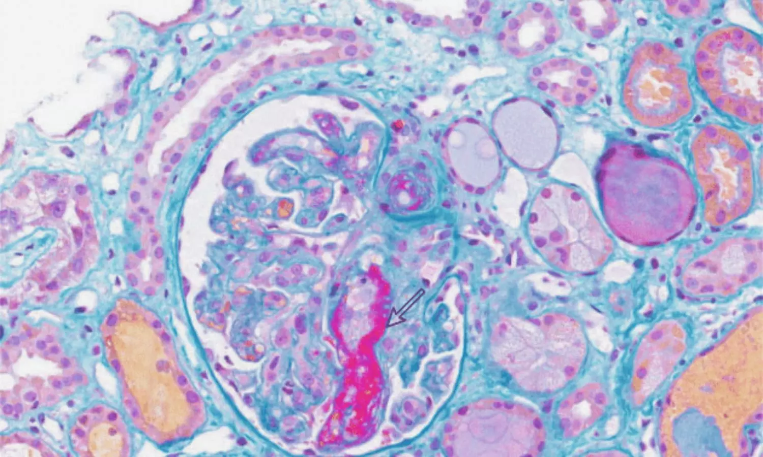
Renal biopsy provided definitive evidence of Bmab-GMA. Microscopic examination revealed mesangial expansion, double-contour formation of the glomerular basement membrane, and pseudothrombotic deposits. Immunofluorescence showed deposition of multiple immunoglobulins and complement components, while electron microscopy identified subendothelial and mesangial electron-dense deposits, widening of the subendothelial space, and partial loss of podocyte foot processes.

These findings confirmed the diagnosis of bevacizumab-induced glomerular microangiopathy, a condition that can cause both acute kidney injury and nephrotic syndrome. Notably, the case mirrors previous reports linking anti-VEGF agents, such as bevacizumab, to unique patterns of renal injury.

The patient was managed with antihypertensive medication (amlodipine), diuretics for edema control, and statin therapy for lipid abnormalities. Cancer treatment was switched to pralatrexate and anlotinib. At follow-up in July 2024, his hypertension persisted, kidney function remained impaired, and proteinuria had worsened to nearly 8 g/day.

According to the authors, the case highlights several important clinical lessons:

  • Oncologists and nephrologists should maintain a high index of suspicion for renal complications in patients receiving bevacizumab.
  • Early detection through regular monitoring of blood pressure, kidney function, and urinary protein can help mitigate irreversible damage.
  • Complement activation, as indicated by elevated C5b-9, may play a central role in the disease process and could represent a target for future therapies.

While Bmab-GMA remains rare, its potential severity makes awareness crucial. As anti-VEGF therapies continue to be widely used in oncology, further research is needed to define their pathogenesis, risk factors, and optimal management strategies.

Reference:

Jiang, R., Yu, Rz., Yang, Hf. et al. Bevacizumab-associated glomerular microangiopathy: a case report and literature review. BMC Nephrol 26, 445 (2025). https://doi.org/10.1186/s12882-025-04385-9

Powered by WPeMatico

FDA Approves Brensocatib as First Treatment for Non-Cystic Fibrosis Bronchiectasis

The FDA has approved oral brensocatib (Brinsupri) for adults and children 12+ with non-cystic fibrosis bronchiectasis. As a first-in-class reversible DPP-1 inhibitor, it targets the disease’s underlying inflammation, which can cause permanent lung damage and affects about 500,000 people in the U.S.

BRINSUPRI is the first and only FDA-approved treatment for NCFB, giving hundreds of thousands of patients and clinicians across the U.S. an option to manage this chronic and progressive disease that can lead to permanent lung damage and lung function decline.

“This FDA approval represents a potential paradigm shift in how we approach non-cystic fibrosis bronchiectasis,” said Doreen Addrizzo-Harris, M.D., FCCP, the Fiona and Stanley Druckenmiller Professor of Pulmonary, Critical Care and Sleep Medicine at NYU Grossman School of Medicine and Director of the NYU Langone Health Bronchiectasis and NTM Program, and ASPEN investigator. “For the first time, we have a treatment that directly targets neutrophilic inflammation and addresses a root cause of bronchiectasis exacerbations. Based on the strength of the data and the impact we’ve seen in patients, I believe this could become the new standard in non-cystic fibrosis bronchiectasis care.”

There are approximately 500,000 people in the U.S. diagnosed with NCFB, and it is estimated that millions more are living with this disease globally. Unlike other respiratory diseases that are characterized by airway narrowing, bronchiectasis causes airways to permanently widen, making it harder to clear mucus and bacteria, leading to persistent inflammation and infection. A hallmark of bronchiectasis is frequent exacerbations, or flares, when symptoms worsen, such as coughing, increased mucus, shortness of breath and fatigue.

“Non-cystic fibrosis bronchiectasis deeply affects the lives of people living with this chronic lung condition, impacting both their physical health and emotional well-being,” added Elisha Malanga, Executive Director of the Bronchiectasis and NTM Association. “Many patients experience frequent flares, which can disrupt daily life and potentially lead to disease progression. The FDA approval of brensocatib represents a significant and long-awaited advancement as the first approved therapy for non-cystic fibrosis bronchiectasis. Our hope is that treatments like this will enable people with bronchiectasis to manage their condition.”

This approval is based on data from the Phase 3 ASPEN and Phase 2 WILLOW studies, which were both published in the New England Journal of Medicine. In ASPEN, patients taking BRINSUPRI 10 mg or 25 mg had a 21.1% and 19.4% reduction in annual rate of exacerbations respectively, as compared to placebo. Both dosage strengths of BRINSUPRI also met several exacerbation-related secondary endpoints, including significantly prolonging the time to first exacerbation and significantly increasing the proportion of patients remaining exacerbation-free over the treatment period. Patients who received BRINSUPRI 25 mg experienced statistically significant less decline in lung function, as measured by forced expiratory volume in one second (FEV₁) after using a bronchodilator, at week 52. The safety of BRINSUPRI was also evaluated in both studies. The most common adverse reactions ≥2% in the ASPEN trial included upper respiratory tract infection, headache, rash, dry skin, hyperkeratosis, and hypertension. The safety profile for adult patients with NCFB in WILLOW was generally similar to ASPEN, except for a higher incidence of gingival and periodontal adverse reactions in WILLOW.

“The FDA approval of the first-ever treatment for non-cystic fibrosis bronchiectasis is a historic milestone for patients and for Insmed,” said Martina Flammer, M.D., MBA, Chief Medical Officer of Insmed. “By keeping patients at the center of everything we do, we have once again delivered a first-in-class medicine for a disease with no prior approved treatments. This is an incredible achievement in medicine. We’re deeply grateful to the patients, providers, and advocates who made this possible – this is just the beginning of what we can accomplish together for this community.”

BRINSUPRI is a first-in-class dipeptidyl peptidase 1 (DPP1) inhibitor, designed to inhibit the activation of enzymes (neutrophil serine proteases) in neutrophils that are key drivers of chronic airway inflammation in NCFB. BRINSUPRI is the first approved therapy to address the underlying inflammatory process of NCFB.

In parallel, applications for brensocatib with the European Medicines Agency (EMA) and the Medicines and Healthcare products Regulatory Agency (MHRA) have been accepted, and the Company plans to file in Japan in 2025. Commercial launches are anticipated in 2026, pending approval in each territory.

BRINSUPRI is now available in the U.S. by prescription through a comprehensive specialty pharmacy network.

About BRINSUPRI™ (brensocatib)

BRINSUPRI™ (brensocatib) is a small molecule, once-daily, oral, reversible inhibitor of dipeptidyl peptidase 1 (DPP1) indicated for the treatment of non-cystic fibrosis bronchiectasis (NCFB) in adult and pediatric patients 12 years of age or older. Brensocatib is designed to inhibit the activation of enzymes (neutrophil serine proteases) in neutrophils that are key drivers of chronic airway inflammation in NCFB. Brensocatib is also being evaluated for its potential role in other neutrophil-mediated diseases.

Powered by WPeMatico

MIND Diet Linked to Lower Dementia Risk via Reduced Hippocampal Sclerosis: Study

An autopsy study published in the Journal of American Medical Association revealed that higher adherence to the Mediterranean–Dietary Approaches to Stop Hypertension Intervention for Neurodegenerative Delay (MIND) diet was associated with lower odds of hippocampal sclerosis and less dementia at death, suggesting the diet’s protective effect may be partly mediated through reduced hippocampal damage.

Severe neuronal loss and astrogliosis in coronal sections of the mid-hippocampus cornu ammonis 1 or subiculum is known as hippocampal sclerosis (HS). This pathology is linked to dementia, Alzheimer’s disease, and limbic-predominant age-related transactive response DNA-binding protein 43 encephalopathy neuropathological change (LATE-NC). In humans, nothing is known about the relationship between food and HS or HS and LATE-NC. Thus, this study examined the relationship between HS and HS with LATE-NC and the MIND diet.

Autopsied patients from the ongoing Rush Memory and Aging Project cohort research were included in this investigation. Researchers included participants with neuropathological and nutritional data from 2004 to 2024. Analysis of the data was place between April 3, 2024, and May 13, 2025. The average MIND diet scores were calculated using all verified food frequency questionnaires that were given every year for up to 18 years before to death.

Hematoxylin and eosin staining was used to determine the presence of HS, while TDP-43 immunohistochemistry was used to identify the presence of LATE-NC in eight different brain areas. A semiquantitative measure was used to evaluate the degree of hippocampus neuronal loss in a group of 300 patients. The scale ranged from 0, which indicates no neuronal loss, to 5, which is classified as none to mild, moderate, and severe neuronal loss.

Of the 809 participants (mean [SD] age at death, 91.3 [6.1] years; mean [SD] follow-up, 7.2 [4.4] years; 538 [72%] female; mean [SD] follow-up, 7.2 [4.4] years), 82 (10.1%) had HS; 71 (9%) had both HS and LATE-NC; and in the scored subset, 35 (12%) and 43 (14%) had severe hippocampus neuronal loss.

After adjusting for age at death, sex, education, total calories, APOE-ε4 status, AD, and vascular pathologies, higher MIND diet scores were linked to decreased risks of HS (odds ratio [OR], 0.78; 95% CI, 0.65 to 0.95) and HS with LATE-NC (OR, 0.79; 95% CI, 0.64 to 0.97).

The MIND diet was linked to a 21% indirect connection through HS (β = -0.05; 95% CI, -0.10 to -0.01; P =.02) and a lower incidence of dementia at the time of death (β = -0.26; 95% CI, -0.36 to -0.15; P <.001) in mediation analyses. Less hippocampus neuronal loss was linked to a higher MIND diet score (P for trend =.01).

Overall, MIND diet adherence throughout follow-up was linked to a decreased risk of HS, HS with LATE-NC, and hippocampus neuronal loss in this cohort analysis of autopsied subjects. According to these results, the MIND diet may lessen negative effects on brain health.

Reference:

Agarwal, P., Agrawal, S., Wagner, M., Cherian, L. J., Aggarwal, N. T., James, B. D., Holland, T. M., Bennett, D. A., Barnes, L. L., Leurgans, S. E., & Schneider, J. A. (2025). MIND diet and hippocampal sclerosis among community-based older adults. JAMA Network Open, 8(8), e2526089. https://doi.org/10.1001/jamanetworkopen.2025.26089

Powered by WPeMatico

Early Postpartum Contraceptive Implant Placement Safe for Breastfeeding females: Study Finds

USA: Researchers have found in a new study that inserting a contraceptive implant within the first 24 hours postpartum is safe for breastfeeding individuals. This finding supports users, healthcare providers, and hospitals in offering placement at a time that best meets the user’s needs.

The study was published online in the American Journal of Obstetrics and Gynecology on 20 March 2025.

The researchers note that breastfeeding and contraception are vital in patient and public health. The etonogestrel contraceptive implant, a widely used long-acting progestin contraceptive, is highly effective and has few medical restrictions. Although there are concerns that early exposure to exogenous progestin might affect lactation, previous studies have shown that immediate placement does not hinder milk production or long-term breastfeeding. However, no studies have directly compared implant placement within 24 hours of delivery to placement at a later postpartum visit.

To address this, Jamie W. Krashin, Department of Obstetrics & Gynecology, University of New Mexico, Albuquerque, New Mexico, 1 University of New Mexico, 4ACC, Albuquerque, New Mexico, USA, and colleagues examined whether inserting the implant within 24 hours of childbirth affects breastfeeding continuation compared to placement at least two weeks later. Breastfeeding rates were assessed at eight weeks postpartum to determine if timing influenced lactation success.

For this purpose, the researchers conducted a non-inferiority randomized controlled trial at two university hospitals in the Mountain West, enrolling postpartum participants planning to breastfeed and use the etonogestrel implant. Eligible participants, aged 13 or older with term deliveries and no contraindications, were randomly assigned to receive the implant within 24 hours or at least two weeks postpartum. Baseline data were collected through chart reviews and questionnaires, while breastfeeding status and implant use were tracked via electronic surveys at multiple postpartum intervals. The study assessed a 15% non-inferiority margin for breastfeeding at 8 weeks in the per-protocol population.

The study led to the following findings:

  • One-hundred-fifty participants were enrolled (78 in the immediate group and 72 in the delayed group).
  • After excluding 8 participants who declined implant placement, withdrew, or were ineligible, and 16 who lacked primary outcome data, the modified intention-to-treat analysis included 126 participants (69 immediate, 57 delayed).
  • The per-protocol analysis included 115 participants (62 immediate, 53 delayed) after excluding 11 who received the implant outside the designated timeframes.
  • Both groups were similar in age, race, ethnicity, previous breastfeeding experience, delivery mode, and epidural use, but prior implant use was less common in the delayed group (29% vs. 43%).
  • At eight weeks postpartum, 77.4% in the immediate group and 81.1% in the delayed group reported any breastfeeding. The -3.7% difference exceeded the predefined non-inferiority margin, with a lower confidence interval of -16%.
  • In the modified intention-to-treat analysis, the difference between groups was smaller (-0.6%) and within the non-inferiority margin (-12.8% lower confidence limit), with 78.3% in the immediate group and 78.9% in the delayed group reporting any breastfeeding.
  • Implant continuation at 24 weeks was slightly higher in the immediate group (96%) compared to the delayed group (85%), though the difference was not statistically significant.
  • Exclusive or any breastfeeding and implant continuation through 24 weeks were similar between both groups.

The study found no significant differences in breastfeeding or implant continuation between immediate and delayed placement, despite the primary outcome slightly exceeding the non-inferiority margin.

“These findings support the safety and flexibility of initiating the etonogestrel implant at any preferred time postpartum for breastfeeding individuals,” the authors concluded.

Reference:

Krashin, J. W., Rivera-Montalvo, M., Leeman, L., Trujillo, V. Y., Petersen, T. R., Sanchez, D., Bender, E., Carroll, S., Bulleit, E., Alcantara, J., Turok, D. K., Black, P., & Espey, E. (2025). Breastfeeding after immediate versus delayed postpartum contraceptive implant placement: A non-inferiority randomized controlled trial. American Journal of Obstetrics and Gynecology. https://doi.org/10.1016/j.ajog.2025.03.019

Powered by WPeMatico

For eye infections, delayed doctor visits and extent of damage affect vision outcomes: Study

Even though microbial keratitis is a preventable cause of blindness, it’s a complex infection to treat, and many patients have lasting vision impairment.

Previous studies have identified several risk factors for reduced vision after treatment, including delays in symptom onset, longer duration of symptoms and existing eye disease.

However, these studies were restricted to one geographic area or a single type of organism that causes the infection.

In a study published in Ophthalmology, physicians followed 562 patients in the United States and India over a three-month period to identify risk factors that could be easily managed with early intervention.

Maria A. Woodward, M.D., an associate professor of ophthalmology and visual sciences and a member of the Institute for Healthcare Policy and Innovation, answers questions about what causes microbial keratitis, symptoms and preventative measures.

What are the common causes for microbial keratitis?

Woodward: We are surrounded by microorganisms, including Staphylococcus aureus, Streptococcus and fungi, which can get into our eyes through small injuries.

This is a common outcome in patients who do not have good contact lens hygiene or have an underlying eye condition that makes them prone to infections.

Although we don’t know the prevalence rates in the U.S., worldwide it affects over 2 million people per year.

What are symptoms of microbial keratitis?

Woodward: It depends on the infection. Some types of bacterial infections can progress incredibly quickly while fungal infections are slower.

Early symptoms include eye pain, severe eye redness and blurry vision. Some patients also experience light sensitivity.

Unfortunately, many ignore these warning signs and continue to use contact lenses, which can make the problem worse.

If left untreated, the condition causes eye pain and can ultimately penetrate deeper into the eye resulting in scarring and vision loss.

What factors affect treatment for microbial keratitis?

Woodward: The majority of infections in the U.S. are usually treated with antibiotic eye drops.

In our study, we found that if a patient had reduced vision or waited for symptoms to worsen, treatments did not work as well.

Interestingly, we saw that patients in the U.S. who wore contact lenses had slightly better outcomes.

We suspect that their physicians could easily identify what kind of infections are associated with wearing lenses and were able to treat them earlier.

In India, we observed a similar effect in patients with a history of eye trauma.

In rural areas, it’s common for people to get something in their eye that can lead to an infection.

It’s possible that the physicians there knew what kind of infection they might have, given their environment and history, and were able to administer treatments sooner.

What future directions are you excited to pursue?

Woodward: Although this work doesn’t represent everyone globally, we found some commonalities regardless of where the participants were from.

It is important to expand these studies to include factors such as access to medications and adherence to eyedrop regimens.

As part of this work, we took serial photographs of patients over time to monitor how their eye characteristics change over time.

We can use this to help physicians distinguish between mild and severe cases and treat patients effectively.

Reference:

Woodward, Maria A. Factors Associated with Vision Outcomes in Microbial Keratitis, Ophthalmology, DOI: 10.1016/j.ophtha.2025.02.004 

Powered by WPeMatico

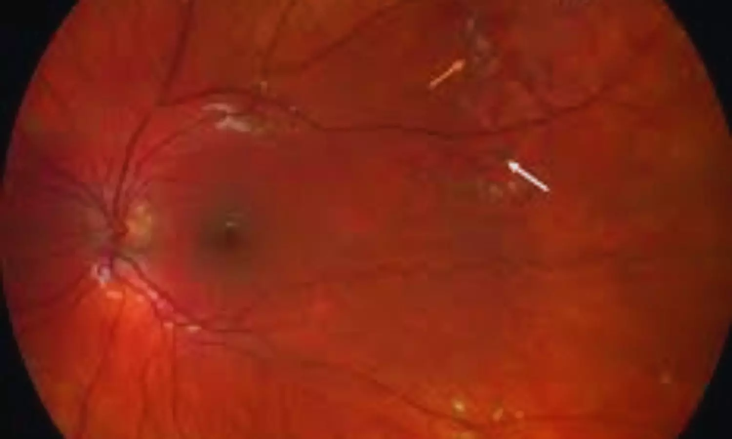
Dexamethasone Implant Promising as Adjunctive Therapy in Coats’ Disease: Study Finds

Turkey: A retrospective single-center study published in BMC Ophthalmology has highlighted the potential role of intravitreal dexamethasone implants as an adjunctive therapy in managing Coats’ disease alongside conventional ablative treatments. The research, conducted by Ozlem Ural Fatihoglu and colleagues from the Department of Ophthalmology at Dokuz Eylül University, İzmir, Turkey, assessed the efficacy and safety of this approach over a long-term follow-up period.

The study evaluated patients treated between February 2012 and January 2024 who received a dexamethasone (Dex) implant in addition to standard ablative therapy, with a follow-up duration exceeding 12 months. Out of 15 enrolled patients, the analysis included 11 patients. The cohort primarily consisted of males (90.9%) with unilateral disease, while the only female participant presented with bilateral involvement. The mean age at diagnosis was 14.8 years, spanning a wide age range from 2 to 60 years, highlighting the diverse presentation of the disease.

The following were the notable findings of the study:

  • At baseline, 58.3% of the eyes were classified as stage 3a1, 25% as stage 3a2, and 16.7% as stage 2b.
  • Over an average follow-up of nearly 66 months, visual acuity remained stable in 50% of the treated eyes.
  • One-third of the eyes showed measurable improvement of at least one Snellen line.
  • Two eyes demonstrated a two-line gain, while the other two eyes achieved a single-line gain in visual acuity.
  • Vision deteriorated in both eyes during the study period.
  • Transient intraocular pressure (IOP) elevation was observed in 66.7% of treated eyes.
  • Cataract progression occurred in 58.3% of eyes, with one-third requiring cataract surgery.
  • One eye developed a secondary vasoproliferative tumor, necessitating CyberKnife therapy and vitrectomy.
  • No severe implant-related complications were reported, indicating a favorable safety profile for the treatment.

The findings suggest that intravitreal dexamethasone implants may serve as a useful adjunctive option, particularly in pediatric patients, to reduce the need for repeated general anesthesia and in adults with extensive macular exudation, where conventional ablative therapy alone may be insufficient. While the outcomes are promising, the authors acknowledge key limitations, including the retrospective design, small sample size, absence of a control group, and the study’s setting in a tertiary referral center, which may have resulted in a higher proportion of advanced cases.

Despite these limitations, the research highlights the value of dexamethasone implants in achieving anatomical and functional stabilization in Coats’ disease. With half of the treated eyes maintaining stable vision and one-third showing improvement, the study adds evidence supporting the integration of steroid implants into multimodal treatment strategies,” the authors wrote.

They recommend further studies to validate these findings and better define patient subgroups that may benefit most from this adjunctive approach.

Reference:

Ural Fatihoglu, O., Ayhan, Z., Ozturk, T. et al. Place of dexamethasone implant as an adjunctive treatment in Coats’ disease: a retrospective single-center study. BMC Ophthalmol 25, 435 (2025). https://doi.org/10.1186/s12886-025-04279-2

Powered by WPeMatico

Imposter Syndrome among Orthopaedic Surgery residents extremely common and disproportionately affects female residents: Study

Imposter syndrome is a psychological phenomenon in which individuals experience persistent self-doubt and feelings of inadequacy despite achieving objective measures of success. This phenomenon is associated with lower job satisfaction, increased rates of burnout, and reduced productivity.

Harsh Wadhwa et al conducted a study to determine the prevalence of imposter syndrome among orthopaedic surgery residents and determine whether specific individual or residency characteristics predict higher rates of imposter syndrome.

The study was performed at the Department of Orthopaedic Surgery, Stanford University Medical Center, Stanford, California.

An anonymous survey was distributed to residents at 7 ACGME-accredited orthopaedic surgery residencies by the authors. Respondents provided deidentified demographic data and completed the Clance Imposter Phenomenon Scale (CIPS) assessment. CIPS scores were used to categorize imposter syndrome severity as none to mild, moderate, significant, or intense. Differences were calculated and compared using t-test and x2 analyses. Multivariable regression analysis was used to identify predictors of increased symptom severity.

The key findings of the study were:

• One hundred orthopaedic surgery residents completed the survey.

• Forty percent of respondents were female, and the mean age was 30.6 ± 2.8 years.

• The prevalence of significant or intense imposter syndrome was 73%.

• CIPS scores and significant or intense imposter syndrome did not vary by age, gender, sexual orientation, race/ethnicity, level of training, program region, research year, non-MD degrees, and Step 1, Step 2, or Orthopaedic In-Training Examination (OITE) scores on univariable analyses.

• On multivariable analysis, female residents were 5.64 (OR = 5.64 [1.04-30.63]) times more likely to have significant or intense imposter syndrome (p = 0.045).

• Residents at western programs were 0.13 (OR = 0.13 [0.02-0.90]) times as likely to experience significant or intense imposter syndrome (p = 0.039).

• Women (estimate = 8.72 [1.41-16.04]; p = 0.0201) and those with OITE scores in the 0 to 25th (estimate = 11.92 [0.29- 23.54]; p = 0.0446) and 51st-75th (estimate = 12.73 [2.79-22.67]; p = 0.0128) percentiles had higher CIPS scores.

The authors concluded that – “Imposter syndrome is highly prevalent among orthopaedic surgery residents, with significant or intense symptoms affecting 73% of those surveyed. Female gender and lower OITE scores were associated with increased imposter syndrome severity, while training at an orthopaedic surgery residency program in the West was associated with decreased imposter syndrome. Additional research is needed to identify effective interventions to address imposter syndrome and minimize its effects on individuals’ careers. Implementing focused initiatives to address imposter syndrome may help alleviate burnout and promote both career fulfillment and professional growth.”

Further reading:

Imposter Syndrome Among Orthopaedic Surgery Residents is Extremely Common and Disproportionately Affects Female Residents

Harsh Wadhwa et al

JBJS Open Access 2025:e24.00132. http://dx.doi.org/10.2106/JBJS.OA.24.00132

Powered by WPeMatico

125 MBBS seats at AIIMS Kalyani Check 2025 admission guidelines, fee, complete details here

Kalyani- All India Institute of Medical Sciences (AIIMS) Kalyani has released a prospectus for admission into the MBBS course for the academic year 2025. The Course of studies leading to the award of Bachelor of Medicine and Bachelor of Surgery (MBBS) of the AIIMS shall last for a minimum of five and a half academic years, including one year’s compulsory internship.

As per the prospectus, a total of 125 seats for Indian nationals are available for admission to the MBBS course. The reservation matrix would be as per the Medical Counselling Committee (MCC) directives. AIIMS Kalyani has an MBBS course divided into three professional divisions, i.e. First, Second and Third Professional MBBS are of one, one and a half and two years duration respectively.

Admitted candidates must attend the Orientation Program and join classes on the stipulated date. The admission of the candidates, who fail to join the Course by the date stipulated in the letter of intimation, shall automatically be cancelled and such seats shall then be offered to the candidates on the waiting list in order of merit in the same category

The candidates selected for admission to the MBBS course at the AIIMS Kalyani, after verification of their eligibility and being offered admission, should bring the following original certificates/ testimonials/documents at the time of admission-

1 Certificate of having passed the 10+2 or equivalent Examinations, showing the subject offered by him/her in the Examination.

2 Certificate from the Board from which he/she passed the High School/Higher Secondary Examination, showing his/her date of birth.

3 Certificate/Mark sheet from the Board from which he/she passed the 10+2 or equivalent Examination showing that he/she has secured 60% or more marks (50% in case of SC/ST/Persons with Benchmark Disability) in aggregate in English, Physics, Chemistry and Biology.

4 Migration Certificate from the University/Board last attended by him/her.

COMPULSORY ROTATORY INTERNSHIP TRAINING

After passing the Final MBBS Examination, all the candidates will be required to do a compulsory internship for a period of one calendar year (12 months) in the hospital and rural health centres recognised by the AIIMS. During the internship period, the interns are allowed a total of 15 days leave in the full term of one year. The degree of MBBS shall be awarded after satisfactory completion of one year’s internship.

FEES & OTHER PAYMENTS

The dues are to be paid in cash immediately, after the candidates have been declared medically fit in the Medical Board of the Institute, as per details given below-

S.NO

ACADEMIC & OTHER FEES

AMOUNT IN RS

1

Registration Fee

25

2

Caution Money

100

3

Tuition Fee

1350

4

Laboratory Fee

90

5

Student Union Fee

63

TOTAL

1628

S.NO

HOSTEL & OTHER FEES

AMOUNT IN RS

1

Hostel Rent*

990

2

Gymkhana Fee

220

3

Pot Fund

1320

4

Electricity Charges

198

5

Mess Security (Refundable)

500

6

Hostel Security (Refundable)

1000

TOTAL

4228

To view the prospectus, click the link below

https://medicaldialogues.in/pdf_upload/aiims-kalyani-mbbs-2025-125-seats-offered-know-complete-admission-rules-fee-structure-297700.pdf

Powered by WPeMatico