Gluconolactone with multiple skin benefits ideal for use in cosmetic and dermatologic procedures

Poland: A recent study published in the Journal of Cosmetic Dermatology has shown a significant impact of gluconolactone (GLA) chemical peel treatment on lowering skin pH and transepidermal water loss (TEWL). GLA was also observed to have seboregulatory properties.

“GLA chemical peel treatment significantly lowered skin pH at all measurement points. Also, there was a significant reduction in TEWL around eyes, right cheek, and left forehead after GLA treatment,” the researchers reported. “No significant differences were observed between different GLA concentrations.”

The skin is a barrier to potentially harmful external factors, with a particular focus on protection against infection and water loss. The barrier’s quality can be assessed using objective instrumental measurements that determine skin parameters such as pH, transepidermal water loss, sebum level, and hydration. Barrier disorders are affected by the use of irritants, chemicals, excessive degreasing (which can affect TEWL increase), internal factors, hormonal disorders, allergies, dermatological diseases, and dietary deficiencies.

Gluconolactone is an oxidized glucose lactone derivative. In nature, GLA is found in cheese, honey, wine, tofu, and fruit juices. GLA exhibits moisturizing and antioxidant effects. It also presents soothing effects, improves the function of the skin barrier, and protects elastin fibres from UV-induced degradation.

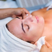
Sylwia Jarząbek-Perz, Medical University of Lódź, Lódź, Poland, and colleagues, therefore, aimed to evaluate skin parameters such as transepidermal water loss, pH, sebum levels before, during, and after a series of applications of 10% and 30% GLA chemical peel in a split-face model.

The study included 15 female subjects. Three split-face procedures were conducted using two GLA solution concentrations applied on two sides of the face. The skin parameters were measured before treatments and 7 days after the last procedure at four measurement sites on either side of the face, that is, on the forehead, on the cheek, around the eye, and on the nose wing.

The study led to the following findings:

  • Measurement of sebum demonstrated some statistically significant changes between sebum levels in the cheeks after a series of treatments.
  • The pH measurement showed a reduction in pH value after each treatment at all measurement points.
  • The level of TEWL after treatments was significantly lower around the eyes, on the left forehead, and on the right cheek.
  • There were no significant differences between the use of different concentrations of the GLA solution.

“The findings reveal the multiple skin benefits of gluconolactone, making it an ideal ingredient for use in cosmetology, cosmetic, and dermatologic procedures,” the researchers concluded.

Reference:

Jarząbek-Perz, S., Dziedzic, M., Rotsztejn, H., & Kołodziejczak, A. (2023). Evaluation of the effects of 10% and 30% gluconolactone chemical peel on sebum, pH, and TEWL. Journal of Cosmetic Dermatology, 22(12), 3305-3312. https://doi.org/10.1111/jocd.15864

Powered by WPeMatico

Benidipine calcium channel blocker may reduce cigarette smoke-induced senescent cells and improves emphysema

France: In a recent study published in Aging, the researchers performed a chemical screen, using an FDA-approved drug library, to identify compounds selectively promoting the death of CS-induced senescent lung cells. 

In the study, researchers Alberta Palazzo, and colleagues from France revealed that benidipine calcium channel blocker promotes the death of cigarette smoke-induced senescent cells and improves lung emphysema.

Smoking is the main risk factor for many lung diseases including chronic obstructive pulmonary disease. Cigarette smoke (CS) contains carcinogenic and reactive oxygen species that favor DNA mutations and perturb the homeostasis and environment of cells. CS induces lung cell senescence resulting in a stable proliferation arrest and a senescence-associated secretory phenotype. It was recently reported that senescent cell accumulation promotes several lung diseases.

“Here, our aim was to identify senolytic compounds in the context of CS-induced senescence and to assess whether they improved lung emphysema.”

Aside from the well-known senolytic, ABT-263, the researchers identified other potentially new senescence-eliminating compounds, including a new class of molecules, the dihydropyridine family of calcium voltage-gated channel (CaV) blockers. Among these blockers, Benidipine decreased senescent lung cells and ameliorates lung emphysema in a mouse model. The dihydropyridine family of CaV blockers thus constitutes a new class of senolytics that could improve lung diseases.

“Hence, our work paves the way for further studies on the senolytic activity of CaV blockers in different senescence contexts and age-related diseases.”

Reference:

Palazzo A, Makulyte G, Goerhig D, Médard J, Gros V, Trottein F, Adnot S, Vindrieux D, Flaman J, Bernard D. Benidipine calcium channel blocker promotes the death of cigarette smoke-induced senescent cells and improves lung emphysema. Aging (Albany NY). 2023 Dec 12; 15:13581-13592 . https://doi.org/10.18632/aging.205259

Powered by WPeMatico

Antioxidants after post-space irrigation increase bond strength favouring glass fiber post-cementation: Study

Brazil: A recent study published in the Journal of Esthetic and Restorative Dentistry has shed light on the effect of antioxidants after post-space irrigation on the adhesive interface of glass fibre post-cementation.

The researchers revealed that the antioxidants (10% sodium ascorbate and 20% alpha-tocopherol) used after post-space irrigation with sodium hypochlorite appear to increase the bond strength favouring the glass fibre post-cementation.

Teeth treated endodontically are characterized by decreased mechanical strength of the intraoral chewing forces and loss of coronal structure. A recent meta-analysis showed that fibre posts were associated with higher overall survival rates than metal posts when used for restoring endodontically treated teeth with extensive coronal destruction. Several characteristics favour the clinical choice of fibre posts; they include the aesthetic advantages, the more conservative removal of dentin, and greater treatment agility since their cementation does not require a laboratory step.

Beatriz Dansini Cândido, São Paulo State University – UNESP, Araraquara, SP, Brazil, and colleagues conducted the study to evaluate the effects of antioxidants, 10% sodium ascorbate (SA) or 20% alpha-tocopherol (AT), after post-space irrigation with 2.5% sodium hypochlorite +17% EDTA (SH) or 1% peracetic acid (PA) on the adhesive interface after glass fibre post cementation.

Sixty bovine roots were treated endodontically. Following the preparation, the post-space was irrigated with PA or SA followed or not by antioxidants (AT or SA; n=10) use.

Failure mode, push-out bond strength test, and dentin penetrability analysis were performed using a confocal laser microscope in the cervical, middle, and apical thirds. One-way ANOVA and Tukey post hoc test were used to evaluate data from dentinal penetrability and bond strength.

The researchers reported the following findings:

  • SH showed the lowest bond strength regardless of the third.
  • In the apical third, mixed failure was the most incident in all groups. Only in the cervical third of the post-space, SH-AT provided the greatest tag extension of the cementation system into dentin.
  • In the middle and apical thirds, SH-AT, SH-SA, and PA-SA provided the largest tag extensions, but similar to each other.

“Antioxidants only favoured bond strength when 2.5% sodium hypochlorite +17% EDTA was used and dentin penetrability of the adhesive and conventional resin cementation, irrespective of the solution used to irrigate the post-space,” the researchers concluded.

Reference:

Cândido, B. D., Manzoli, T. M., Zaniboni, J. F., Besegato, J. F., Godoy, E. F., Kuga, M. C., & Rached Dantas, A. A. (2023). Effect of antioxidants after post-space irrigation on the adhesive interface of glass fiber post cementation. Journal of Esthetic and Restorative Dentistry, 35(8), 1293-1300. https://doi.org/10.1111/jerd.13081

Powered by WPeMatico

Rare case of Left fourth and sixth costovertebral dislocation abutting the aorta: a report

While rib fractures are common in blunt thoracic trauma, dislocations of the costovertebral joints (CVJs) are extremely rare and typically involve the first, eleventh, or twelfth rib.

Natalia Gorelik et al reported a rare case of dislocation of the left fourth and sixth CVJs in a 36-year-old man who was run over by a car. It has been published in “Skeletal radiology” journal.

A 36-year-old man was admitted to the emergency department of a level I trauma center after he was run over by a car at approximately 40 km per hour. On arrival, his level of consciousness was fluctuating, and his vital signs were unstable. He was complaining of chest pain and difficulty breathing and had a respiratory rate of 32 breaths per minute with an oxygen saturation of 88%. Physical examination revealed a lip laceration, a road rash covering 40% of his anterior abdominal surface, a large hematoma on the right flank, and pelvic tenderness. Portable radiographs of the chest and pelvis revealed multiple bilateral displaced rib fractures, a left clavicular fracture, small bilateral pneumothoraces, subcutaneous emphysema, and left lung patchy opacities concerning for pulmonary contusions, as well as multiple pelvic fractures with a left sacroiliac joint diastasis. Bilateral chest tubes were placed, and the patient was intubated. The focused assessment with sonography in trauma (FAST) was positive for intra-abdominal free fluid.

The patient was taken to the operating room for an emergency exploratory laparotomy. He was found to have a large retroperitoneal zone 3 hematoma and a grade 3 splenic injury. A splenectomy and preperitoneal packing were performed. He also underwent angioembolization of bilateral internal iliac arteries. Post-operatively, he was transferred to the intensive care unit. A CT of the head, facial bones, cervical spine, thorax, abdomen, and pelvis obtained later the same day revealed multiple injuries, including bilateral hemopneumothoraces, pneumomediastinum, pulmonary contusions, grade 3 splenic injury, left adrenal hematoma, retroperitoneal hematoma, Morel-Lavallée lesions at bilateral hips, right acromioclavicular joint separation, left sacroiliac joint diastasis, and multiple fractures, namely, at the nasal bones, scapulae, clavicles, ribs, pelvis, right distal radius, and right L1 and bilateral C7 and L5 transverse processes.

Regarding the thoracic cage injuries, on the right, there were fractures at the neck and at the costochondral junction of the first rib, and comminuted fractures of the second through seventh ribs posteriorly. On the left, there were fractures of the head and neck of the first rib; costal cartilages of the first, second, and fourth ribs; costochondral junctions of the fifth and sixth ribs; and body of the second through ninth ribs at more than one site except for the eight rib (flail chest). There was subluxation of the left fourth costotransverse joint and a fracture/dislocation of the left fourth costocentral joint; there was a tiny intra-articular chip fracture of the posterior aspect of the head of the rib. There was also a subluxation of the left sixth costocentral joint. The heads of both the left fourth and sixth ribs abutted the posterior aspect of the proximal descending aorta. These costovertebral joint injuries and their proximity to the aorta were not described on the initial CT report.

The patient underwent multiple subsequent chest CTs for ongoing fever. The last scan performed 54 days post trauma demonstrated persistent costovertebral joints malalignment and no aortic injury. A transverse fracture through the superior endplate of T4 with no significant loss of height or involvement of the posterior wall was occult on the initial CT and became apparent on subsequent CTs. Over the course of his hospitalization, the patient underwent surgical fixation of the pelvic fractures. His recovery was complicated by infarctions at the left posterior inferior cerebellar artery and right posterior cerebral artery territories. The patient was discharged to a rehabilitation center after a 75-day hospitalization.

The authors commented – “In conclusion, this case report presented dislocations of non-consecutive left fourth and sixth CVJs where the rib head abutted the aorta. It documents an unusual presentation of a rare entity and suggests that conservative management may be acceptable in some of these cases. It also raises awareness among radiologists about these injuries, which can be easily overlooked, but are important for the stability of the thoracic spine. Finally, this report emphasizes the association between CVJ dislocations and spine injuries, which can sometimes be occult on initial imaging. Future studies with larger sample sizes and long-term follow-up could help determine the optimal management of CVJ dislocations.” 

Further reading:

Left fourth and sixth costovertebral dislocation abutting the aorta

Natalia Gorelik, Dany Croteau et al

Skeletal Radiology (2024) 53:187–192

https://doi.org/10.1007/s00256-023-04415-3

Powered by WPeMatico

Inflow control may safely reduce blood loss in laparoscopic subsegmentectomy of liver

A recent study scrutinized various inflow control methods used during laparoscopic subsegmentectomy in liver resections to determine their impact on patient outcomes. This study was published in BMC Surgery by Wang and colleagues. The study offers clinicians substantial evidence that both the half-Pringle and Pringle maneuvers are safe and effective methods for inflow control during laparoscopic liver resections. Conducted between October 2010 and December 2020, the study evaluated 133 patients who underwent laparoscopic subsegmentectomy under the care of a single surgeon at a specific institute. The study aimed to compare perioperative and long-term outcomes associated with three different inflow control techniques.

The 133 patients were categorized into three groups based on the inflow control method used: no inflow control (49 patients), half-Pringle maneuver (46 patients), and Pringle maneuver (38 patients). Analysis revealed differences among the groups, notably a lower proportion of patients with cirrhosis in the half-Pringle maneuver group, fewer previous abdominal or liver surgeries in the same group, and more anterolateral segment tumors in the no inflow control group.

  • Cirrhosis Rates: Half-Pringle group: Lower proportion (P = .02).
  • Previous Surgeries: Fewer abdominal and liver surgeries in the half-Pringle group (P = .01 and P = .02 respectively).
  • Tumor Location: More tumors in anterolateral segments in the no inflow control group (P = .001).
  • Operative Outcomes: Shorter operation time in the no inflow control group (P < .001). Less blood loss observed in the no inflow control group (P = .03).
  • Postoperative Measures: No significant differences in the need for blood transfusion, morbidity, or hospital stay among the three groups.
  • Long-Term Survival: Overall survival did not significantly differ among the groups (P = .89).

The study reported that the operation time was shorter and blood loss was less in the no inflow control group compared to the other groups. However, there were no significant differences among the groups regarding the need for blood transfusion, postoperative complications, or length of hospital stay. Additionally, the overall survival rates did not differ significantly among the three groups. The findings suggest that both the half-Pringle and Pringle maneuvers showed comparable perioperative and long-term outcomes in laparoscopic subsegmentectomy.

Reference:

Wang, H.-P., Hou, T.-Y., Li, W.-F., & Yong, C.-C. Inflow control can be safely used in laparoscopic subsegmentectomy of the liver: a single-center 10-year experience. BMC Surgery,2023;23(1). https://doi.org/10.1186/s12893-023-02282-2

Powered by WPeMatico

Lower HDL-C, total cholesterol and apolipoprotein A-I levels encountered in multiple myeloma

Cyprus: A recent study published in Atherosclerosis Plus has reported a significant reduction in HDL-C, total cholesterol (TC), and apolipoprotein A-I (apoA-I) levels in patients with multiple myeloma (MM).

Hence, the researchers suggest considering lipid profile parameters when assessing such patients.

Multiple myeloma, also called plasma cell myeloma, is the second most common haematological malignancy, characterized by uncontrolled monoclonal proliferation of plasma cells in the bone marrow. In multiple myeloma, variations are seen in several laboratory and clinical parameters, including those in lipid profiles, particularly in high-density lipoprotein cholesterol (HDL-C).

HDL have many protective properties, including anti-apoptotic, anti-oxidative, and anti-inflammatory effects. HDL and their major constituent, apolipoprotein A-I, inhibit hematopoietic stem and progenitor cell proliferation. Consequently, low levels of apoA-I and/or HDL-C are associated with an increased risk of haematological cancers like MM, while they may also promote resistance to therapy and cancer aggression.

To date, the association between hypoalphalipoproteinemia and MM has not been studied well, despite relevant supportive evidence. Therefore, Anastasios Makris, School of Medicine, European University Cyprus, Nicosia, Cyprus, and colleagues performed a systematic review and meta-analysis to determine the value of HDL-C and other lipid parameters as biomarkers in multiple myeloma disease.

For this purpose, the researchers searched online databases for studies with evidence on HDL-C in MM patients. A qualitative synthesis was performed of published retrospective and prospective studies for the role of HDL-C and other lipid profile parameters in MM. Additionally, a meta-analysis was performed on HDL-C mean differences (MD) between MM cases and controls.

The study led to the following findings:

  • Fourteen studies (3 prospective, 11 retrospective) comprising 895 MM patients were eligible for the systematic review.
  • Ten studies compared HDL-C levels in MM patients with healthy controls. In these ten studies (n = 17,213), pooled analyses showed that MM patients had significantly lower HDL-C levels compared to healthy controls (MD: −13.07 mg/dl).
  • Regarding secondary endpoints, total cholesterol (MD: −22.19 mg/dl) and apolipoprotein A-I (−40.20 mg/dl) demonstrated significant decreases, while differences in low-density lipoprotein cholesterol (LDL-C) (MD: −11.33 mg/dl) and triglycerides (MD: 9.93 mg/dl) were not significant.

The meta-analysis provides clarity that HDL-C and, apoA-I and total cholesterol do not only reflect cardiovascular risk but also represent important features in MM.

“Our findings point towards a need for implementing larger clinical studies that will provide more robust data on the clinical significance of the association between HDL-C and MM, and clinical value of lipid panel addition in assessing MM patients,” the researchers wrote.

“Finally, future research perspectives in multiple myeloma include investigating the role of lipids as potential biomarkers of treatment response and disease burden,” they concluded.

Reference:

Makris, A., Pagkali, A., Nikolousis, E., Filippatos, T. D., & Agouridis, A. P. (2023). High-density lipoprotein cholesterol and multiple myeloma: A systematic review and meta-analysis. Atherosclerosis Plus, 54, 7-13. https://doi.org/10.1016/j.athplu.2023.09.003

Powered by WPeMatico

High-dose vitamin D During pregnancy Fails to Reduce Autism and ADHD risk in kids

Denmark: Researchers from the University of Copenhagen reported in their original research article that their study revealed a reduced risk of autism, a lower load of autistic symptoms, and a decreased risk of ADHD diagnosis for mothers with higher levels of 25(OH)D before starting the intervention. However, high-dose vitamin D3 supplementation during pregnancy had no impact on the risk of autism or ADHD.

This investigation is published in the American Journal of Clinical Nutrition.

During pregnancy, a deficiency of vitamin D may lead to a higher likelihood of developing autism or attention deficit hyperactivity disorder (ADHD).

This study aimed to determine the impact of vitamin D3 supplementation during pregnancy on the risk of developing autism and ADHD.

This randomized clinical trial was conducted as part of the COPSYCH project, which is nested within the COPSAC2010 cohort, a population-based sample of 700 healthy mother-child pairs (enrolled at week 24 of pregnancy). The maternal 25-hydroxy-vitamin D (25(OH)D) levels were measured, and 623 mothers were randomly assigned to receive either high-dose (2800 IU/d) or standard-dose (400 IU/d) vitamin D3 until one week after giving birth (315 received high-dose, 308 received standard-dose). When the children reached the age of 10, the diagnoses and symptom load of autism and ADHD were assessed using the Kiddie Schedule for Affective Disorders and Schizophrenia for School-Age Children-Present and Lifetime Version (K-SADS-PL).

Key summary points are:

At ten years of age, a total of 591 children completed the psychopathological evaluation.

2.7 % (16 children) were diagnosed with autism.

11 % (65 children) were diagnosed with ADHD.

Of the 496 children in the vitamin D3 trial, 246 received high doses, and 250 received standard doses. Twelve children with autism and 58 with ADHD were diagnosed among them.

Higher maternal pre-intervention 25(OH)D levels were linked to lower risk of autism (OR per 10 nmol/L 0.76), reduced autistic symptom load (β per 10 nmol/L -0.03), and reduced ADHD diagnosis risk (OR per 10 nmol/L 0.88).

There was no association between High-dose vitamin D3 supplementation and autism or ADHD risk.

They said that higher maternal pre-intervention 25(OH)D reduces autism risk, ADHD diagnosis and autistic symptom load, but high-dose vitamin D3 does not affect these outcomes in pregnancy.

Lundbeck Foundation, the Ministry of Health, and the Danish Council for Strategic Research funded the study.

Reference:

Aagaard, K et al. High-dose vitamin D3 supplementation in pregnancy and risk of neurodevelopmental disorders in the children at age 10 – A randomized clinical trial. The American Journal of Clinical Nutrition.

Powered by WPeMatico

Transcranial direct current stimulation may boost mental processes in patients wit Alzheimer’s disease: Study

USA: Findings from a small clinical trial showed that twice-daily non-invasive electrical stimulation of the brain may boost mental processes (cognitive function) in patients with Alzheimer’s disease. The findings were published online in the journal General Psychiatry.

The technology, known as transcranial direct current stimulation, or tDCS for short, may help to fire up the brain’s plasticity, enabling ‘rewiring’ through the formation of new neural networks, the findings suggest.

tDCS comes in the form of a device with two electrodes, placed over specific areas of a person’s head, which deliver a constant low intensity electrical current. It is starting to be used in many areas of medicine, including for the treatment of depression.

The researchers wanted to find out if tDCS might improve cognitive function in people with Alzheimer’s disease, and if so, whether this might be linked to their recovering some level of cortical plasticity–the brain’s capacity to form new neural networks.

Some 140 patients with mild to moderate Alzheimer’s disease from four different hospitals were randomly allocated to receive either 2 daily sessions of active (constant low intensity 1-2 mA current) or sham tDCS, for 5 days of the week for a maximum period of 6 weeks.

The currents were applied to the prefrontal cortex, the region of the brain involved in higher order activities, such as planning, decision-making, working memory, moderating social behaviours, and controlling certain aspects of speech and language.

Participants were all aged over 65, had had their disease for more than 6 months, and had all scored below 26 on the Mini-Mental State Exam (MMSE). A score of 24 is regarded as abnormal and indicative of cognitive impairment. Both groups were comparable in terms of age, sex, and educational attainment.

The MMSE and the Alzheimer’s Disease Assessment Scale-Cognitive (ADAS-Cog) Test, which focuses on language and memory, were used to evaluate cognitive performance at the start of the trial, after 2 weeks, and again after 6 weeks.

Readings of electrical signalling through the motor pathways of the nervous system, known as motor evoked potential or MEP, were used to indicate changes in neural plasticity.

In all, 133 patients completed the 2-week intervention and 124 completed the 6-week intervention. Reasons for withdrawal varied, but no one withdrew because of discomfort.

The results showed that after 2 weeks there was no change in either group, but compared with baseline, 30 daily 20-minute sessions of tDCS significantly improved the cognitive function of those in the tCDS group, particularly their word recall, recall of test instructions, and word recognition. No such improvements were seen in the sham group.

The results also showed that in people with Alzheimer’s disease cortical plasticity is impaired, but that this improved after 6 weeks of tDCS.

In tandem with a decrease in MEP, word recall and word recognition got better among those in the tDCS group, but not among those in the sham group, suggesting that the improvement in cortical plasticity may reflect the degree of cognitive improvement, say the researchers.

But it’s still not clear how tDCS might exert its effects, they add, citing previous research, indicating that the technology may alter ion activity, neurotransmitter release, and electrical activity in various areas of the brain.

The researchers acknowledge various limitations to their findings, including the small study size, the lack of MRI or electroencephalography scans to chart changes in brain structure, and the absence of cerebrospinal fluid and blood samples to monitor neurotransmitter changes.

But they nevertheless conclude: “The results of this study strongly indicate that tDCS treatment is a significant and promising intervention for improving cognitive function in [Alzheimer’s disease]. In addition, plasticity plays a vital role in cognitive change.”

Reference:

Li X, Chen L, Yu K, et alImpact of twice-a-day transcranial direct current stimulation intervention on cognitive function and motor cortex plasticity in patients with Alzheimer’s diseaseGeneral Psychiatry 2023;36:e101166. doi: 10.1136/gpsych-2023-101166.

Powered by WPeMatico

Hyperosmolar hyperglycaemic state higher among type 1 diabetes patients compared to type 2 diabetes patients

Hyperosmolar hyperglycaemic state is  higher among patients with type 1 diabetes compared with type 2 diabetes suggests a new study published in the Diabetes Care

The hyperosmolar hyperglycemic state (HHS) is a rare and life-threatening complication of diabetes. We aimed to estimate the incidence of HHS and describe the clinical and biomarker profiles of patients with HHS, including subgroups with acidosis and acute kidney injury.

This nationwide, descriptive cohort study used Danish registry data during years 2016–2018 to identify acutely admitted patients fulfilling the hyperglycemia and hyperosmolarity criteria of HHS (glucose ≥33 mmol/L and osmolarity [2 × sodium + glucose] ≥320 mmol/L).

RESULTS

They identified 634 patients (median age, 69 years (first quartile; third quartile: 58; 79) who met the criteria of HHS among 4.80 million inhabitants aged ≥18 years. The incidence rates were 16.5 and 3.9 per 10,000 person-years among people with known type 1 (n = 24,196) and type 2 (n = 251,357) diabetes, respectively. Thirty-two percent of patients with HHS were not previously diagnosed with diabetes. Patients were categorized as pure HHS (n = 394) and combined HHS and diabetic ketoacidosis (HHS-DKA; n = 240). The in-hospital mortality rate for pure HHS was 17% and 9% for HHS-DKA.

The incidence of HHS was higher among patients with type 1 diabetes compared with type 2 diabetes. HHS is a spectrum of hyperglycemic crises and can be divided in pure HHS and HHS-DKA. In one-third of patients, HHS was the debut of their diabetes diagnosis.

Reference:

Emilie V. Rosager, Amalia Lærke K. Heltø, Cathrine U. Fox Maule, Lennart Friis-Hansen, Janne Petersen, Finn E. Nielsen, Steen B. Haugaard, Rasmus Gregersen; Incidence and Characteristics of the Hyperosmolar Hyperglycemic State: A Danish Cohort Study. Diabetes Care 2023; dc230988. https://doi.org/10.2337/dc23-0988

Keywords:

Hyperosmolar, hyperglycaemic, state, higher, among, patients, type 1, diabetes, compared, type 2 diabetes, Diabetes Care, Emilie V. Rosager, Amalia Lærke K. Heltø, Cathrine U. Fox Maule, Lennart Friis-Hansen, Janne Petersen, Finn E. Nielsen, Steen B. Haugaard, Rasmus

Powered by WPeMatico

Breakthrough Treatment with finerenone for Advanced Kidney Disease and Diabetes Cost-Effective

In the Netherlands, where the healthcare system grapples with the staggering burden of over one million patients combating type 2 diabetes (T2D), a significant breakthrough has emerged. A study evaluating the cost-effectiveness of integrating finerenone, a promising therapeutic addition to standard care, offers hope for patients with advanced chronic kidney disease (CKD) and T2D. The study results were published in the journal Cardiovascular Diabetology. 

T2D often brings along the ominous companion of CKD, affecting approximately 36% of patients in the Netherlands. The annual medical costs associated with T2D and CKD are a considerable financial strain, totaling around €1.3 billion and €805 million, respectively. The FIDELIO-DKD trial, a landmark investigation, showcased that the inclusion of finerenone in the standard of care (SoC) regimen significantly reduces the risk of CKD progression and cardiovascular (CV) events in patients dealing with CKD stages 2–4 linked to T2D.

To delve deeper into the potential impact of finerenone on healthcare economics, the study employed the validated FINE-CKD model. This Markov cohort model simulates the lifelong journey of patients, focusing on outcomes related to CV events and renal replacement therapy. The researchers tailored the model to reflect the Dutch societal perspective, considering both clinical effectiveness and economic implications.

Findings: 

  • Results from the study paint a promising picture. When finerenone is integrated into the SoC, patients experience an extension in the time free from CV events and renal replacement therapy, translating to 0.20 additional quality-adjusted life years (QALYs).
  • Perhaps even more compelling is the cost-saving aspect: finerenone led to a €6136 decrease in total lifetime costs per patient compared to SoC alone.
  • This financial benefit stems from the significant reduction in renal and CV events, showcasing finerenone as a dominant treatment option.
  • The study found that finerenone, when added to SoC, has an 83% probability of being a dominant strategy and a 93% probability of being cost-effective, particularly at a willingness-to-pay threshold of €20,000.

The implications of this study are groundbreaking, not just for patients facing the challenging intersection of T2D and advanced CKD but also for the broader healthcare system. By demonstrating that the integration of finerenone not only improves patient outcomes but also yields substantial cost savings, this research paves the way for more widespread adoption of this innovative treatment approach. In essence, the study offers a glimmer of hope for those navigating the complexities of diabetes and advanced kidney disease, showcasing that cutting-edge treatments can not only enhance lives but also contribute to the economic sustainability of healthcare systems. 

Further reading: 

Quist, S.W., van Schoonhoven, A.V., Bakker, S.J.L. et al. Cost-effectiveness of finerenone in chronic kidney disease associated with type 2 diabetes in The Netherlands. Cardiovasc Diabetol 22, 328 (2023). https://doi.org/10.1186/s12933-023-02053-6

Powered by WPeMatico