FDA Approves First Test to predict Elevated Risk of Developing Opioid Use Disorder

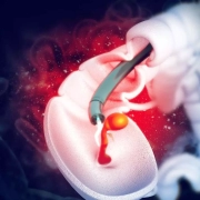
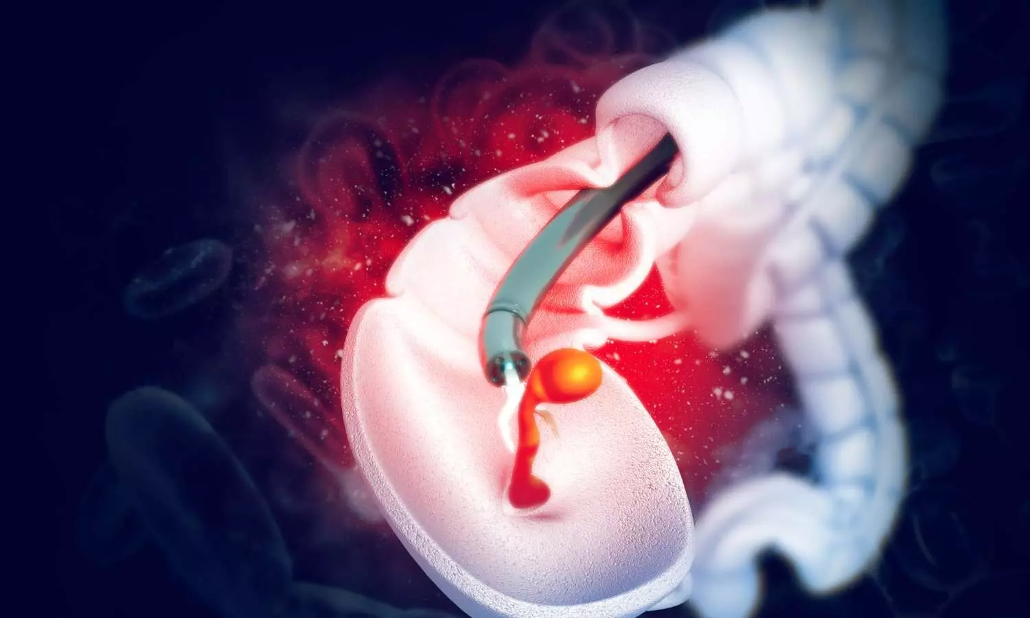
Today, the U.S. Food and Drug Administration approved the first test that uses DNA in assessing whether certain individuals may have an elevated risk of developing opioid use disorder (OUD). As part of a clinical evaluation, the AutoGenomics, Inc. AvertD test is intended to be used prior to first exposure to oral opioid pain medications in patients being considered for a 4-30 day prescription for the treatment of acute pain, such as in patients scheduled to undergo a planned surgical procedure.

The AvertD test, a prescription-use only genetic laboratory test for patients 18 years and older, is to be used only with patients who consent to the test and have no prior use of oral opioid analgesics. The test is administered by a health care provider by swabbing the cheek of a patient to collect a DNA sample that will be used to determine if a patient has a combination of genetic variants that may be associated with an elevated risk of developing OUD. This information should be used as part of a complete clinical evaluation and risk assessment; it should not be used alone to make treatment decisions. The test is not intended to be used in patients being treated for chronic pain. AvertD may help patients who are concerned about being treated with an opioid for acute pain make better informed decisions.

In August 2022, the FDA introduced the FDA Overdose Prevention Framework. Through the Framework, the agency has taken steps to address the drug overdose crisis and substance use disorder, including the approvals of the first nonprescription naloxone nasal spray product and the first generic nonprescription naloxone nasal spray product in March 2023 and July 2023, respectively. The FDA also published draft guidance on Clinical Considerations for Studies of Devices Intended to Treat Opioid Use Disorder in July 2023.

As part of the approval order, AutoGenomic, Inc. must provide training to health care providers to help ensure appropriate use of the test and conduct a large post-market study assessing device performance in patients, regularly reporting progress made toward completion of that study to the FDA.

An earlier version of the AvertD test was the focus of an October 2022 advisory committee meeting, in which a panel discussed data and information related to the test, provided recommendations and voted on the De Novo request for the test. Following the advisory committee meeting, FDA worked with AutoGenomics, Inc. as it modified its test. AutoGenomics subsequently submitted a Premarket Approval application (PMA) for the modified test. The advisory committee’s feedback helped inform the FDA’s evaluation of the test and today’s decision, including the conditions for approval.

The primary risks associated with AvertD, as with any in vitro diagnostic test, are false negative and false positive results. A false negative result could lead to a false sense of security for a patient who is at increased risk of OUD, and/or a health care provider considering prescribing an opioid analgesic which could result in a provider prescribing an opioid analgesic to a patient to whom they would otherwise not do so. The risks of a false positive result (i.e., incorrectly identifying an individual as having a higher risk of OUD) include inadequate pain management due to avoidance of opioids, which may prevent patients from receiving optimal medical care to treat their acute pain. The risks of false negative and false positive results can be mitigated, in part, through accurate, transparent product labeling and a health care provider training program. It is critical that users of the test (health care providers and patients) understand how to interpret the test result and use it not in isolation, but as part of a comprehensive clinical evaluation and risk assessment.

The FDA recognizes that in premarket decision-making for devices, there generally exists some uncertainty around benefits and risks. Given the totality of available evidence and the urgent need for medical devices that can make a positive impact on the overdose crisis, and specifically devices that can help assess the risk of developing OUD, the FDA determined that there is a reasonable assurance of AvertD’s safety and effectiveness, taking into consideration available alternatives, patients’ perspectives, the public health need and the ability to address uncertainty through the collection of post-market data. The PMA incorporates very precise conditions of approval, including mandating a post-approval study and the monitoring of test performance.

The opioid crisis, one of the most profound public health issues facing the United States, calls for innovative measures to prevent, diagnose and treat opioid use disorder, including to assess the risk of developing the disorder. The FDA also has previously granted marketing authorization to several medical devices intended to help individuals reduce the need to use opioid analgesics. The FDA will continue to offer support and expertise to help with the development of medical devices that address the evolving needs of the overdose crisis. This approval represents another step forward in the FDA’s efforts to prevent new cases of OUD, support the treatment of those with the disorder and decrease the misuse of opioid analgesics.

Powered by WPeMatico

Modified INFIX combined with sacroiliac joint screws safe and effective for pelvic instable injuries

C-INFIX safe with low complication rates among patients with pelvic unstable injuries suggests a new study published in the BMC Surgery.

The INFIX technique is becoming one of the most commonly performed surgical procedures for anterior pelvic ring instability injuries. The purpose of this article is to compare the clinical outcomes of modified anterior subcutaneous internal fixation (M-INFIX) with conventional anterior subcutaneous internal fixation (C-INFIX) for anterior pelvic ring instability injuries.

A retrospective analysis of 36 cases of unstable pelvic injuries treated operatively at our institution, 20 of which were treated with C-INFIX and 16 with M-INFIX. Data collected included age, gender, ISS score, fracture typing, operative time, operative bleeding, postoperative complications, fracture healing time, Matta score, Majeed score, and follow-up time. Statistical sub-folding of each variable between the two groups was performed.

Results

There was no statistical difference between the C-INFIX and M-INFIX groups in terms of age, gender, ISS (Injury Severity Score), follow-up time, fracture typing, fracture healing time, and Majeed score (P > 0.05). the M-INFIX had a significantly lower incidence of postoperative complications than the C-INFIX group, especially in the incidence of Lateral femoral cutaneous nerve (LFCN) injury (P < 0.05). In contrast, the M-INFIX group had statistically higher operative time, intraoperative bleeding, and Matta score than the C-INFIX group (P < 0.05).

This study was based on a modified application of the surgical experience with C-INFIX and showed better clinical outcomes in terms of complication rates and quality of repositioning than the conventional surgical approach. These findings indicate that further analytical studies of this study would be valuable.

Reference:

Zhao, P., Li, R., Liu, L. et al. Clinical study of modified INFIX combined with sacroiliac joint screws for pelvic instable injuries. BMC Surg 23, 350 (2023). https://doi.org/10.1186/s12893-023-02205-1

Keywords:

C-INFIX, safe, low, complication, rates, among, patients, pelvic, instable, injuries,BMC Surgery, Zhao, P., Li, R., Liu, L.

Powered by WPeMatico

Estrogen-only use was associated with increased dementia rate among women after hysterectomy

A recent comprehensive study examining the correlation between estrogen-only therapy and dementia risk among women has unveiled compelling insights into the association. The study, conducted using extensive Danish registers, offers significant evidence regarding the potential impact of estrogen-only use on dementia rates, particularly in women nearing or experiencing menopause.

This study was published in the journal JAMA Network by Nelsan Pourhadi and colleagues. The study encompassed 29,104 women who underwent hysterectomy, tracking their health over 500,000 person-years. Among these women, 541 were diagnosed with dementia during the follow-up period, with Alzheimer’s disease accounting for 92 cases. Notably, estrogen-only users constituted a significant portion, representing 53.2% of dementia cases and 45.0% of controls. The median age at diagnosis stood at 70 years, with a median treatment duration of approximately 5.4 years among users.

The study revealed a concerning trend, showcasing a substantial link between estrogen-only use and an increased risk of dementia. Compared to never users, those who opted for estrogen-only therapy exhibited a notable elevation in dementia rates, with a hazard ratio (HR) of 1.55. This association persisted even among women who initiated therapy closer to menopause, with an HR of 1.58 among those treated until a maximum age of 55 years.

Furthermore, the study highlighted a dose-dependent relationship between estrogen dosage and dementia risk. An increasing daily estradiol dose corresponded to higher hazard ratios, indicating a potential dose-response effect.

While the findings shed light on a concerning association, the study acknowledges several limitations, including potential residual confounding and underregistration of specific dementia diagnoses, notably Alzheimer’s disease. Additionally, the study’s reliance on primarily hospital memory clinics might have missed cases diagnosed and treated exclusively in primary care settings.

Reference:

Pourhadi, N., Mørch, L. S., Holm, E. A., Torp-Pedersen, C., & Meaidi, A. Dementia in women using estrogen-only therapy. JAMA: The Journal of the American Medical Association,2023. https://doi.org/10.1001/jama.2023.23784

Powered by WPeMatico

Minocycline Effectively Prevents Delirium in Critical Patients: CHEST

In a recent study, Filipe Dal-Pizzol and colleagues explored the potential of minocycline in preventing delirium among critically ill patients. Delirium, a severe form of acute encephalopathy, poses significant risks to patients in Intensive Care Units (ICUs). The findings were published in CHEST Journal.

This randomized, placebo-controlled, double-blind trial conducted across four ICUs and aimed to determine if minocycline could be a potential drug in managing this occurrence of delirium. This research included 159 patients and revealed a significant decrease in delirium incidence among those treated with minocycline. Compared to the placebo group, the minocycline recipients showed a lower occurrence of delirium (20% vs. 35%), marking a small but statistically significant improvement.

Beyond delirium incidence, the study explored various secondary outcomes such as delirium/coma free days, length of mechanical ventilation, and mortality rates. Intriguingly, minocycline treatment unexpectedly correlated with a significant decrease in hospital mortality (23% vs. 39%). This unanticipated finding adds a layer of complexity to the potential benefits of minocycline in critical care.

Exploratory outcomes involved monitoring inflammatory and brain-related biomarkers. While the study found a significant decrease in plasma levels of C-reactive protein after minocycline treatment, the broader implications of these changes are yet to be fully understood.

This study opens a promising avenue for future research into the use of minocycline as a neuroprotective agent in critical care settings. The potential to mitigate delirium, coupled with unexpected reductions in mortality, underscores the urgency of more extensive studies to validate these findings and explore the broader applications of minocycline in critical care protocols. Larger and well-structured studies are crucial to confirm the potential benefits of minocycline as a preventive measure against delirium in critically ill patients.

Reference:

Dal-Pizzol, F., Coelho, A., Simon, C. S., Michels, M., Corneo, E., Jeremias, A., Damásio, D., & Ritter, C. (2023). Prophylactic minocycline for delirium in critically ill patients: a randomized controlled trial. In CHEST. Elsevier BV. https://doi.org/10.1016/j.chest.2023.11.041

Powered by WPeMatico

Incidence of Spontaneous coronary artery dissection on the rise, finds study

Incidence of Spontaneous coronary artery dissection is on the rise finds study published in the International Journal of Cardiology.

Spontaneous coronary artery dissection (SCAD) has been described as an infrequent cause of acute coronary syndrome (ACS). Knowledge about the disease is still limited and SCAD might still be underdiagnosed.

Patients with SCAD between 1997 and 2021 at the University Hospital Zurich, Switzerland, were included. Incidences were assessed as total numbers and proportions of ACS cases. Clinical data were collected from medical records and angiographic findings were reviewed. Major adverse cardiac events (MACE) were defined as the composite of all-cause death, cardiac arrest, SCAD recurrence or progression, other myocardial infarction, and stroke.

Results

One hundred fifty-six SCAD cases were included in this study. The incidence increased significantly in total (p < 0.001) and relative to ACS cases (p < 0.001). This was based on an increase of shorter lesions (p = 0.004), SCAD type 2 (p < 0.001), and lesions in side branches (p = 0.014), whereas lesions in the left main coronary artery and proximal segments were decreasing (p-values 0.029 and < 0.001, respectively). There was an increase in conservative therapy (p < 0.001). The rate of MACE (24%) was stable, however, there was a reduced proportion of patients with a need for intensive care treatment (p = 0.017).

SCAD represents an important entity of ACS that still might be underappreciated. The increasing incidence of SCAD is likely based on better awareness and familiarity with the disease. A lower need for intensive care treatment suggests positive effects of the increasing implementation of conservative management.

Reference:

Michael Würdinger, Victor Schweiger, Thomas Gilhofer, Victoria L. Cammann, Annika Badorff, Iva Koleva, Davide Di Vece, David Niederseer, Alessandro Candreva, Jonathan Michel, Alexander Gotschy, Julia Stehli, Barbara E. Stähli, Jelena R. Ghadri, Christian Templin. Twenty-five-year trends in incidence, angiographic appearance, and management of spontaneous coronary artery dissection, International Journal of Cardiology,Volume 395,

2024,131429,ISSN 0167-5273,https://doi.org/10.1016/j.ijcard.2023.131429.

(https://www.sciencedirect.com/science/article/pii/S0167527323014596)

Keywords:

Incidence, Spontaneous, coronary artery, dissection, rise, International Journal of Cardiology, Spontaneous coronary artery dissection; SCAD; Trends; Incidence; Types; Management, Michael Würdinger, Victor Schweiger, Thomas Gilhofer, Victoria L. Cammann, Annika Badorff, Iva Koleva, Davide Di Vece, David Niederseer, Alessandro Candreva, Jonathan Michel, Alexander Gotschy, Julia Stehli, Barbara E. Stähli, Jelena R. Ghadri, Christian Templin

Powered by WPeMatico

Temporomandibular Disorders may Contribute to Primary Headaches

A study published in the Journal of Oral and Facial Pain and Headache reveals that individuals with painful myogenous temporomandibular disorders (TMDs) often experience a concurrent presence of fibromyalgia and chronic widespread pain (CWP).

Fibromyalgia, recognized as a subtype within chronic widespread pain (CWP), is defined by pervasive musculoskeletal pain. The coexistence of temporomandibular disorders (TMD) and fibromyalgia is commonly acknowledged as a duo of chronic overlapping pain conditions (COPCs). Additionally, conditions such as migraine, chronic tension-type headache, chronic lower back pain, chronic fatigue syndrome, and irritable bowel syndrome (IBS) are also classified as COPCs. 

Despite numerous studies exploring the coexistence of fibromyalgia and temporomandibular disorders (TMDs), the connection between these two conditions remains unclear. Clarifying the epidemiological aspect of this association is crucial for the accurate diagnosis and management of patients experiencing these conditions. Hence, this study was conducted with the recognition of this need for understanding.

To ascertain the prevalence of chronic widespread pain and fibromyalgia in individuals with temporomandibular disorders and vice versa, a thorough search was systematically carried out across electronic databases. The quality of the studies was evaluated using the Newcastle-Ottawa Scale, and meta-analyses employing specific diagnostic criteria were employed to assess pooled prevalence estimates.

The assessment incorporated 19 studies of moderate to high quality, comprising 9 studies examining the prevalence of chronic widespread pain or fibromyalgia in individuals with temporomandibular disorders, and 10 studies evaluating the prevalence of TMDs in those with CWP or fibromyalgia. Among these studies, 10 were case-control, 6 were cross-sectional, and 3 were cohort studies. Heterogeneity across the pooled studies was noted, attributed to variations in criteria guidelines, protocols, subjective patient and clinician assessments, and differences in application over time.

The key findings of this study were:

As indicated by the meta-analyses, three-quarters of individuals with fibromyalgia concurrently had temporomandibular disorders (76.8% [69.5% – 83.3%]), and around one-third of those with temporomandibular disorders had comorbid fibromyalgia (32.7%, 4.5% – 71.0%).

In comparison to disc displacement disorders, myogenous temporomandibular disorders were more prevalent in this patient group (63.1% [47.7% – 77.3%] vs. 24.2% [19.4% – 39.5%], respectively). Additionally, comorbid inflammatory degenerative temporomandibular disorders were observed in 41.8% (21.9% – 63.2%) of individuals with fibromyalgia.

The highest percentages of patients with temporomandibular disorders exhibiting symptoms of fibromyalgia were noted in a study of individuals with painful disorders of the masticatory muscles lasting ≥6 months (63.2%) and in a study where patients were referred to a physiatrist for the evaluation of potential fibromyalgia (52.4%).

These results imply that clinicians should take into account the intersection of temporomandibular disorders with chronic widespread pain and fibromyalgia when providing care to affected individuals. Additionally, in suitable cases, adopting multidisciplinary approaches to treatment may be beneficial.

Source:

Dibello, V., Lozupone, M., Sardone, R., Ballini, A., Lafornara, D., Dibello, A., Vertucci, V., Santarcangelo, F., Maiorano, G., Stallone, R., Petruzzi, M., Daniele, A., Solfrizzi, V., & Panza, F. (2023). Temporomandibular Disorders as Contributors to Primary Headaches: A Systematic Review. In Journal of Oral & Facial Pain and Headache (Vol. 37, Issue 2, pp. 91–100). Quintessence Publishing. https://doi.org/10.11607/ofph.3345

Powered by WPeMatico

3D elastography displays better performance in differentiating benign and malignant breast lesions

Ultrasound imaging is an alternative option for breast cancer detection, especially for women with dense breasts. However, using hand-held transducers makes screening dependent on the operator and time-consuming, leading to increased costs. Automated breast volume scanners (ABVSs) were introduced to address this issue. ABVSs use a large linear transducer to acquire ultrasound data and reconstruct 3-D images of the breast, which can be visualized in multiple planes.

In their recent study, Gijs A.G.M. Hendriks and colleagues concluded that three-dimensional strain imaging was successfully implemented on a clinically used ABVS to obtain, visualize and analyze in vivo strain images in three dimensions. Results showed significantly increased maximal principal strain ratios in malignant lesions compared to benign lesions.

This study was published in Ultrasound in Medicine and Biology.

Studies show that adding 2-D quasi-static elastography to B-mode ultrasound improves malignant lesion detection specificity. Malignant lesions are typically stiffer than benign lesions. However, this method has user dependency issues, making it unsuitable for breast screening. To overcome this, researchers implemented quasi-static elastography in an ABVS, a 3-D ultrasound system that is operator-independent and useful for screening women with dense breasts. The present study aimed to determine if 3-D quasi-static elastography in a clinically used ABVS can differentiate between benign and malignant breast lesions.

Volumetric breast ultrasound radiofrequency data sets from 82 patients were collected before and after automated transducer lifting for breast examinations. Lesions were marked, and strain was calculated using a custom algorithm. Two strain ratios were calculated for each lesion based on axial and maximal principal strain.

The study results are:

  • Forty-four lesions were detected, including 9 carcinomas, 23 cysts and 12 other benign lesions.
  • There was a significant difference between malignant and benign using maximal principal strain ratios.
  • The axial strain ratio did not reveal a significant difference between benign and malignant lesions.

Concluding further, they said that our study results revealed that maximal principal strain ratios are increased in malignant lesions compared to benign lesions.

This research is supported by Siemens Healthineers and the Dutch Technology Foundation, which is partly funded by the Ministry of Economic Affairs, they acknowledged.

Reference:

Hendriks GAGM, et al. Automated 3-D Ultrasound Elastography of the Breast: An In Vivo Validation Study. Ultrasound Med Biol. 2023 Dec 15:S0301-5629(23)00367-8. doi: 10.1016/j.ultrasmedbio.2023.11.006. Epub ahead of print. PMID: 38103946.

Powered by WPeMatico

Cholesterol-lowering therapy could help prevent and treat serrated-type colorectal tumours

USA: A recent preclinical study from researchers at Weill Cornell Medicine points to the possibility of using cholesterol-lowering drugs to prevent or treat hard-to-detect colorectal pre-cancerous lesions known as serrated polyps, and the aggressive tumours that develop from them. 

According to the study, published in Nature Communications, such tumors depend heavily on the ramped-up production of cholesterol. 

In the study, the researchers analyzed mice that develop serrated polyps and tumours, detailing the chain of molecular events in these tissues that leads to increased cholesterol production.

They confirmed their findings in analyses of human serrated polyps and tumours, and showed in mouse models that replicate the human cancer that blocking cholesterol production prevented the progression of these types of intestinal tumors.

“Serrated-type polyps and tumors currently are not treated differently from other colorectal neoplasias, but as our work shows, they have this specific metabolic vulnerability that can be targeted,” said study co-senior author Dr. Jorge Moscat, a Homer T. Hirst III Professor of Oncology in Pathology, Vice-Chair for Cell and Cancer Pathobiology in the Department of Pathology and Laboratory Medicine and a member of the Sandra and Edward Meyer Cancer Center at Weill Cornell Medicine.

The other co-senior author is Dr. Maria Diaz-Meco, also a Homer T. Hirst III Professor of Oncology in Pathology and a member of the Meyer Cancer Center at Weill Cornell Medicine. The study’s first author is Dr. Yu Muta, a postdoctoral associate in the Moscat/Diaz-Meco laboratories.

Cholesterol is generally considered a pro-growth molecule, being a building block for cell membranes and having other growth-supporting functions. Prior studies have linked high blood cholesterol levels to various cancers, including colorectal cancers. However, it hasn’t been clear that lowering cholesterol, for example with common statin drugs, can prevent colorectal cancers.

“Trials of statins to prevent colorectal cancer have had conflicting results,” Dr. Diaz-Meco said. “Our findings suggest that this is because targeting cholesterol has a preventive but selective effect only against polyps and tumors of this serrated type.”

Serrated polyps are so-called because of their sawtooth appearance under a microscope. They are flatter than ordinary colorectal polyps and can often be missed during colonoscopies. Yet the tumors into which they develop, which account for roughly 15 to 30 percent of colorectal cancers, contain many “metaplastic” cells that are particularly invasive and resistant to treatments.

Several years ago, the Moscat/Diaz-Meco team linked serrated polyps and tumors to low levels of two enzymes known as aPKCs. They showed that mice engineered to lack these aPKC enzymes in their gut linings reliably form serrated polyps and then aggressive tumors.

In the new study, the scientists found that in serrated-type tumors in these mice, and even in intestinal tissue poised to develop these types of cancerous lesions, cholesterol synthesis was strikingly upregulated, suggesting that cholesterol may be an early driver of tumor development.

The researchers revealed how the absence of aPKC enzymes, especially in metaplastic tumor cells, unleashes the activation of a transcription factor called SREBP2, which switches on cholesterol production. Tests on colorectal polyp and tumor samples from human patients dovetailed with the mouse findings. They found, for example, that only serrated-type tumors had low aPKC levels concomitant with the accumulation of SREBP2, a driver and a marker of upregulated cholesterol biosynthesis in the serrated cancer cells.

Lastly, the researchers tested a combination of two cholesterol synthesis-blocking drugs, including the widely used atorvastatin. The treatment, delivered when the low-aPKC mice were still quite young, significantly lowered the rate at which both serrated polyps and tumors later formed—and the serrated-type tumors that did form were less aggressive than those normally arising in the untreated mice.

The results indicate that targeting cholesterol could be a viable strategy for treating and preventing serrated-type colorectal tumors. The Moscat and Diaz-Meco labs are now hoping to set up an initial clinical trial of a cholesterol-lowering intervention in patients from whom serrated colorectal polyps have been removed.

“Currently when these polyps are detected early with colonoscopy, they are removed and patients have to hope that they don’t come back,” Dr. Moscat said. “In the future, we hope to have a more active method to prevent this very aggressive form of cancer before it is fully developed and more difficult to treat.”

Powered by WPeMatico

Pitolisant effectively reduces daytime sleepiness among patients of sleep apnea

A recent study published in the CHEST Journal unveiled promising long-term effects of Pitolisant in curbing excessive daytime sleepiness. Even in patients adhering to the standard continuous positive airway pressure (CPAP) therapy, daytime drowsiness can persist, posing a significant challenge to effective obstructive sleep apnea (OSA) management.

Initial outcomes of two 12-week randomized controlled trials (HAROSA 1 and HAROSA 2) demonstrated the efficacy of Pitolisant in reducing daytime sleepiness, with one trial focusing on CPAP-compliant patients and the other on those resistant or intolerant to CPAP. 

The subsequent open-label cohort study provided a resounding affirmative. Adults who participated in the initial trials underwent both Pitolisant and placebo arms and were offered Pitolisant for an additional 40 weeks (from week 13 onwards). The primary measure of success was the change in the Epworth Sleepiness Score (ESS) over the course of 52 weeks.

The results found the pooled mean difference in ESS from baseline to one year was -8.0 [-8.3, -7.5], indicating a significant reduction in daytime sleepiness. Importantly, this effect was consistent across patients using CPAP and those who were not.

In addition to its efficacy, the safety profile of Pitolisant over the year-long study was encouraging. The overall occurrence of treatment-emergent adverse events (TEAE) was 35.1%, with only 2.0% deemed serious. Special interest TEAE, including cardiovascular parameters, did not raise significant concerns.

This study suggests that Pitolisant not only tackles daytime sleepiness but does so sustainably over an extended period. Further exploration and validation are anticipated to solidify the role of Pitolisant in enhancing the quality of life for those navigating the challenges of OSA.

Source:

Pépin, J.-L., Attali, V., Caussé, C., Verbraecken, J., Hedner, J., Lecomte, I., Tamisier, R., Lévy, P., Lehert, P., & Dauvilliers, Y. (2023). Long-term efficacy and safety of pitolisant for residual sleepiness due to Obstructive Sleep Apnea. In CHEST. Elsevier BV. https://doi.org/10.1016/j.chest.2023.11.017

Powered by WPeMatico

Higher BMI associated with higher prevalence of thin-cap fibroatheroma in Younger T2D Patients with CAD

Japan: Masahiro Katamine et and colleagues, in a recent study, said that higher BMI is linked to a higher prevalence of TCFA in younger type 2 diabetes (T2D) patients with a history of coronary artery disease (CAD), especially those who have HbA1c ≥7.0%.

This study was published in Nutrition Metabolism and Cardiovascular Diseases.

The relationship between BMI and coronary plaque characteristics in younger T2D patients with CAD is yet to be explored.

One hundred thirty-eight younger T2D patients (<65 yrs) with CAD underwent OCT (optical coherence tomography) imaging of the culprit lesion. Patients were divided into higher BMI (n=68) and lower BMI (n=70) groups according to the median BMI (25.9 kg/m2.

Key findings from this investigation are:

  • The high BMI group had a higher prevalence of thin-cap fibroatheroma (TCFA) (35.3 vs. 17.1%) than the lower BMI group.
  • The prevalence of TCFA was higher in patients with higher BMI compared to lower BMI among patients with haemoglobin A1c (HbA1c) ≥7.0% with an odds ratio of 5.40, although a significant difference was not observed among patients with HbA1c <7.0% with OR 0.89.

Researchers from Kitasato University School of Medicine concluding further said, “Our study highlighted that Higher BMI in younger diabetes patients with coronary artery disease is linked to vulnerable coronary plaques. In patients with HbA1c ≥7.0%, higher BMI is associated with a higher prevalence of thin-cap fibroatheromas. However, in patients with HbA1c <7.0%, higher BMI is not associated with a higher prevalence of thin-cap fibroatheromas.”

Reference:

Katamine, M. et al. Body mass index and characteristics of coronary plaque in younger patients with type 2 diabetes. Nutrition Metabolism and Cardiovascular Diseases. https://doi.org/10.1016/j.numecd.2023.12.009

Powered by WPeMatico