New Study Reveals Comparable Postoperative Risks of Tofacitinib and Biologics in Ulcerative Colitis Patients

In a significant breakthrough, a recent multicenter retrospective study has shed light on the postoperative outcomes of ulcerative colitis (UC) patients treated with tofacitinib versus biologics prior to total colectomy. Researchers found that Preoperative tofacitinib was as safe as biologics in patients with UC undergoing colectomy. The findings, published in The American Journal of Gastroenterology offers valuable insights into the comparative safety profiles of these treatment modalities, potentially reshaping clinical decision-making in UC management.

Patients with ulcerative colitis (UC) who are treated with immunosuppressive drugs face a significant risk of colectomy, a surgical procedure to remove the colon. Hence, researchers set out to examine the risk of postoperative complications associated with exposure to tofacitinib, prior to colectomy, and compare it with the risk associated with biologics, commonly used to treat UC in patients undergoing colectomy for medically refractory disease by conducting a multicenter, retrospective, observational study
Notably, the patient pool comprised 301 individuals across multiple centers treated with tofacitinib, anti-tumor necrosis factor-α agents, vedolizumab, and ustekinumab. Primary outcomes focused on the occurrence of any complication within both early (30 days) and late (90 days) postoperative periods, while secondary outcomes examined specific complications such as infections, sepsis, surgical site issues, venous thromboembolic events (VTE), hospital readmissions, and redo surgeries.
Findings:
  • The study revealed no significant disparities in outcomes between patients treated with tofacitinib and those receiving biologics.
  • Although there were slight variations, such as a marginally higher incidence of early VTE with anti-tumor necrosis factor-α agents and late VTE with vedolizumab, these differences did not hold significance upon multivariate analysis.
  • Of particular note, urgent colectomy emerged as a notable risk factor for early complications, hospital readmission, and the necessity for redo surgery, underscoring the critical importance of timely intervention in the management of UC.
  • Additionally, patients receiving high steroid doses were found to be at increased risk of early complications, surgical site issues, and the need for repeat surgery.
  • On a positive note, the study highlighted the benefits of laparoscopic surgery, which was associated with a reduced risk of early complications, infections, and late hospital readmissions.
These findings underscore the potential advantages of minimally invasive approaches in improving postoperative outcomes for UC patients undergoing colectomy. Overall, the study’s findings represent a significant step forward in elucidating the postoperative risks of different treatment modalities in UC management. With further research and validation, these insights have the potential to inform personalized treatment approaches tailored to the individual needs of UC patients, ultimately enhancing their quality of life and long-term outcomes.

Further reading: Dragoni G, Innocenti T, Amiot A, et al. Rates of Adverse Events in Patients With Ulcerative Colitis Undergoing Colectomy During Treatment With Tofacitinib vs Biologics: A Multicenter Observational Study. Am J Gastroenterol. Published online March 19, 2024. doi:10.14309/ajg.0000000000002676

Powered by WPeMatico

FDA approves Benralizumab as Add-On Maintenance Therapy for kids aged 6 to 11 with severe asthma

AstraZeneca’s FASENRA® (benralizumab) is now approved by the US Food and Drug Administration (FDA) for add-on maintenance treatment for patients with severe asthma aged 6 to 11 with an eosinophilic phenotype.1 FASENRA was first approved in 2017 as an add-on maintenance for the treatment of severe eosinophilic asthma (SEA) in patients aged 12 and older.1

This additional indication for FASENRA was supported by evidence from TATE, an open-label, multinational, non-randomized, parallel assignment Phase III trial, as well as adequate and well-controlled trials in adult and adolescent populations.2 In the TATE study, FASENRA met the primary endpoints, demonstrating pharmacokinetics (PK) and pharmacodynamics (PD) in children aged 6 to 11 years old with SEA were consistent with those seen in prior trials.

The safety and tolerability of FASENRA in the trial was also consistent with the known profile of the medicine.2 The recommended dose for FASENRA is 30 mg for patients 6 years and older who weigh 35 kg or more. For patients aged 6 to 11 who weigh less than 35 kg, a new 10 mg dose will be available.1 FASENRA is administered by subcutaneous injection every 4 weeks for the first 3 doses, and then every 8 weeks.

Lynda Mitchell, MA, CAE, CEO, of the Allergy & Asthma Network, said: “We welcome additional treatment options for children living with severe asthma, a condition that remains complicated to manage, further helping to address the unmet need in this patient population and reducing the burden of disease for the broader asthma community.”

Asthma is the most common chronic childhood disease and can cause serious symptoms such as coughing, wheezing and difficulty breathing.3 Children with severe asthma and their families face a significant burden, including impaired school performance, substantially higher healthcare resource use and a poorer quality of life.4 Severe asthma is a debilitating type of asthma that can be complicated and challenging to treat.4

Liz Bodin, Vice President, US Respiratory & Immunology, AstraZeneca said: “We’re proud that FASENRA has helped more than 100,000 patients in the US to date. Expanding options for children whose quality of life has been drastically impacted by severe eosinophilic asthma with the help of FASENRA is an exciting step in our mission to revolutionize asthma care.”

FASENRA is currently approved as an add-on maintenance treatment for patients aged 6 and older with SEA in the US.1

IMPORTANT SAFETY INFORMATION

CONTRAINDICATIONS

Known hypersensitivity to benralizumab or excipients.

WARNINGS AND PRECAUTIONS

Hypersensitivity Reactions

Hypersensitivity reactions (e.g., anaphylaxis, angioedema, urticaria, rash) have occurred after administration of FASENRA. These reactions generally occur within hours of administration, but in some instances have a delayed onset (i.e., days). Discontinue in the event of a hypersensitivity reaction.

Acute Asthma Symptoms or Deteriorating Disease

FASENRA should not be used to treat acute asthma symptoms, acute exacerbations, or acute bronchospasm.

Reduction of Corticosteroid Dosage

Do not discontinue systemic or inhaled corticosteroids abruptly upon initiation of therapy with FASENRA. Reductions in corticosteroid dose, if appropriate, should be gradual and performed under the direct supervision of a physician. Reduction in corticosteroid dose may be associated with systemic withdrawal symptoms and/or unmask conditions previously suppressed by systemic corticosteroid therapy.

Parasitic (Helminth) Infection

It is unknown if FASENRA will influence a patient’s response against helminth infections. Treat patients with pre-existing helminth infections before initiating therapy with FASENRA. If patients become infected while receiving FASENRA and do not respond to anti-helminth treatment, discontinue FASENRA until infection resolves.

ADVERSE REACTIONS

The most common adverse reactions (incidence ≥ 5%) include headache and pharyngitis.

Injection site reactions (e.g., pain, erythema, pruritus, papule) occurred at a rate of 2.2% in patients treated with FASENRA compared with 1.9% in patients treated with placebo.

USE IN SPECIFIC POPULATIONS

A pregnancy exposure registry monitors pregnancy outcomes in women exposed to FASENRA during pregnancy. To enroll call 1-877-311-8972 or visit www.mothertobaby.org/Fasenra.

The data on pregnancy exposure from the clinical trials are insufficient to inform on drug-associated risk. Monoclonal antibodies such as benralizumab are transported across the placenta during the third trimester of pregnancy; therefore, potential effects on a fetus are likely to be greater during the third trimester of pregnancy.

INDICATION

FASENRA is indicated for the add-on maintenance treatment of patients with severe asthma aged 6 years and older, and with an eosinophilic phenotype.

FASENRA is not indicated for treatment of other eosinophilic conditions

FASENRA is not indicated for the relief of acute bronchospasm or status asthmaticus

Powered by WPeMatico

Youth with mood disorders less likely to acquire driver’s license and have higher rates of road accidents: JAMA

USA: A recent cohort study of 1879 youths with and 84 294 youths without a mood disorder revealed that youth with mood disorders are 30% less likely to acquire a driver’s license than those without. Also, they had a significantly higher rate of moving violations, crashes, and license suspensions.

The findings published in JAMA Network Open suggest that opportunities may exist to improve driving autonomy among youths with mood disorders while concurrently ensuring safe mobility.

Driving is a rite of passage for many youths, symbolizing independence and freedom. However, for those living with mood disorders, the journey to obtaining a driver’s license and navigating the responsibilities of driving can be fraught with unique challenges.

Mood disorders, such as bipolar disorder and depression, affect millions of youths worldwide. These conditions can significantly affect several aspects of daily life, including decision-making, impulse control, and emotional regulation—essential skills for safe driving. The understanding of how mood disorders are associated with youth driving outcomes is limited. To widen the knowledge about the same, Christopher E. Gaw, Division of Emergency Medicine, Nationwide Children’s Hospital, Columbus, Ohio, and colleagues aimed to examine the association between the presence of a mood disorder and rates of licensing, crashes, violations, and suspensions among adolescents and young adults.

For this purpose, the researchers undertook a cohort study among New Jersey residents born between 1987 to 2000, age eligible to acquire a driver’s license from 2004 to 2017, and patients of the Children’s Hospital of Philadelphia network within 2 years of licensure eligibility at age 17 years.

Among 1879 youths with and 84 294 youths without a current mood disorder from 2004 to 2017, rates of licensure and driving outcomes among youths who were licensed were compared.

The investigators assessed the acquisition of a driver’s license and first involvement as a driver in a police-reported crash and rates of other adverse driving outcomes. Adjusted rate ratios (aRRs) were estimated for driving outcomes 12 and 48 months following licensure. Survival analysis was used to estimate adjusted hazard ratios (aHRs) for driving and licensing outcomes.

The study led to the following findings:

  • Among 86 173 youths (median age at the end of the study, 22.8 years; 49.8% were females), there were 1879 youths with and 84 294 youths without a mood disorder.
  • A greater proportion of youths with mood disorders were female (65.2%) compared with those without mood disorders (49.4%).
  • At 48 months after licensure eligibility, 75.5% and 83.8% of youths with and without mood disorders, respectively, had acquired a license.
  • Youths with mood disorders were 30% less likely to acquire a license than those without a mood disorder (aHR, 0.70).
  • Licensed youths with mood disorders had higher overall crash rates than those without mood disorders over the first 48 months of driving (137.8 vs 104.8 crashes per 10,000 driver-months; aRR, 1.19); licensed youths with mood disorders also had higher rates of moving violations (aRR, 1.25) and license suspensions (aRR, 1.95).

In conclusion, the study found that youths with mood disorders were less likely to be licensed and had higher rates of adverse driving outcomes than those without mood disorders.

“These findings suggest that opportunities may exist to enhance driving mobility in this population and elucidate the mechanisms by which mood disorders are associated with crash risk,” the researchers wrote.

Reference:

Gaw CE, Metzger KB, Pfeiffer MR, et al. Driver’s Licensure and Driving Outcomes Among Youths With Mood Disorders. JAMA Netw Open. 2024;7(4):e245543. doi:10.1001/jamanetworkopen.2024.5543

Powered by WPeMatico

New Vision Training Method Shows Promise in Slowing Myopia Progression in Children: JAMA

In a notable study conducted across three hospitals, researchers have discovered a potentially game-changing approach to combatting the alarming rise of myopia in children. The findings, published in a recent issue of JAMA Pediatrics, highlight the efficacy and safety of a novel vision training technique known as naked-eye 3-dimensional vision training (NVT). Researchers found that naked-eye 3-dimensional vision training (NVT) is safe and hopeful in preventing pediatric myopia progression. 

Myopia, commonly known as nearsightedness, has become a growing concern globally, particularly with its early onset in childhood. The condition, characterized by difficulty seeing distant objects clearly, poses significant risks when left unchecked, including the development of high myopia, which can lead to irreversible retinal damage and even loss of central vision. To address this pressing issue, the research team embarked on a randomized clinical trial involving children aged 6 to 18 years diagnosed with myopia within a certain range of severity.
Over the course of the study, which spanned from May 2022 to February 2023, participants were divided into two groups: one receiving daily 20-minute sessions of NVT and the other continuing with their usual activities without any vision training. The primary goal of the study was to assess whether NVT could effectively halt the progression of myopia, as measured by changes in axial length—a key indicator of eye elongation associated with myopia—over a period of six months. Secondary outcomes included evaluating changes in spherical equivalent refraction (SER), another crucial measure of visual impairment.
Findings:
  • Remarkably, the results revealed a significant difference between the two groups.
  • Children who underwent NVT exhibited less change in axial length (0.18 mm) and spherical equivalent refraction (-0.25 D) compared to their counterparts in the control group.
  • In contrast, those in the control group experienced greater changes in both axial length (0.23 mm) and spherical equivalent refraction (-0.35 D) over the same period.
  • One of the most encouraging aspects of the study was the absence of any adverse reactions related to the NVT treatment during the follow-up period.
This underscores the safety profile of the intervention, further supporting its potential as a viable option for managing myopia in children. While the results are promising, researchers acknowledge the need for further investigation to confirm the long-term benefits and optimal implementation of NVT. Nonetheless, the findings represent a significant step forward in the quest to combat myopia and safeguard children’s eye health. As the prevalence of myopia continues to rise worldwide, with profound implications for public health, innovative approaches like NVT offer hope for stemming its progression and preserving vision for future generations. With continued research and collaboration, the prospect of a world where myopia is no longer a looming threat to children’s eye health grows ever closer.

Further reading: Xie R, Zhao F, Yu J, et al. Naked-Eye 3-Dimensional Vision Training for Myopia Control: A Randomized Clinical Trial. JAMA Pediatr. Published online April 08, 2024. doi:10.1001/jamapediatrics.2024.0578

Powered by WPeMatico

Poorly Controlled Type 1 Diabetes in children linked to MASLD irrespective of overweight: Study

Recent research study has highlighted the intricate connection between metabolic dysfunction associated with steatotic liver disease (MASLD) and diabetes, particularly type 2 diabetes (T2D). MASLD, characterized by liver steatosis, presents a significant health concern, particularly in individuals with diabetes. Effective management of T1D is essential to mitigate the risk of liver injury in pediatric patients.

This study aimed to explore the relationship between poorly controlled T1D and liver injury in children and adolescents. The study was published in the Journal Of Pediatric Gastroenterology and Nutrition. The study was conducted by Koutny F. and colleagues.

The prevalence of diabetes, including T1D, among children and adolescents is a growing public health concern. While MASLD is well-established in individuals with T2D, its association with T1D requires further investigation. Elevated alanine aminotransferase (ALT) levels serve as a proxy for MASLD, highlighting liver injury in diabetic patients. Effective glycemic control is crucial to prevent complications such as MASLD in pediatric patients with T1D.

The study analyzed clinical and laboratory data from pediatric patients (aged 2-17 years) with T1D across multiple centers in Germany, Austria, Switzerland, and Luxembourg. Researchers examined the association between poorly controlled T1D, indicated by hemoglobin A1C (HbA1C) levels, and elevated ALT levels, a marker of MASLD.

The key findings of the study were as follows:

  • Among 32,325 participants, 14.4% presented with elevated ALT levels at baseline.

  • Factors such as overweight status (BMI ≥ 90th percentile) and poorly controlled T1D (HbA1C ≥ 11%) were associated with higher odds of elevated ALT levels, indicating liver injury.

  • Long-term follow-up data revealed that inadequately controlled T1D increased the risk of elevated ALT levels over a period of up to 5.5 years.

  • Children with HbA1C ≥ 11% and overweight status had the highest risk of liver injury, emphasizing the importance of glycemic control and weight management.

The study underscores the critical role of effective diabetes management in pediatric patients with T1D to prevent liver injury associated with MASLD. Regular monitoring of liver enzymes is essential for early detection and intervention. These findings emphasize the complex interplay between glycemic control, weight status, and liver health in children and adolescents with T1D, highlighting the need for comprehensive care strategies to optimize patient outcomes.

Reference:

Koutny F, Wiemann D, Eckert A, et al. Poorly controlled pediatric type 1 diabetes mellitus is a risk factor for metabolic dysfunction associated steatotic liver disease (MASLD): An observational study. Journal of Pediatric Gastroenterology and Nutrition. https://doi.org/10.1002/jpn3.12194

Powered by WPeMatico

Gestational Hypertension Linked to Cognitive Decline in new research

Hypertensive disorders of pregnancy, including gestational hypertension, preeclampsia, and eclampsia, are known to have significant implications for maternal and fetal health during pregnancy. However, their potential long-term impact on cognitive function later in life remains unclear, particularly among Hispanic/Latina individuals in the United States.

A recent study conducted as part of the Hispanic Community Health Study/Study of Latinos (HCHS/SOL) aimed to address this gap in knowledge. The study was published in the journal Obstetrics and Gynecology. The study was conducted by Elfassy T. and colleagues.

Hypertensive disorders of pregnancy are prevalent and can have serious consequences for both maternal and infant health. Understanding their association with cognitive decline later in life is essential for identifying potential long-term health risks among affected individuals, particularly in diverse populations such as Hispanic/Latina communities in the United States.

The study analyzed data from the HCHS/SOL, a population-based study of Hispanic/Latino individuals aged 18–74 years from four U.S. communities. Participants included parous individuals aged 45 years or older who underwent neurocognitive assessments at two study visits: visit 1 (2008–2011) and visit 2 (2015–2018). Hypertensive disorders of pregnancy were retrospectively assessed, and cognitive functioning was measured using standardized tests. Linear regression models were used to assess the association between hypertensive disorders of pregnancy and cognitive decline, adjusting for various factors.

The key findings of the study were as follows:

  • The analysis included 3,554 individuals with a mean age of 56.2 years, among whom 13.4% reported at least one hypertensive disorder of pregnancy.

  • Gestational hypertension was associated with a 0.17-standard deviation (SD) decline in Digit Symbol Substitution scores (95% CI, −0.31 to −0.04) after an average of 7 years of follow-up.

  • However, neither preeclampsia nor eclampsia was associated with neurocognitive differences.

  • Individuals with hypertensive disorders of pregnancy had higher mean systolic blood pressure, fasting glucose, and body mass index compared to those without.

The study concludes that among the U.S. Hispanic/Latina individuals, gestational hypertension alone is associated with decreased processing speed and executive functioning later in life. However, there was no significant association observed between preeclampsia or eclampsia and cognitive decline. These findings highlight the importance of identifying and managing gestational hypertension to potentially mitigate the risk of cognitive impairment in later years. Further research is needed to elucidate the underlying mechanisms and develop targeted interventions for at-risk individuals.

Reference:

Elfassy, T., Kulandavelu, S., Dodds, L., Mesa, R. A., Rundek, T., Sharashidze, V., Paidas, M., Daviglus, M. L., Kominiarek, M. A., Stickel, A. M., Perreira, K. M., Kobayashi, M. A., Garcia, T. P., Isasi, C. R., Lipton, R. B., & González, H. M. (2024). Association between hypertensive disorders of pregnancy and interval neurocognitive decline: An analysis of the Hispanic Community Health Study/study of Latinos. Obstetrics and Gynecology, 10.1097/AOG.0000000000005571. https://doi.org/10.1097/aog.0000000000005571

Powered by WPeMatico

CT Fractional Flow Reserve imaging may predict which CAD patients would benefit from PCI: Study

A recent retrospective observational study has unveiled a transformative approach to managing coronary artery disease (CAD) utilizing CT fractional flow reserve (CT-FFR) analysis. The study, conducted without heart rate control, showcases the potential of this technique to significantly reduce the need for invasive procedures in patients with significant stenosis, revolutionizing the landscape of cardiac care. The study results were published in the journal Radiology: Cardiothoracic Imaging. 

Conducting CT fractional flow reserve (CT-FFR) analysis on dual-source CT scans, administered without the need for heart rate regulation, among patients presenting significant stenosis, demonstrated a notable reduction in the incidence of invasive procedures such as coronary angiography and percutaneous coronary intervention (PCI), all while maintaining safety standards. Hence, researchers conducted a retrospective observational study to analyze the clinical outcomes of CT fractional flow reserve (CT-FFR) analysis using dual-source coronary CT angiography (CTA) scans performed without heart rate control. 
Spanning from August 2020 to August 2021, the study included all patients undergoing clinically indicated coronary CT angiography (CTA). Notably, scans were performed in the late systolic to early diastolic period without heart rate control and analyzed based on the interpreting physician’s discretion. Through meticulous chart review, researchers assessed demographic data, coronary CTA features, and clinical outcomes at 3 months post-procedure, including rates of invasive coronary angiography (ICA), percutaneous coronary intervention (PCI), myocardial infarction, and all-cause death. Out of 3098 patients undergoing coronary CTA, a subset of 292 individuals (9.7%) was referred for CT-FFR analysis.
Findings:
  • Throughout the study duration, 3098 patients underwent coronary computed tomography angiography (CTA), with 113 individuals having undergone coronary bypass grafting being excluded from the analysis.
  • Among the remaining 2985 patients, 292 (9.7%) were referred for CT fractional flow reserve (CT-FFR) analysis.
  • A small proportion, comprising two studies (0.7%), was disqualified from CT-FFR analysis, while six (2.1%) analyses did not assess the target lesion.
  • Notably, 160 patients (56.3%) exhibited CT-FFR values exceeding 0.80.
  • Among patients diagnosed with significant stenosis via coronary CTA, those who underwent CT-FFR analysis demonstrated markedly reduced rates of invasive coronary angiography (ICA) (74.5% vs 25.5%, P = .04) and percutaneous coronary intervention (PCI) (78.9% vs 21.1%, P = .05).
The study’s findings challenge traditional approaches to CAD management, particularly in patients with significant stenosis and an average heart rate of 65 beats per minute, where heart rate control may not be deemed necessary. By leveraging dual-source coronary CTA acquisition, CT-FFR analysis emerges as a game-changing tool, guiding clinicians towards more precise and tailored treatment strategies. By harnessing the power of CT-FFR analysis, clinicians can now navigate the complexities of CAD with greater precision and confidence, sparing patients unnecessary invasive procedures while ensuring timely and effective treatment. As further research and validation efforts unfold, the widespread adoption of CT-FFR analysis promises to revolutionize the standard of care for CAD patients worldwide. 
Further reading: Randhawa, Mangun K et al. “Selective Use of CT Fractional Flow at a Large Academic Medical Center: Insights from Clinical Implementation after 1 Year of Practice.” Radiology. Cardiothoracic imaging vol. 6,2 (2024): e230073. doi:10.1148/ryct.230073

Powered by WPeMatico

AI-assisted breast-cancer screening may reduce unnecessary testing, finds study

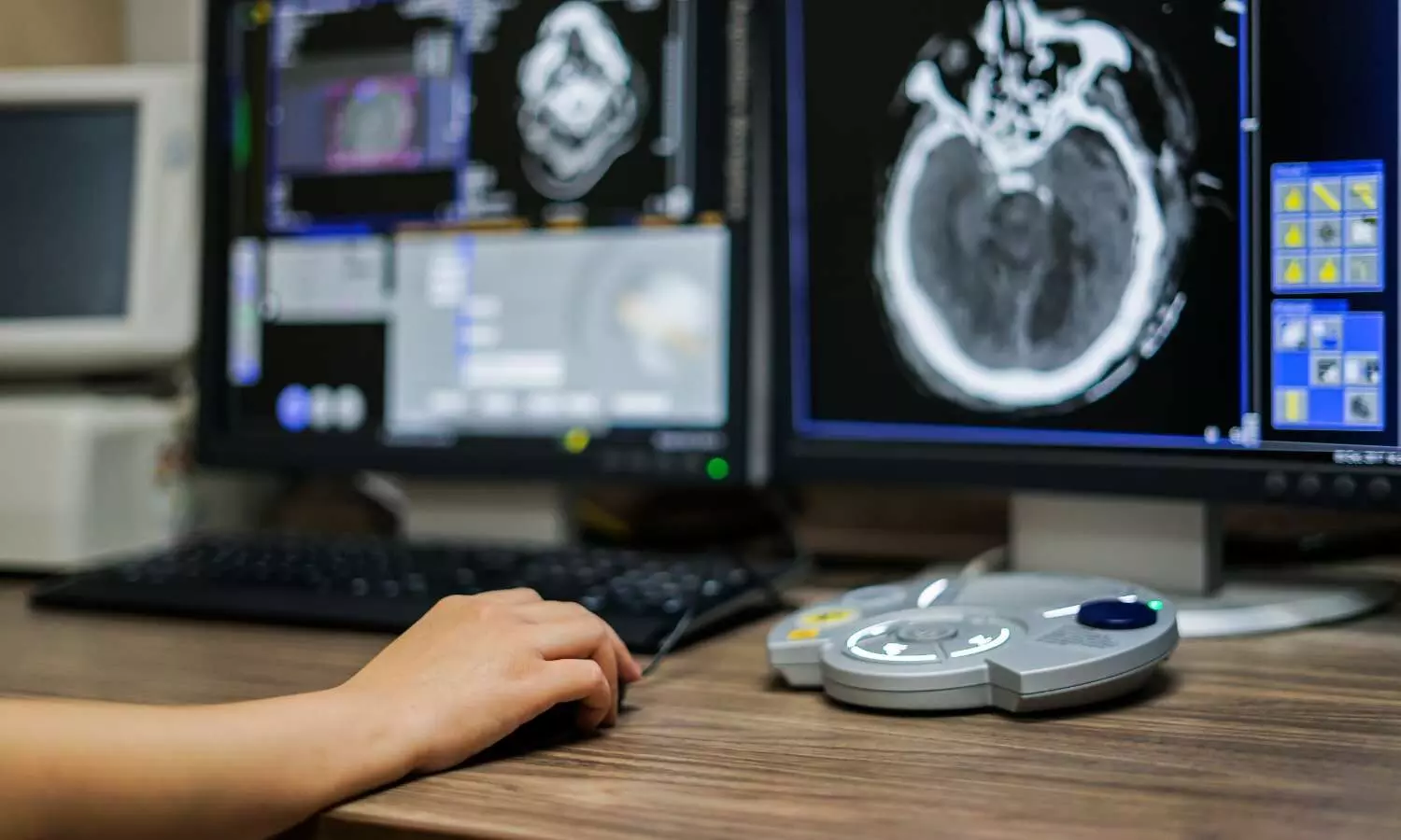
The researchers developed an algorithm that identified normal mammograms with very high sensitivity.

They then ran a simulation on patient data to see what would have happened if all of the very low-risk mammograms had been taken off radiologists’ plates, freeing the doctors to concentrate on the more questionable scans.

The simulation revealed that fewer people would have been called back for additional testing but that the same number of cancer cases would have been detected.

“False positives are when you call a patient back for additional testing, and it turns out to be benign,” explained senior author Richard L. Wahl, MD, a professor of radiology at Washington University’s Mallinckrodt Institute of Radiology (MIR) and a professor of radiation oncology.

“That causes a lot of unnecessary anxiety for patients and consumes medical resources. This simulation study showed that very low-risk mammograms can be reliably identified by AI to reduce false positives and improve workflows.”

The study is published April 10 in the journal Radiology: Artificial Intelligence.

Wahl previously collaborated with Whiterabbit.ai on an algorithm to help radiologists judge breast density on mammograms to identify people who could benefit from additional or alternative screening.

That algorithm received clearance from the Food and Drug Administration (FDA) in 2020 and is now marketed by Whiterabbit.ai as WRDensity.

In this study, Wahl and colleagues at Whiterabbit.ai worked together to develop a way to rule out cancer using AI to evaluate mammograms.

They trained the AI model on 123,248 2D digital mammograms (containing 6,161 showing cancer) that were largely collected and read by Washington University radiologists.

Then, they validated and tested the AI model on three independent sets of mammograms, two from institutions in the U.S. and one in the United Kingdom.

First, the researchers figured out what the doctors did: how many patients were called back for secondary screening and biopsies; the results of those tests; and the final determination in each case.

Then, they applied AI to the datasets to see what would have been different if AI had been used to remove negative mammograms in the initial assessments and physicians had followed standard diagnostic procedures to evaluate the rest.

For example, consider the largest dataset, which contained 11,592 mammograms.

When scaled to 10,000 mammograms (to make the math simpler for the purposes of the simulation), AI identified 34.9% as negative.

If those 3,485 negative mammograms had been removed from the workload, radiologists would have made 897 callbacks for diagnostic exams, a reduction of 23.7% from the 1,159 they made in reality.

At the next step, 190 people would have been called in a second time for biopsies, a reduction of 6.9% from the 200 in reality.

At the end of the process, both the AI rule-out and real-world standard-of-care approaches identified the same 55 cancers.

In other words, this study of AI suggests that out of 10,000 people who underwent initial mammograms, 262 could have avoided diagnostic exams, and 10 could have avoided biopsies, without any cancer cases being missed.

“At the end of the day, we believe in a world where the doctor is the superhero who finds cancer and helps patients navigate their journey ahead,” said co-author Jason Su, co-founder and chief technology officer at Whiterabbit.ai. “The way AI systems can help is by being in a supporting role. By accurately assessing the negatives, it can help remove the hay from the haystack so doctors can find the needle more easily. This study demonstrates that AI can potentially be highly accurate in identifying negative exams. More importantly, the results showed that automating the detection of negatives may also lead to a tremendous benefit in the reduction of false positives without changing the cancer detection rate.”

Reference: Stefano Pedemonte, Trevor Tsue, Brent Mombourquette, Yen Nhi Truong Vu, Thomas Matthews, Rodrigo Morales Hoil, Meet Shah, Nikita Ghare, Naomi Zingman-Daniels, Susan Holley, Catherine M. Appleton, Jason Su, Richard L. Wahl. A Semiautonomous Deep Learning System to Reduce False-Positive Findings in Screening Mammography. Radiology: Artificial Intelligence, 2024; DOI: 10.1148/ryai.230033

Powered by WPeMatico

Endoscopic Assisted Percutaneous Fixation of Anterior Inferior Iliac Spine Avulsion Fracture: Surgical Technique

Avulsion fractures of the anterior inferior iliac spine rarely occur in adolescent athletes during rectus femoris contractions or eccentric muscle lengthening while the growth plate is still open. Currently, there are no official guidelines in the literature on the treatment indications of this type of fracture or the type of surgical technique to be used. Nowadays, young and athletic patients desire a quick return to their previous activities, which makes surgical treatment a reasonable choice. Open reduction and internal fixation with an anterior approach are usually recommended when the avulsion fragment has more than 1.5–2 cm displacement on plain radiographs. However, ORIF is associated with a higher risk of heterotopic ossifications and increases the risk of damage to the Lateral femoral cutaneous nerve (LFCN).

Alessandro Aprato et al designed an endoscopic technique to reduce these complications. The technical note describes a procedure of percutaneous fixation to AIIS through 3 endoscopic portals that could potentially minimize complications associated with an open surgical dissection, allowing anatomic reduction under direct visualization. It has been published in ‘Indian Journal of Orthopaedics.’

Technical note:

On a radiolucent bed, the limb to be operated on is left free so that the hip can be flexed during surgery if necessary. Through intraoperative x-rays, the AIIS is identified so the three endoscopic portals can be located:

(1)Lateral Portal (LP): 2 cm lateral to the AIIS;

(2)Direct Portal (DP): directly on the AIIS;

(3)Inferior Portal (IP): 1 cm inferior to the AIIS.

Starting from the LP and DP portals, nitinol wires are introduced, and then the 30° optic and the radiofrequency are placed. The water pump is activated until a pressure of 40 mmHg is reached. Approaching the LP it is crucial to avoid being too lateral to the AIIS as there is a risk of entering the territory of the LFCN; in fact, it emerges under the inguinal ligament, just medial to the ASIS and descends along the surface of the sartorius muscle, in case of injury there is a risk of causing paresthetic meralgia.

On the medial side, femoral vascular-nervous structures are at risk, so care must be taken to locate the DP and IP portals. By endoscopic vision, the fracture is identified; through the use of a shaver and radiofrequency, an accurate debridement of the fracture’s interface is performed, allowing it to be mobilized. Through the IP, a switching stick or a shaver is inserted, and it can be used for the reduction maneuver, with the aim of reducing the fragment as anatomically as possible. If there is difficulty in retracting the avulsed fragment, flexion of the hip can be helpful, detending the rectus femoris muscle. Once anatomical reduction is achieved, a 2 mm k-wire is inserted perpendicular to the fracture by the same portal, and an intraoperative x-ray check is necessary to confirm adequate temporary reduction. Finally, the definitive fixation can be performed with a partially threaded cannulated inter fragmentary screw 4.5 mm in diameter inserted by the IP.

Limitation of the proposed technique is the cost associated with the arthroscopic instrumentation, a possible extension of surgical time and the demand for excellent arthroscopic skills.

Further reading:

Endoscopic Assisted Percutaneous Fixation of Anterior Inferior Iliac Spine Avulsion Fracture, Surgical Technique

Alessandro Aprato et al

Indian Journal of Orthopaedics (2024) 58:433–438

https://doi.org/10.1007/s43465-024-01107-5

Powered by WPeMatico

Non-invasive ventilation and early mobilization improves VAP prevention: Study

A recent study published in the Journal of Hospital Infection found selective decontamination of the digestive tract (SDD) and the non-invasive ventilation (NIV) use to protect against Ventilator-Associated Pneumonia (VAP) which is a significant threat to critically ill patients on mechanical ventilation.

The comprehensive analysis reviewed a total of 34 interventions across 31 studies and identified the key strategies to reduce the incidence of VAP in intensive care practices. VAP poses a formidable challenge in critical care, with its incidence directly impacting the patient outcomes and healthcare costs. The meticulous review of randomized and quasi-randomized controlled trials revealed that 19 of the total interventions studied markedly reduced VAP incidents. This study highlighted the effectiveness of selective decontamination of the digestive tract and the use of non-invasive ventilation in weaning patients from mechanical ventilation as the most promising preventive measures.

SDD involved the application of topical antibiotics in the oropharynx and gastrointestinal tract and was backed by highly suggestive (Class II) evidence showing a risk ratio (RR) of 0.439. This indicates a significant reduction in VAP rates among the patients receiving SDD when compared to the individuals who did not. Also, NIV showed a compelling reduction in VAP incidence, with its evidence strength upgraded to highly suggestive (Class II) based on a risk ratio of 0.32 that highlights its efficacy in safely reducing the duration of mechanical ventilation.

The study points to early mobilization of patients as a beneficial strategy that is supported by suggestive (Class III) evidence. This approach significantly shortens the length of stay in the ICU and reduces the duration of mechanical ventilation in contributing to better outcomes for critically ill patients. However, this study also underscores that none of the preventive interventions demonstrated robust evidence for reducing mortality. This emphasizes the complexity of VAP management and the need for multifaceted strategies to prevent the condition and also to improve the overall patient survival.

The beneficial results of this study suggests that incorporating SDD, NIV and early mobilization into the ventilator care bundle could revolutionize the efforts of VAP prevention. These strategies enhance the patient care by reducing the burden of VAP in critical care settings and ultimately improving the prognosis of critically ill patients.

Reference:

Zhu, D., Zhao, Q., Guo, S., Bai, L., Yang, S., Zhao, Y., Xu, Y., & Zhou, X. (2024). Efficacy of preventive interventions against ventilator-associated pneumonia in critically ill patients: an umbrella review of meta-analyses. In Journal of Hospital Infection (Vol. 145, pp. 174–186). Elsevier BV. https://doi.org/10.1016/j.jhin.2023.12.017

Powered by WPeMatico