Newly detected diabetes patients with acute coronary syndrome experience more extensive ischemic myocardial insult: Study

Greece: A recent study has shown that acute coronary syndrome (ACS) patients with newly detected diabetes mellitus (NDDM) experience heightened cardiometabolic challenges similar to those with established diabetes mellitus. The findings were published online in Diabetes Research and Clinical Practice on April 08, 2024.

NDDM patients showed lower HDL-C and higher triglyceride levels versus non-DM counterparts, alongside elevated waist circumference and body mass index (BMI). Moreover, they experienced more extensive necrosis compared to patients with established DM.

In the realm of cardiovascular health, the interplay between diabetes mellitus and acute coronary syndrome presents a multifaceted challenge for patients and clinicians alike. Recent studies have shed light on a concerning trend: newly detected diabetes mellitus in ACS patients not only parallels the adverse cardiometabolic profile of those with established diabetes but also signifies a potentially more extensive ischemic myocardial insult. This revelation underscores the critical need for tailored management strategies and heightened vigilance in clinical practice.

The convergence of diabetes mellitus and ACS marks a pivotal intersection of two formidable health concerns. ACS, encompassing conditions such as myocardial infarction and unstable angina, represents acute manifestations of coronary artery disease (CAD), a leading cause of morbidity and mortality worldwide. Diabetes mellitus, meanwhile, exerts its insidious influence through mechanisms of insulin resistance, hyperglycemia, and systemic inflammation, fostering a milieu ripe for cardiovascular complications.

The impact of newly detected diabetes mellitus on metabolic parameters and the extent of myocardial necrosis in patients with ACS is not fully explored. Loukianos S Rallidis, National and Kapodistrian University of Athens, University General Hospital ATTIKON, Athens, Greece, and colleagues examined the impact of NDDM on cardiometabolic characteristics and myocardial necrosis in ACS patients.

CALLINICUS-Hellas Registry is an ongoing prospective multicenter observational study assessing adherence to lipid-lowering therapy (LLT) among ACS patients in Greece. Three groups were created: patients with NDDM (abnormal fasting glucose, HbA1c ≥ 6.5 % and no previous history of DM), b) patients without known diabetes and HbA1c < 6.5 % (non-DM), and c) patients with prior diabetes.

The study led to the following findings:

  • The prevalence of NDDM among 1084 patients was 6.9 %. NDDM patients had lower HDL-C [38 versus 42 mg/dL] and higher triglyceride levels [144 vs 115 mg/dL] versus non-DM patients.
  • NDDM patients featured both higher BMI [29.5 vs 27.1 kg/m2] and waist circumference [107 vs 98 cm] compared to non-DM patients.
  • NDDM patients had more extensive myocardial necrosis than patients with prior DM.

“Acute coronary syndrome patients with newly detected diabetes mellitus have an adverse cardiometabolic profile similar to patients with prior DM and have more extensive myocardial insult,” the researchers concluded.

Reference:

Rallidis LS, Papathanasiou KA, Tsamoulis D, Bouratzis V, Leventis I, Kalantzis C, Malkots B, Kalogeras P, Tasoulas D, Delakis I, Lykoudis A, Daios S, Potoupni V, Zervakis S, Theofilatos A, Kotrotsios G, Kostakou PM, Kostopoulos K, Gounopoulos P, Mplani V, Zacharis E, Barmpatzas N, Kotsakis A, Papadopoulos C, Trikas A, Ziakas A, Skoularigis I, Naka KK, Tziakas D, Panagiotakos D, Vlachopoulos C. Newly detected diabetes mellitus patients with acute coronary syndrome have an adverse cardiometabolic profile similar to patients with prior diabetes and a more extensive ischemic myocardial insult. Diabetes Res Clin Pract. 2024 Apr 9;211:111664. doi: 10.1016/j.diabres.2024.111664. Epub ahead of print. PMID: 38604446.

Powered by WPeMatico

Thane Hospital doctors succesfully treat Ano Rectal Malformation in 1-year-old boy

Thane: In a remarkable case, doctors at Wockhardt Hospitals in Thane have successfully treated a one-year-old boy born with multiple complications, including the absence of an anus, officials announced on Wednesday.

Child specialists Bhavesh Doshi and Nitu Mundhra addressed the child’s condition known as ‘Ano Rectal Malformation’ (ARM), which meant the baby lacked a rectal opening. Additionally, the child faced challenges such as tongue-tie, where the tongue was stuck to the floor of the mouth, and Hypospadias, a condition where the urinary outlet is misplaced.

The newborn’s parents — identified as the Salian couple — were shocked to learn of these issues in their first child, whom the doctors admitted to the hospital’s neonatal intensive care unit (NICU), and put on parenteral nutrition as breastfeeding was not feasible, reports IANS.

Also Read:Complex surgery by Wockhardt Hospitals doctors saves eyes of patient with Multiple Facial Fractures

Mundhra said ARM is rare, seen just once in about a lakh deliveries, and in the delivery room, all the newborn babies are thoroughly examined from head to toe to detect any abnormalities, especially if they are born with a problem.

“In this baby, when we detected ARM, we checked him thoroughly and found the tongue-tie which could affect his feeding and lead to speech difficulties and also Hypospadias. After counselling the parents, the baby underwent multiple surgeries,” said Mundhra.

In this case, after detecting ARM, the medical team identified additional concerns like tongue-tie and Hypospadias. The child underwent five procedures including colostomy, anal reconstruction, and colostomy closure to address ARM issues, alongside procedures for Hypospadias and tongue-tie.

The ARM correction was done in three surgical steps, comprising a temporary opening to allow passage of stool and enable the baby to feed. Two months later, a four-hour-long operation was performed to create a new anus at the normal position, and after two more months, another surgery was performed for colostomy closure.

During this surgery, the opening created in the abdominal wall was closed and the baby now passes motions normally from the opening, said the medicos.

According to an IANS report, Eight months after the surgery, the child underwent a major surgery for Hypospadias to get the urinary opening in the normal position, which was also successful and the child can now pass urine without any difficulty.

Now the child is one year old and functions like other kids of his age after undergoing five surgeries in the first 12 months of his life.

Also Read:Mumbai doctors successfully treat patient with a Rare Complication of DVT

Powered by WPeMatico

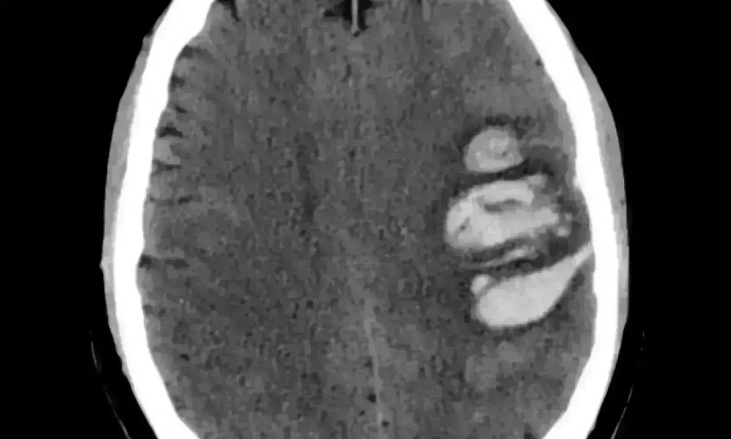
Early minimally invasive hematoma evacuation improves outcomes in acute intracerebral hemorrhage patients: NEJM

A recent study published in The New England Journal of Medicine found that early minimally invasive surgery to remove brain hemorrhages may lead to better recovery than traditional medical management alone. 

This multicenter, randomized trial focused on patients who experienced an acute intracerebral hemorrhage, specifically the individuals with lobar or anterior basal ganglia hemorrhages and had hematoma volumes between 30 to 80 milliliters. The participants were divided into a group receiving minimally invasive surgical removal of the hematoma along with standard medical treatment and another the other group receiving only medical management according to the existing guidelines.

The primary goal of this study was to evaluate the effectiveness of surgical intervention in improving the patient outcomes which was measured by the utility-weighted modified Rankin scale. This scale ranges from 0 to 1, where the higher scores signify better functional outcomes according to the patient assessments. A total of 300 patients were enrolled, with the trial noticing an adaptation part-way through after 175 patients had joined where this shift limited further enrollments to only the individuals with lobar hemorrhages due to an observed trend in the data.

The results from the study indicated a notable difference in recovery outcomes. The surgery group had an average score of 0.458 on the modified Rankin scale when compared to 0.374 in the control group 6 months after treatment. This difference was statistically significant and suggested a clear benefit of surgical intervention among the patients with lobar hemorrhages. In this subgroup, the mean score difference was more pronounced at 0.127 in favor of surgery. However, for the individuals with anterior basal ganglia hemorrhages, surgery did not show a notable benefit.

Only 9.3% of patients in the surgery group died within 30 days of enrollment, when compared to 18.0% in the control group which points to the reduction in early mortality rates. This significant decrease illuminates the additional benefit of surgical intervention in managing acute intracerebral hemorrhages. However, the surgical approach evidenced a 3.3% incidence of postoperative complications like rebleeding and neurological deterioration in the surgery group. Overall, these findings suggest that for certain patients with specific types of brain hemorrhages, early minimally invasive surgery could be a beneficial treatment option that offers improved functional outcomes and lower mortality rates.

Reference:

Pradilla, G., Ratcliff, J. J., Hall, A. J., Saville, B. R., Allen, J. W., Paulon, G., McGlothlin, A., Lewis, R. J., Fitzgerald, M., Caveney, A. F., Li, X. T., Bain, M., Gomes, J., Jankowitz, B., Zenonos, G., Molyneaux, B. J., Davies, J., Siddiqui, A., Chicoine, M. R., … Barrow, D. L. (2024). Trial of Early Minimally Invasive Removal of Intracerebral Hemorrhage. In New England Journal of Medicine (Vol. 390, Issue 14, pp. 1277–1289). Massachusetts Medical Society. https://doi.org/10.1056/nejmoa2308440

Powered by WPeMatico

First trimester Use of Hydroxychloroquine in lupus or RA may not increase risk of birth defects, finds study

First trimester Use of Hydroxychloroquine in lupus or RA not linked to higher risk of birth defects suggests a new study published in the Rheumatology.

A study was done to assess the infant risk of major congenital malformations (MCM) associated with first-trimester exposure to hydroxychloroquine (HCQ) among mothers with systemic lupus erythematosus (SLE) or rheumatoid arthritis (RA). This population-based cohort study utilised Swedish nationwide registers and included all singleton births (2006-2021) among individuals with prevalent SLE or RA in Sweden. The exposure was filling ≥1 HCQ prescription during the first trimester. The outcome was infant MCM within one year of birth. Inverse probability of treatment weighting was applied to adjust for potential confounders (e.g. maternal smoking, body mass index, pregestational diabetes, and corticosteroids). Modified Poisson regression models with robust variance estimated risk ratios and 95% confidence intervals (RR 95%CI).

Results: They included 1,007 births (453 exposed) and 2,500 births (144 exposed) in the SLE and RA cohorts, respectively. The MCM risks in the SLE overall cohort, exposed, and unexposed groups were 3.6%, 3.7%, and 3.4%, respectively. The corresponding figures in the RA cohort were 4.4%, 5.6%, and 4.3%, respectively. The adjusted RRs (95%CI) were 1.29 (0.65-2.56) in the SLE cohort, 1.32 (0.56-3.13) in the RA cohort, and 1.30 (0.76-2.23) in the pooled analysis. The adjusted risk difference (exposed vs unexposed) was small (0.9% in SLE and 1.3% in RA). Sensitivity analyses examining different exposure and outcome windows yielded similar findings. First-trimester exposure to HCQ was not associated with a significantly increased risk of MCM. HCQ’s benefits may outweigh the risks in managing SLE or RA during pregnancy.

Reference:

Nguyen NV, Svenungsson E, Dominicus A, Altman M, Hellgren K, Simard JF, Arkema EV. Hydroxychloroquine in lupus or rheumatoid arthritis pregnancy and risk of major congenital malformations: a population-based cohort study. Rheumatology (Oxford). 2024 Mar 13:keae168. doi: 10.1093/rheumatology/keae168. Epub ahead of print. PMID: 38479815.

Powered by WPeMatico
DNA analysis of saliva may predict treatment response and relapse in oral cancer patients: Study

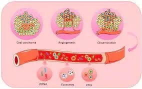
DNA analysis of saliva may predict treatment response and relapse in oral cancer patients suggests a new study published in the Oral Oncology.

The incidence of head and neck squamous cell carcinoma (HNSCC) continues to increase and although advances have been made in treatment, it still has a poor overall survival with local relapse being common. Conventional imaging methods are not efficient at detecting recurrence at an early stage when still potentially curable. The aim of this study was to test the feasibility of using saliva to detect the presence of oral squamous cell carcinoma (OSCC) and to provide additional evidence for the potential of this approach. Fresh tumor, whole blood and saliva were collected from patients with OSCC before treatment. Whole exome sequencing (WES) or gene panel sequencing of tumor DNA was performed to identify somatic mutations in tumors and to select genes for performing gene panel sequencing on saliva samples.

Results: The most commonly mutated genes identified in primary tumors by DNA sequencing were TP53 and FAT1. Gene panel sequencing of paired saliva samples detected tumor derived mutations in 9 of 11 (82%) patients. The mean variant allele frequency for the mutations detected in saliva was 0.025 (range 0.004 – 0.061). Somatic tumor mutations can be detected in saliva with high frequency in OSCC irrespective of site or stage of disease using a limited panel of genes. This work provides additional evidence for the suitability of using saliva as liquid biopsy in OSCC and has the potential to improve early detection of recurrence in OSCC. Trials are currently underway comparing this approach to standard imaging techniques.

Reference:

Ahmed A. Ahmed, Mateja Sborchia, Hannah Bye, Maria Roman-Escorza, Ariella Amar, Rhonda Henley-Smith, Edward Odell, Mark McGurk, Michael Simpson, Tony Ng, Elinor J. Sawyer, Christopher G. Mathew. Mutation detection in saliva from oral cancer patients, Oral Oncology, Volume 151, 2024, 106717, ISSN 1368-8375, https://doi.org/10.1016/j.oraloncology.2024.106717. (https://www.sciencedirect.com/science/article/pii/S1368837524000356)

Powered by WPeMatico
Extracorporeal shockwave therapy improves quality of life in patients with claudication: JAMA

UK: A randomized clinical trial has shown that extracorporeal shockwave therapy (ESWT) may be considered a safe and effective alternative therapy for patients with intermittent lower limb claudication. Also, there were comparable improvements in quality of life to supervised exercise.

The study, published in JAMA Surgery comprising 138 patients, revealed that patients receiving ESWT had statistically higher quality of life measures than patients receiving a placebo.

An estimated 237 million people worldwide are affected with lower limb peripheral arterial disease (PAD), due to population aging this number is expected to rise. Intermittent claudication is the most common symptomatic manifestation of PAD and limits walking distances, physical function, and quality of life.

Current first-line recommendations for intermittent claudication management consist of smoking cessation, supervised exercise, cardiovascular risk reduction, and best medical therapy. Supervised exercise programs are not readily available, and a noninvasive alternative is required. Considering this, Paris Cai, Hull York Medical School, Hull, United Kingdom, and colleagues aimed to assess extracorporeal corporeal shockwave therapy in improving the quality of life in patients with claudication.

For this purpose, the researchers conducted a double-blind, placebo-controlled randomized clinical trial, including patients in the outpatient setting at a single tertiary center for vascular surgery. They were randomized in a ratio of 1:1 to ESWT or placebo therapy with no shockwaves delivered.

Patients were recruited between 2015 and 2020, with a 12-week follow-up ending in March 2020. A convenience sample of patients with claudication and conservative treatment requirements who were unable or refused to participate in supervised exercise were eligible. Patients with active cancer or receiving anticoagulation therapy were excluded.

Of 522 patients screened, 389 were eligible, 138 were enrolled, and 110 completed follow-up and were included in the primary analysis. Of 138 patients recruited and randomized, 67% were male, and the mean age of the study population was 67 years.

In the intervention group, patients received 100 impulses of 0.1mJ/mm/cm2 in an area of the gastrocnemius muscle three times weekly for 3 weeks. For the control group, the steps for treatment were replicated without delivering the treatment.

The study’s primary outcome was the Physical Functioning domain of the 36-item Short-Form Quality of Life Questionnaire at the follow-up of 12 weeks. Secondary outcomes were ankle brachial pressure index, walking distances, and other quality-of-life measures.

The study led to the following findings:

  • The intervention group had a significantly higher physical function score at 12 weeks (estimated median difference 3.8). However, this significance did not remain when adjusting for covariates.
  • At 12 weeks, the intervention group had significantly longer pain-free and maximum walking distances (pain-free estimated median difference, 34.1; maximum estimated median difference, 51.4).

“To our knowledge, this is the first double-blind, placebo-controlled, randomized clinical trial to consider extracorporeal shockwave therapy for intermittent claudication management,” the researchers wrote.

“It demonstrated efficacy for walking distances, may positively impact the quality of life, and may provide a safe, non-invasive alternative therapy for intermittent claudication patients.”

Reference:

Cai P, Pymer S, Ibeggazene S, et al. Extracorporeal Shockwave for Intermittent Claudication and Quality of Life: A Randomized Clinical Trial. JAMA Surg. Published online April 10, 2024. doi:10.1001/jamasurg.2024.0625

Powered by WPeMatico

Oral minoxidil vs topical minoxidil: Choosing the right treatment for male androgenetic alopecia

Brazil: For treating men with androgenetic alopecia (AGA), low-dose low-dose oral minoxidil (5 mg per day) has similar efficacy to topical minoxidil, 5%, and can be an option for patients who are intolerant to topical treatment or prefer oral therapy, a recent study has claimed.

The double-blind, placebo-controlled randomized clinical trial (RCT) comprising 90 men with androgenetic alopecia found that daily oral minoxidil, 5 mg, was well tolerated and did not show superiority over topical minoxidil, 5% after 24 weeks of treatment. The findings were published online in JAMA Dermatology on April 10, 2024.

Androgenetic alopecia is the main cause of hair loss in men. It occurs due to progressive miniaturization of the hair follicles and shortening of the anagen phase. This form of hair loss causes low self-esteem and hurts the quality of life. There has been an increased interest in low-dose oral minoxidil for AGA treatment. However, the effectiveness of oral minoxidil for male AGA has not yet been evaluated in comparative therapeutic trials.

Against the above background, Mariana Alvares Penha, São Paulo State University (UNESP), Botucatu, Brazil, and colleagues aimed to compare the safety, efficacy, and tolerability of daily oral minoxidil, 5 mg, versus twice-daily topical minoxidil, 5%, for 24 weeks in male AGA treatment.

For this purpose, they conducted a double-blind, placebo-controlled randomized clinical trial at a single specialized clinic in Brazil. Eligible men with AGA aged 18 to 55 years, were included and classified using the Norwood-Hamilton scale as 3V, 4V, or 5V.

Participants were randomized in a ratio of 1:1 into two groups: oral minoxidil, 5 mg, daily, and topical placebo solution; or 1 mL of topical minoxidil, 5%, twice daily, and oral placebo for 24 weeks. Sixty-eight completed the study among 90 enrolled participants, the mean age was 36.6 years. Thirty-three participants were enrolled in the oral minoxidil group and 35 in the topical treatment group.

The study’s primary outcome was a change in terminal hair density on the vertex and frontal scalp regions. The secondary outcomes were changes in total hair density and photographic evaluation.

The study led to the following findings:

  • Both groups were homogenous in terms of demographic data and AGA severity.
  • For the frontal area, the mean change from baseline to week 24 between the groups was 3.1 hairs per cm2 for terminal hair density and 2.6 hairs per cm2 for total hair density.
  • For the vertex area, the mean change from baseline to week 24 was 23.4 hairs per cm2 for terminal density and 5.5 hairs per cm2 for total hair density.
  • According to the photographic analysis, oral minoxidil was superior to topical minoxidil on the vertex (24%) but not on the frontal scalp (12%).
  • The most common adverse effects in the oral minoxidil group were hypertrichosis (49%) and headache (14%).

In conclusion, oral minoxidil did not demonstrate superiority over topical minoxidil in treating male AGA after 24 weeks. However, in the oral minoxidil group, the overall photographic improvement in the vertex was superior.

“Low-dose oral minoxidil has shown to be well tolerated and, therefore, is an option for patients intolerant to topical treatment or those who prefer oral therapy,” the researchers wrote.

“By weighing the pros and cons of each treatment approach, patients can make informed decisions to address their hair loss concerns effectively,” they concluded. 

Reference:

Penha MA, Miot HA, Kasprzak M, Müller Ramos P. Oral Minoxidil vs Topical Minoxidil for Male Androgenetic Alopecia: A Randomized Clinical Trial. JAMA Dermatol. Published online April 10, 2024. doi:10.1001/jamadermatol.2024.0284

Powered by WPeMatico

Deeper sedation may help find difficult-to-detect polyps during colonoscopy, reveals study

In patients undergoing colonoscopy to screen for colorectal cancer, deeper sedation using the anesthetic drug propofol may improve detection of “serrated” polyps-a type of precancerous lesion that can be difficult to detect, reports a study in the Online First edition of Anesthesiology, the peer-reviewed journal of the American Society of Anesthesiologists (ASA).

“Our study provides the first evidence that monitored anesthesia care with propofol might increase detection of serrated polyps, which are more likely to be missed than adenomatous polyps during colonoscopy,” said lead author Aurora N. Quaye, M.D., member of the Acute Pain and Regional Anesthesia Service at Maine Medical Center, Portland. “Propofol-based anesthesia may contribute to a more effective screening process for colorectal cancer, especially in patients with risk factors for serrated polyps.”

Nearly all colorectal cancers start off as small growths called polyps. Identifying and removing polyps during colonoscopy screening can prevent them from developing into cancer. Compared to a more common type of polyp called adenoma, serrated polyps may be harder to see, because they are often flatter and blend into the folds of the colon tissue.

Propofol is an alternative to moderate sedation-sometimes called “conscious” sedation-for colonoscopy. “Propofol results in deeper sedation, and also begins to work and wears off more quickly, compared to conscious sedation,” Dr. Quaye explained. Research has shown that propofol-based anesthesia is more efficient and improves patient and provider satisfaction ratings. The new study is the first to assess whether propofol may be associated with improved detection of serrated polyps.

The analysis included detailed information on more than 54,000 completed colonoscopies drawn from the New Hampshire Colonoscopy Registry. The procedures were performed between 2015 and 2020; all patients were older than 50 years. Rates of polyp detection, including serrated polyps and adenomas, were compared for patients receiving moderate sedation versus propofol-based anesthesia.

The overall polyp detection rate was higher when colonoscopy was performed using propofol: 34%, compared to 24.5% with moderate sedation. The results were similar on analysis of a “restricted” sample of about 19,000 colonoscopies performed at facilities that did not predominantly use one form of sedation over the other: overall polyp detection rate was 30.3% with propofol versus 25.7% with moderate sedation.

After adjustment for other confounding factors among these 19,000 colonoscopies, propofol was still associated with a clinically and statistically significant 13% higher likelihood of serrated polyp detection, although other types of polyps did not demonstrate a difference in detection.

The conclusions are strengthened by the use of systematically collected clinical registry data, the researchers note. However, the study cannot provide any information on how propofol might improve detection of serrated polyps. “It may be that propofol increases patient comfort and relaxation, optimizing detection of polyps that are more difficult to see,” said Dr. Quaye. “Additionally, propofol may cause smooth muscle relaxation in the colon, allowing more careful inspection and improved visualization.”

The researchers emphasize the need for further studies to clarify the possible advantages of propofol for polyp detection. “The finding that propofol-based anesthesia might improve the detection of precancerous polyps may bring us closer to our goal of further optimizing the use of colonoscopy for the prevention and early detection of colorectal cancer,” said Dr. Quaye.

The modest but significant reported association between propofol use and the detection of serrated polyps illustrates the “promise and peril” of studies using clinical registry data, according to an accompanying editorial by Douglas A. Colquhoun, MB, ChB, MSc, MPH, University of Michigan, Ann Arbor, and colleagues. While warning that the results must be interpreted with caution due to limitations in analyzing registry data, the editorial authors highlight the importance of preserving access to propofol and call for further, “rigorously conducted” studies focusing on the value of anesthesia care for patients undergoing colonoscopy.

Reference:

Aurora N. Quaye, William M. Hisey, Todd A. Mackenzie, Christina M. Robinson, Janelle M. Richard, Joseph C. Anderson, Robert D. Warters, Lynn F. Butterly, Association between Colonoscopy Sedation Type and Polyp Detection: A Registry-based Cohort Study,Anesthesiology, https://doi.org/10.1097/ALN.0000000000004955

Powered by WPeMatico

Hydronephrosis area useful predictor of impacted ureteral stones, finds study

Hydronephrosis area useful predictor of impacted ureteral stones, finds study published in the BMC Urology.

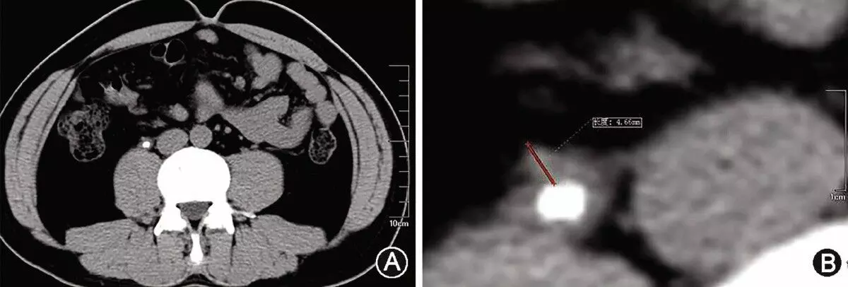
Ureteroscopic lithotripsy for impacted stones is challenging, but it is important to predict impacted stones preoperatively. Hydronephrosis, which is evaluated by a grading system, is often apparent in impacted stones. However, the currently used grading system is a qualitative evaluation. We, therefore, focused on a quantitative evaluation: the area of hydronephrosis. The aim of this study was to investigate whether the area of hydronephrosis could predict impacted stones more accurately than Society for Fetal Urology grade. We retrospectively identified 160 patients who underwent ureteroscopic lithotripsy for ureteral stones at our hospital between January 2014 and April 2022. Impacted stones were defined as stones fixed to the ureteral wall that could not be moved by means of ureteroscopic manipulation or water pressure. Of the 160 patients, 54 (33.8%) had impacted stones. Comparing patient characteristics, there were significant differences in stone size, ureteral wall thickness, Society for Fetal Urology grade, renal pelvic width and area of hydronephrosis (all P < 0.01). Receiver operating characteristic analysis showed that area of hydronephrosis was the more significant predictive value (area under the curve 0.781) compared with Society for Fetal Urology grade (area under the curve 0.676, P < 0.01). Multivariate analysis revealed that significant independent predictive factors of impacted stones were thicker ureteral wall thickness and larger area of hydronephrosis (both P < 0.01). The area of hydronephrosis and ureteral wall thickness were significant predictors of impacted stones in patients undergoing ureteroscopic lithotripsy for ureteral stones. These factors may be useful for selecting the treatment and preoperative settings.

Reference:

Iwahashi Y, Kohjimoto Y, Deguchi R, Wakamiya T, Yamashita S, Hara I. Area of hydronephrosis is a useful predictive factor of impacted ureteral stones. Urolithiasis. 2024 Apr 2;52(1):56. doi: 10.1007/s00240-023-01526-3. PMID: 38564028; PMCID: PMC10987368.

Keywords:

Hydronephrosis area, useful predictor, impacted ureteral stones, BMC urology, Iwahashi Y, Kohjimoto Y, Deguchi R, Wakamiya T, Yamashita S, Hara I, Area of hydronephrosis, Ureteral wall thickness, Impacted stone, Ureteroscopic lithotripsy

Powered by WPeMatico

Exposure to air pollution during first two years of life may worsen attention capacity in children: Study

A growing body of research shows that exposure to air pollution, especially during pregnancy and childhood, may have a negative impact on brain development. Now a study led by the Barcelona Institute for Global Health (ISGlobal), a centre supported by the “la Caixa” Foundation, has found that exposure to nitrogen dioxide (NO2) during the first two years of life is associated with poorer attention capacity in children aged 4 to 8, especially in boys. NO2 is a pollutant that comes mainly from traffic emissions.

The study, published in Environment International, shows that higher exposure to NO2 was associated with poorer attentional function in 4- to 6-year-olds, with increased susceptibility to this pollutant observed in the second year of life. This association persisted at an age of 6 to 8 years of age only in boys, with a slightly greater susceptibility period from birth to 2 years of age.

The researchers used data from 1,703 women and their children from the INMA Project birth cohorts in four Spanish regions. Using the home address, the researchers estimated daily residential exposure to NO2 during pregnancy and the first 6 years of childhood. In parallel, they assessed the attentional function (the ability to choose what to pay attention to and what to ignore) at 4-6 years and 6-8 years, and working memory (the ability to temporarily hold information) at 6-8 years, using validated computerised tests.

Periods of higher susceptibility to air pollution

A previous INMA study reported that exposure to NO2 during pregnancy and childhood was associated with impaired attentional function in children at 4-5 years of age. The present study found that:

  • Higher exposure to NO2 between 1.3 and 1.6 years of age was associated with higher hit reaction time standard error, an indicator of speed consistency, in the attentional function test at 4–6 years of age.
  • Higher exposure to NO2 between 1.5 and 2.2 years of age was associated with more omission errors.
  • Higher exposure to NO2 between 0.3 and 2.2 years was associated with higher hit reaction time standard error at 6–8 years only in boys.
  • No associations were found between higher exposure to NO2 and working memory in children aged 6 to 8 years.

“These findings underline the potential impact of increased traffic-related air pollution on delayed development of attentional capacity and highlight the importance of further research into the long-term effects of air pollution in older age groups”, explains Anne-Claire Binter, last author of the study and postdoctoral researcher at ISGlobal.

As the brain matures

Attentional function is crucial for the development of the brain’s executive functions, which manage and control actions, thoughts and emotions to achieve a goal or purpose. “The prefrontal cortex, a part of the brain responsible for executive functions, develops slowly and it is still maturing during pregnancy and childhood,” adds Binter. This makes it vulnerable to exposure to air pollution, which has been linked in animal studies to inflammation, oxidative stress, and impaired energy metabolism in the brain.

“In boys, the association between exposure to N02 and attentional function may last longer because their brains mature more slowly, which could make them more vulnerable”, she points out. To understand this better, future studies should follow people over time to see how age and gender affect the relationship between air pollution and attention span, especially in older age groups.

In conclusion, “this study suggests that early childhood, up to the age of 2, seems to be a relevant period for implementing preventive measures,” says Binter. “Even a small effect at the individual level from relatively low levels of exposure, as in this study, can have large consequences at the population level. Exposure to traffic-related air pollution is therefore a determinant of the health of future generations.”

Reference:

Kellie L.H.A. Crooijmans, Carmen Iñiguez, Kristina W. Withworth, Marisa Estarlich, Aitana Lertxundi, Ana Fernández-Somoano, Adonina Tardón, Jesús Ibarluzea, Jordi Sunyer, Mònica Guxens, Anne-Claire Binter, Nitrogen dioxide exposure, attentional function, and working memory in children from 4 to 8 years: Periods of susceptibility from pregnancy to childhood, Environment International, https://doi.org/10.1016/j.envint.2024.108604.

Powered by WPeMatico