Strength training improves knee health, lowers risk of knee osteoarthritis: Study

USA: It has been assumed for a long time that strength training has adverse effects. Now, a recent study published in Arthritis & Rheumatology has counteracted this assumption stating that strength training is beneficial for future knee health and lowers the risk of knee osteoarthritis (OA).

“Participants with a history of strength training had a 17% to 23% lower risk of developing frequent knee pain, radiographic OA, and symptomatic OA,” reported Grace H. Lo, Michael E. DeBakey Medical Center, Houston, Texas, and colleagues.

The research team aimed to evaluate the relationship of a history of strength training with symptomatic and structural outcomes of knee OA. They conducted a retrospective, cross-sectional study within the Osteoarthritis Initiative (OAI), a multicenter prospective longitudinal observational study.

For this purpose, the researchers reviewed data on strength training and knee pain from 2607 adults. The Historical Physical Activity Survey Instrument was used to assess the impact of strength training during four periods (ages 12–18 years, 19–34 years, 3549 years, and 50 years and older). The participants were enrollees of the Osteoarthritis Initiative; 44% were male, the average age was 64.3 years, and the mean body mass index (BMI) was 28.5 kg/m2.

Strength training was defined as those exposed and those not exposed, and divided into low, medium, and high tertiles for those exposed; a total of 818 individuals were exposed to strength training, and 1789 were not exposed to strength training.

The study’s primary outcomes were frequent knee pain, radiographic OA (ROA), and symptomatic radiographic OA (SOA).

The study led to the following findings:

  • Strength training at any point in life was associated with a lower incidence of frequent knee pain, ROA, and SOA, compared with no strength training (odds ratios, 0.82, 0.83, and 0.77, respectively).
  • When separated by tertiles, only the high-exposure group had significantly reduced odds of frequent knee pain, ROA, and SOA, with odds ratios of 0.74, 0.70, and 0.69, respectively; a dose-response relationship appeared for all three conditions, with the lowest odds ratios in the highest strength training exposure groups.
  • Findings were similar for different age ranges, but the association between strength training and less frequent knee pain, less ROA, and less SOA was strongest in the older age groups.

“Our findings support the idea that the medical community should proactively encourage more people to participate in strength training to help lower their risk of osteoarthritis and other chronic conditions,” the researchers write.

This study, however, was limited by its potential selection bias, observational nature, and the inability to rule out reverse causation or confounding.

Reference:

Lo, G. H., Richard, M. J., McAlindon, T. E., Kriska, A. M., Price, L. L., Rockette-Wagner, B., Eaton, C. B., Hochberg, M. C., Kwoh, C. K., Nevitt, M. C., & Driban, J. B. Strength Training Is Associated With Less Knee Osteoarthritis: Data From the Osteoarthritis Initiative. Arthritis & Rheumatology. https://doi.org/10.1002/art.42732

Powered by WPeMatico

Hospital acquired gram positive infections linked to increased mortality in cirrhosis patients admitted to ICU: Study

A recent study published in the recent issue of Annals of Intensive Care shed light on a previously overlooked area of hospital-acquired bloodstream infections in the ICU, especially for patients with cirrhosis.

This study was conducted across 101 centers and analyzed data from the Eurobact-2 international cohort study by encompassing a total of 1059 cases of hospital-acquired bloodstream infections. This study underlines the increased vulnerability of cirrhotic patients to infections contracted within hospital walls. Cirrhotic patients who accounted for 160 of the cases showed a distinctive epidemiological profile when compared to their non-cirrhotic counterparts.

The variance in infection sources was a crucial finding of this study. While non-cirrhotic patients predominantly suffered from pulmonary infections, cirrhotic patients were more susceptible to abdominal sources that hinted at underlying physiological factors that could possibly influence the infection localization.

Cirrhotic patients expressed a predilection for Gram-positive infections, notably Enterococcus faecium. The prevalence of this pathogen was significantly higher among the cirrhotic patients that underlined a potential association between cirrhosis and increased susceptibility to specific pathogens. The association between cirrhosis and Enterococcus faecium infections remained profound even after adjusting for confounding factors which highlighted the distinct microbial landscape within cirrhotic patients.

The study uncovered a concerning trend in mortality rates. Cirrhotic patients afflicted with hospital-acquired bloodstream infections faced a 30% higher risk of mortality when compared to their non-cirrhotic counterparts. This underlines the urgent need for tailored interventions and elevated surveillance protocols to reduce the impact of infections in this vulnerable population.

The findings emphasized the significance of this study in guiding clinical management strategies for critically ill cirrhotic patients. Understanding the unique epidemiological nuances of the infections in cirrhotic patients is critical in improving the patient outcomes and reducing mortality rates

The benefits of this study extend beyond the ICU which prompts a reevaluation of infection prevention and control measures in hospital settings. With increase in the prevalence of cirrhosis, proactive measures that target this high-risk population could yield substantial benefits in reducing the burden of hospital-acquired infections.

Source:

Wozniak, H., Tabah, A., Barbier, F., Ruckly, S., Loiodice, A., Akova, M., Leone, M., Conway Morris, A., Bassetti, M., Arvaniti, K., Ferrer, R., de Bus, L., Paiva, J. A., Bracht, H., Mikstacki, A., Alsisi, A., Valeanu, L., Prazak, J., … Timsit, J.-F. (2024). Hospital-acquired bloodstream infections in critically ill cirrhotic patients: a post-hoc analysis of the EUROBACT-2 international cohort study. In Annals of Intensive Care (Vol. 14, Issue 1). Springer Science and Business Media LLC. https://doi.org/10.1186/s13613-024-01299-x

Powered by WPeMatico

Axillary lymph node dissection effective for breast cancer with sentinel-node macrometastases: Study

A recent clinical trial determined that omitting the routine procedure of completion axillary-lymph-node dissection (ALND) does not compromise the survival rates of patients with certain types of breast cancer by potentially setting the stage for less invasive treatment options. The findings were published in The New England Journal of Medicine.

The study included 2,766 participants across five countries from January 2015 to December 2021 and looked at patients with clinically node-negative primary T1 to T3 breast cancers where the patients had either one or two sentinel-node macrometastases. These patients would undergo ALND, where additional lymph nodes are surgically removed after the sentinel lymph node is found to contain cancer.

The participants were randomly assigned to either proceed with the standard ALND or to undergo only a sentinel-node biopsy (SNB) where only the first few lymph nodes into which a tumor drains are removed and examined. The primary goal was to compare overall survival rates while the secondary outcomes focused on the recurrence-free survival

The omission of ALND did not show inferior results when compared to the traditional approach. In fact, the 5-year recurrence-free survival rates were impressive with 89.7% in the SNB-only group and 88.7% in the dissection group. The hazard ratio was 0.89 (95% CI, 0.66 to 1.19) which was significantly under the preset threshold.

Also, the vast majority of patients in both groups received radiation therapy that included nodal target volumes, with 89.9% in the SNB-only group and 88.4% in the group undergoing dissection. This consistency in post-operative treatment underscores the rigorous standards maintained across the study.

These findings are pivotal as they suggest that for certain patients with node-negative breast cancer, the less invasive procedure of sentinel-node biopsy alone could be just as effective as more extensive surgery to prevent cancer recurrence. This could directly translate to fewer surgical complications, quicker recovery times and potentially lower healthcare costs. Overall, the significant changes in the surgical management of breast cancer offers many patients a less invasive option without compromising their long-term health outcomes.

Reference:

de Boniface, J., Filtenborg Tvedskov, T., Rydén, L., Szulkin, R., Reimer, T., Kühn, T., Kontos, M., Gentilini, O. D., Olofsson Bagge, R., Sund, M., Lundstedt, D., Appelgren, M., Ahlgren, J., Norenstedt, S., Celebioglu, F., Sackey, H., Scheel Andersen, I., Hoyer, U., Nyman, P. F., … Christiansen, P. (2024). Omitting Axillary Dissection in Breast Cancer with Sentinel-Node Metastases. In New England Journal of Medicine (Vol. 390, Issue 13, pp. 1163–1175). Massachusetts Medical Society. https://doi.org/10.1056/nejmoa2313487

Powered by WPeMatico

Vitamin D levels among neonates born after IVF higher compared to neonates from general population: Study

Vitamin D levels among neonates born after IVF are higher compared with neonates from the general population suggests a study published in the Acta Obstetricia et Gynecologica Scandinavica.

Sufficient levels of vitamin D have been associated with higher chances for both clinical pregnancy and live birth among women undergoing assisted reproductive techniques, whereas low levels of maternal vitamin D have been associated with preeclampsia and late miscarriage. In Denmark, subgroups at risk for low vitamin D levels, including neonates and toddlers, are recommended to use supplementation. The aim was to study the level of vitamin D3 among neonates born after in vitro fertilization compared with neonates from the general population. In this cohort study a random sample of 1326 neonates representing the general population and 1200 neonates conceived by in vitro fertilization born in Denmark from 1995 to 2002 were identified from registries covering the whole Danish population. Information on use of assisted reproduction was collected from the Danish In Vitro Fertilization register, ICD-10 code: DZ358F. 25-Hydroxyvitamin D was measured from dried blood spots routinely collected by heel prick 48-72 h after birth and corrected according to the hematocrit fraction for capillary blood of neonates. Linear regression analysis was performed, both crude and adjusted, for predefined putative confounders, identified through directed acyclic graphs. Results: Vitamin D3 analysis could be performed from a total of 1105 neonates from the general population and 1072 neonates conceived by in vitro fertilization that were subsequently included in the study. The median vitamin D3 was 24.0 nmol/L (interquartile range [IQR] 14.1-39.3) and 33.0 nmol/L (IQR 21.3-48.8) among neonates from the general population and neonates conceived by in vitro fertilization, respectively. The adjusted mean difference between neonates from the general population and those conceived by in vitro fertilization was 6.1 nmol/L (95% confidence interval 4.1-8.1). In this study, children born after in vitro fertilization have a higher vitamin D3 than a random sample of neonates in Denmark.

Reference:

Walker KC, Pristed SG, Thorsteinsdottir F, Specht IO, Cohen A, Heitmann BL, Kesmodel US. Vitamin D3 among neonates born after in vitro fertilization compared with neonates from the general population. Acta Obstet Gynecol Scand. 2024 Apr 18. doi: 10.1111/aogs.14819. Epub ahead of print. PMID: 38637997.

Keywords:

Vitamin D levels, neonates, born, IVF higher, neonates, general, population, Acta Obstetricia et Gynecologica Scandinavica, Walker KC, Pristed SG, Thorsteinsdottir F, Specht IO, Cohen A, Heitmann BL, Kesmodel US, in vitro fertilization; infertility; neonate; vitamin D.

Powered by WPeMatico

Plant-Centered Diet May Reduce Emphysema Risk in Young Smokers, reveals study

Researchers have found that adherence to a nutritionally rich, plant-centered diet may significantly reduce the risk of emphysema in young adults who smoke or have smoked in the past. This finding comes from a longitudinal study following young ever-smokers for 30 years, aiming to determine whether dietary habits could impact the development of future radiographic emphysema. This study was published in the Journal Of The COPD Foundation by Mariah K. and colleagues.

Chronic obstructive pulmonary disease (COPD) remains a major public health concern, and preventing the development of emphysema is crucial for managing COPD. Smokers, particularly young ones, represent a high-risk population for emphysema. Although there are limited prevention strategies for emphysema, dietary interventions may offer a promising approach.

The study analyzed data from the Coronary Artery Risk Development in Young Adults (CARDIA) Lung Prospective Cohort Study, which enrolled participants aged 18–30 years and followed them for 30 years. The study focused on 1,706 adults who reported current or former smoking by year 20. Diet history was assessed using A Priori Diet Quality Scores (APDQSs) and divided into quintiles, with higher quintiles indicating greater adherence to a nutritionally rich plant-centered diet. Emphysema was assessed by computed tomography (CT) at year 25.

The key findings of the study were:

  • In the cohort, 13% developed emphysema, with an average age of 50.4 ± 3.5 years.

  • The prevalence of emphysema was 4.5% in the highest APDQS quintile (high nutritional richness) compared to 25.4% in the lowest quintile.

  • After adjusting for several covariates, including smoking, greater adherence to a plant-centered diet was inversely associated with emphysema.

  • Those in the highest quintile had an odds ratio of 0.44 for developing emphysema (95% CI 0.19-0.99, ptrend=0.008).

These findings suggest that a nutritionally rich, plant-centered diet may provide a protective effect against emphysema development in young ever-smokers. This dietary approach could serve as a promising strategy for emphysema prevention among high-risk smoking populations.

The study’s results indicate that following a plant-centered diet over time may decrease the risk of emphysema development in middle adulthood, especially in individuals with a history of smoking. This highlights the potential for dietary changes as a preventive measure for emphysema in at-risk populations.

Reference:

Jackson MK, Choi Y, Eisenberg E, et al. A plant-centered diet is inversely associated with radiographic emphysema: findings from the CARDIA Lung Study. Chronic Obstr Pulm Dis. 2024; 11(2): 164-173. doi: http://doi.org/10.15326/jcopdf.2023.0437

Powered by WPeMatico

No benefit of adjuvant leukotriene receptor antagonists with antihistamines in chronic urticaria: Study

Brazil: A comprehensive systematic review and meta-analysis have shed new light on the potential of leukotriene receptor antagonists (LTAs) as effective adjuvant therapy alongside antihistamines in treating chronic urticaria (CU). The study’s findings, published in the International Journal of Dermatology, offer hope for millions worldwide grappling with this debilitating skin condition.

The study findings suggest that leukotriene receptor antagonists with antihistamines do not have better outcomes than antihistamines alone regarding total symptom score (TSS) and pruritus in patients with chronic urticaria.

Chronic urticaria, characterized by persistent and recurrent hives lasting more than six weeks, significantly burdens patients’ quality of life. While antihistamines serve as the primary treatment, many patients continue to experience symptoms despite adherence to standard therapies. This has spurred interest in exploring alternative or adjunctive treatments for achieving better symptom control and improved outcomes.

Certain guidelines recommend a second-generation H1-antihistamine (AH) as a first-line treatment for CU patients. However, some patients show insufficient response to a standard dose of this therapy and might benefit from leukotriene receptor antagonist addition. Led by Ocílio Ribeiro Gonçalves, Federal University of Piauí (UFPI), Teresina, Brazil, the systematic review and meta-analysis aimed to compare LTA plus antihistamines with antihistamines alone.

For this purpose, the researchers searched online databases for randomized clinical trial (RCT) data comparing LTA plus AH treatment to AH alone in patients with CU.

The meta-analysis included three studies comprising 234 patients with urticaria. The patients’ mean age was 37.23 years in the leukotriene antagonist + antihistamines (LTA + AH) group and 39.14 years in the antihistamines (AH) group. Follow-up ranged from 2 to 18 months between studies.

The study led to the following findings:

  • There was no statistically significant difference between groups in terms of TSS level (SMD: −74.82), nor in terms of pruritus (MD: −0.07).
  • After sensitivity analysis, with the systematic exclusion of each study from the grouped estimates, the result for TSS level did not change.

In conclusion, the researchers scrutinized the efficacy of combining leukotriene receptor antagonists with second-generation H1-antihistamines (AH) versus AH alone in treating chronic urticaria. They found no significant difference in TSS levels or pruritus between the groups. The results were confirmed by further analysis, suggesting that LTA addition to AH does not yield superior results in CU.

As research in chronic urticaria continues to evolve, the insights gleaned from this meta-analysis pave the way for future investigations into novel therapeutic avenues and optimization of patient care. With a growing understanding of the complex immune mechanisms driving chronic urticaria, the prospect of improved treatment strategies offers hope for better outcomes and enhanced quality of life for affected individuals.

Reference:

Gonçalves, O. R., A. Ribeiro, V. E., L. Galvão, M. T., Silva, S., S. Sobral, M. V., A. Kojima, G. S., R. Freitas, J. L., & Soares, V. G. Leukotriene receptor antagonists as adjuvant therapy of antihistamines in chronic urticaria: A systematic review and meta-analysis. International Journal of Dermatology. https://doi.org/10.1111/ijd.17172

Powered by WPeMatico

Anaesthesia overdose during MRI results in cardiac arrest, brain death: Consumer Court slaps Rs 15 lakh compensation on scan centre for negligence

Thiruvallur: The District Consumer Disputes Redressal Forum, Thiruvallur recently held a Scan Centre in Chennai guilty of medical negligence for administrating overdosage of Anesthesia during an MRI Scan resulting in cardiac arrest and brain death of the patient. Accordingly, the Court directed the Scan Centre to pay Rs 15 lakh compensation to the deceased patient’s family along with Rs 10,000 as the cost of litigation.

The history of the case goes back to 2008 when the concerned patient, now deceased, was diagnosed with Cervical Spondylosis, Arnold Chiari Malformation type I, Syrinx C1 –T1. Initially, he was treated at New Hope Medical Centre, where he underwent a minor surgery. Back then, he was advised to repeat the investigation in the form of MRI 3 months and 6 months later to assess the syrinx.

On 19.02.2009, the patient, the complainant’s husband, approached Aarthi Scans for investigation. Allegedly, when the patient was brought to the Scan centre for an MRI scan, he was administered a heavy dose of Anaesthesia which resulted in Cardio-Respiratory Arrest and Brain death. Thereafter, the patient was transferred to St. Isabel’s Hospital for further treatment. 

It was alleged the Scan Centre did not furnish details about the treatment given at their Scan Centre and the drug administered for anaesthesia intentionally. The patient was admitted to the ICCU at the hospital on 19.02.2009 and never regained consciousness for nearly 13 days and was kept under ventilation all the time. On request, the hospital discharged the patient on 04.03.2009. However, he died the next day. Therefore, alleging deficiency in service on the part of both the Scan Centre and the treating hospital, the wife and children of the deceased patient approached the District Consumer Court, Thiruvallur.

Also Read: Anaesthesia Overdose: Mumbai Hospital, Anaesthesiologist, Orthopaedician held guilty of medical negligence, slapped Rs 12 lakh compensation

On the other hand, the Scan Centre submitted that though the negligence was attributed to the administration of excessive anaesthesia to the patient, neither the anaesthetist was named nor impleaded as a party to the complaint and therefore the complaint was not maintainable for non-joinder of necessary parties.

The counsel submitted that contributory negligence was attributed to New Hope Medical Centre where the patient underwent serious and major surgery related to his neuro-genetic deformities named as Arnoid Chiari Malformation Type 1 and Syrinx (Syringomyelia). In this regard, it was alleged that chances for adoption of imperfect surgical procedures and post-operative complications could have been the reason for his sudden cardiac arrest.

The Scan Centre argued that the anaesthesia for MRI scans comes in 5 mg pre-packed and sealed vials and diluted with distilled water before administration and accordingly there could be no administration of lethal dosage to scan patients.

Further, it was submitted that medical consent had also been obtained from the patient’s family. It was argued that in cases of wrong administration of anaesthesia, the patient shall not remain alive even for a minute whereas the complainant’s husband lived for nearly 15 days after the administration of the sedative dosage of anaesthesia.

They submitted that the Scan Centre promptly attended to the complainant’s husband immediately by performing cardiac resuscitation and by quickly transferring him on ET Tube/Ambu ventilation Oxygen to the fully equipped St.Isabel Hospital for further management and suitable Medical intervention.

“All the medical journals and Books on Neuro surgery reiterate the fact that even after surgical intervention such genetic neuro deformities are not completely cured and neurological dysfunction remain more severe and surgical results were very poor since patients exhibited accentuated pre-operative symptoms such as upper-extremity muscle atropy, ambulatory dysfunction and spinal atropy which was discovered in post operative MR imaging along with post-operative complications, leading to neural deficits and increased duration of morbidity,” the scan centre argued.

It was submitted that the cardio respiratory arrest that occurred was purely coincidental that had arisen out of incompletely cured Arnold chiari and Syrinx Deformity and due to post operative complications and it was unfortunate that the same happened in the scan centre.

Further, they pointed out that the patient had been receiving consultation and treatment from the local doctors since the year 1991 for the restricted mobility of the Right shoulder joint and had also been diagnosing the real reason for his serious illness affecting shoulder joint mobility for nearly 18 years thereby allowing his illness to advance and progress to reach a critical stage.

The Scan centre then argued that anaesthesia used in diagnostic centres is 1mg dosages in syringes which was extremely low dosage and was 1/10th of the dose necessary to cause death; such a huge dose was not even given in case of surgeries within operation theatres. Thus it was a non-invasive procedure unlike epidural administration of Anesthesia given during major invasive surgeries, the counsel said.

Meanwhile, the 2nd opposite party, the treating facilty- Isabel Hospital, refuted the medical negligence allegations stating that it had nothing to do with the anesthesia administrated to the patient. “The statement of the complainant that David suffered brain death was wrong and incorrect. Brain death has to be certified by a Neuro Surgeon and a Neuro Physician. David was treated by the specialists of this Hospital from 19.02.2009 to 04.03.2009 and they never made a diagnosis of brain death,” the hospital pointed out.

While considering the matter, the Consumer Forum noted that the Scan Centre did not file any affidavit of Anesthetist to discharge their burden of proof.

“Except producing the referral letter and consent letter, no other evidentiary proof was submitted by the 1st opposite party (the scan centre) to disprove the allegations as to improper or over dosage of administration of Anesthetist drug to the patient resulting in cardiac respiratory arrest and brain death expect medical literatures about Nero Surgery Syringomyelia etc. However, even by the Medical literatures they failed to establish that patient without any pre-history of cardiac respiratory arrest would suffer cardiac arrest and Brain death when Anesthesia was given during MRI Scan…Thus the surgery undergone by the patient could not be considered as the reason for the unfortunate event happened during MRI scan,” noted the Commission.

“In the DIL form (Ex.B16) signed by the family members of the patient/deceased it has been specifically mentioned that the diagnosis was Hypoxic Encephalopathy which clearly established that the patient who went normally for taking MRI scan with the 1st opposite party was made to suffer Hypoxic Encephalopathy due to the act of the 1st opposite party,” it further observed.

The Commission also noted that the expert opinion obtained from the Cardiologist of the treating hospital dated 19.02.2009 clearly provided that the patient did not possess any cardiac problem or brain damage before taking the MRI scan except for the surgery done for Cervical Spondylosis.

“The 1st opposite party failed to establish that the surgery done for Cervical Spondylosis, Arnold Chiari Malformation type –L, Syrinx C1-T1 at New Hope Medical Centre was the roof cause for the cardiac arrest and brain death suffered by the deceased at the time of MRI Scan,” noted the Commission.

Further, the Commission observed that the defence of the treating hospital that non-joinder of the first hospital, where the patient was treated in 2008, was fatal to the case, “could not be appreciated for the reason that no allegations was raised in the complaint against the said Medical Centre in the matter of treatment or surgery provided to the patient. Also no nexus was proved by the opposite parties between the treatment given at the New Hope Medical Centre and the cardiac arrest and brain death suffered by the deceased patient. Therefore non-joinder of New Hope Medical Centre is not fatal to the case.”

Therefore, the consumer court held the scan centre liable for medical negligence. It further opined that there was no negligence by the treating hospital.

“Based on the above reasonings this commission is of the view that the complainants had successfully discharged their burden of proof of negligence against the 1st opposite party for administrating over dosage of Anesthesia during MRI Scan resulting in cardiac arrest and brain death. However, the 1st opposite party failed to discharge their onus. There is no specific allegation against the 2nd opposite party (the treating hospital). Further the 2nd opposite party has also produced the case sheet for the treatment provided for the deceased along with DIL form, expert opinion of Respiratory Physician, cardiologist, Neuro Surgeon which clearly shows that they have followed the standard line of treatment and hence no negligence could be attributable against them,” noted the Commission.

“Thus, we hold that the 1st opposite party was to be held liable for the cardiac arrest and brain death sustained by the patient which resulted in his death as the complaint allegations as to medical negligence in administrating improper or over dosage of Anesthesia by the 1st opposite party has been successfully proved by the complainants against the 1st opposite party. The Principle of res ipsa loqiutor could be very well applied to the facts of the case as the deceased, who went for MRI Scan with the 1st opposite party in a good state, came out with cardiac arrest which resulted in his death. No negligence is proved against the 2nd opposite party. Thus we answer these points accordingly in favour of the complainants and as against the 1st opposite party,” it further observed.

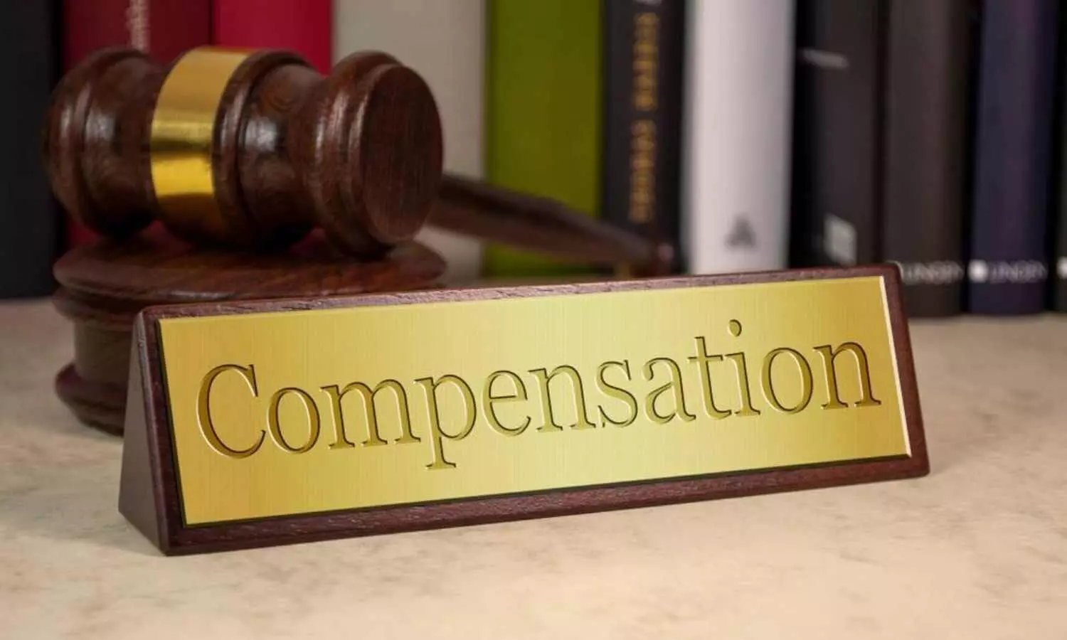
Holding the scan centre liable, the Commission directed it to pay Rs 15 lakh as compensation to the family of the deceased patient. “Taking into consideration of the above aspects we award a compensation of Rs.15,00,000/- to be paid by the 1st opposite party to the complainants along with a cost of Rs.10,000/- towards litigation expenses. As we dismiss the complaint against the 2 nd opposite party we do not order the medical expenses to be reimbursed to the complainants as prayed by the complainants,” the Commission observed.

To view the order, click on the link below:

https://medicaldialogues.in/pdf_upload/dcdrc-thiruvallur-237730.pdf

Also Read: Rs 12 lakh compensation slapped on Hospital, Paediatrician for failure in timely treatment of snakebite

Powered by WPeMatico

Fact Check: Can placing onion on feet and wearing socks cure Pneumonia?

A video on Instagram claiming that placing chopped onions on feet and covering it with socks can cure Pneumonia.

Claim An Instagram video claims that onions can cure pneumonia. The video mentions “ how to use onions to cure pneumonia and bronchitis at home. Two medium-sized onions are chopped, placed under the feet, and covered by socks. Flavonoids, absorbed through the pores of the feet, help to remove phlegm from the lungs and treat pneumonia due to their properties.”

The link to the claim is here.

Fact Check:

The Claim is False.

What is Pneumonia?

Pneumonia is an infection of the lungs that may be caused by bacteria, viruses, or fungi. The infection causes the lungs’ air sacs (alveoli) to become inflamed and fill with fluid or pus. That can make it hard for the oxygen you breathe to enter your bloodstream. The symptoms of pneumonia can range from mild to severe and include cough, fever, chills, and trouble breathing. (1)

Causes of Pneumonia:

Pneumonia can be caused by bacteria, viruses, or fungi. Bacterial pneumonia, common in adults, is often due to Streptococcus pneumoniae, while atypical forms like “walking pneumonia” are caused by Mycoplasma pneumoniae. Viral pneumonia can result from influenza or cold viruses, with Respiratory syncytial virus (RSV) being a frequent cause in children. COVID-19, caused by SARS-CoV-2, also leads to pneumonia. Fungal pneumonia is more common in individuals with weakened immune systems. (2)

Incidence of Pneumonia:

The estimated worldwide incidence of community-acquired pneumonia varies between 1.5 to 14 cases per 1000 person-years. (3) India accounts for 23 per cent of the global pneumonia burden, with case fatality rates between 14 and 30 per cent. (4)

Risk Factors :

Speaking to the Medical Dialogues team, Dr Kamal Kant Kohli, Chest Specialist, explained, ” Pneumonia risk factors can be categorised into medical conditions, health behaviours, and environmental factors. Medical conditions that increase vulnerability include chronic lung diseases like COPD, serious illnesses such as diabetes, and weakened immune systems from conditions like HIV/AIDS. Health behaviours contributing to higher risk include cigarette smoking and substance abuse. Environmental factors involve exposure to chemicals, pollutants, and toxic fumes, including secondhand smoke, which can compromise lung health and increase the likelihood of developing pneumonia.”

Onion and its health  benefits:

Onions are a very common and rich source of dietary flavonoids. They contain three diverse and highly valuable phytochemicals in perfect proportion: flavonoids, fructans, and organosulfur compounds. Onion has many properties that can help with numerous health issues. It is anti-allergenic, anti-inflammatory, cardioprotective, vasodilatory, anti-carcinogenic, antioxidant, antibacterial, and antifungal. Quercetin is the main element that gives onion these properties. (5)

Phytochemicals can potentially promote human health benefits and offer protection from various diseases. Apart from the main sulfur and flavonoid constituents, several other constituents of intact onion, such as lectins (the most abundant proteins in onion), prostaglandins, fructan, pectin, adenosine, vitamins B1, B2, B6, C, and E, biotin, nicotinic acid, fatty acids, glycolipids, phospholipids, and essential amino acids, have been studied for their biological effects over several decades. (6)

Can placing onion on feet and wearing socks cure Pneumonia?

Some studies show that onions have antimicrobial and possible bronchodilatory effects. However, no scientific evidence supports the claim that placing onion on feet and wearing socks can cure Pneumonia.

This study indicates possible bronchodilatory and preventive effects of onion and quercetin (Qt) (derivatives of onion) on asthma and symptoms of virus-infected flu, including cough, headache, and sputum production. The effect of onion on the lung infected with P. aeruginosa, S. aureus, and S. pneumonia was also demonstrated. (7)

Garlic and the variety of onions used in all dilutions were seen to possess antimicrobial properties against various microorganisms causing pneumonia. (8)

Also, the study by Beigoli et al., which the video refers to, Suggests that compounds in onions may help reduce inflammation and improve immune responses, which could be beneficial in managing symptoms associated with respiratory infections like pneumonia. The study emphasises the potential of these extracts in preventative care rather than as a cure for existing conditions. (9) Nowhere does the study that onions can cure pneumonia or the fact that the treatment modality includes placing the onion on feet and wearing socks. 

Despite the studies showing antimicrobial and possible bronchodilatory, the claim lacks scientific and consensus evidence in the medical fraternity to support its effectiveness in treating pneumonia, a serious respiratory infection.

Responding to the claim, Dr Mrigakshi Agarwal, Pulmonologist at Sanjeevan Hospital, New Delhi, clarified, “Placing onions under the feet and covering them with socks does not cure pneumonia. This practice is a folk remedy without any scientific basis or clinical evidence supporting its effectiveness against pneumonia, which is a serious respiratory infection. Onions contain some antioxidants and other compounds that have anti-inflammatory and possibly expectorant properties, which may help alleviate symptoms associated with respiratory conditions that, too, when consumed. Placing onion on feet and covering with socks does not help and lacks scientific evidence. It is essential to consult healthcare professionals for proper diagnosis and treatment rather than relying on unproven home remedies.”

Medical Dialogues Final Take:

Placing onions on the feet and wearing socks does not cure pneumonia. Although it has some antimicrobial and bronchodilatory properties when consumed, no scientific evidence or research in the medical fraternity supports the claim. Hence, the claim that ” placing onion on feet and wearing socks cure Pneumonia” is False.

References:

1. https://www.lung.org/lung-health-diseases/lung-disease-lookup/pneumonia/learn-about-pneumonia.

2. https://www.nhlbi.nih.gov/health/pneumonia/causes

3.https://www.ncbi.nlm.nih.gov/books/NBK430749/#:~:text=The%20estimated%20worldwide%20incidence%20of,%2C%20season%2C%20and%20population%20characteristics.

4. Eshwara VK, Mukhopadhyay C, Rello J. Community-acquired bacterial pneumonia in adults: An update. Indian J Med Res. 2020 Apr;151(4):287-302. doi: 10.4103/ijmr.IJMR_1678_19. PMID: 32461392; PMCID: PMC7371062.

5. Shafiq, S., Shakir, M., & Ali, Q. (2017). Medicinal uses of onion (Allium cepa L.): An overview. Life Science Journal, 14(6).

6. Yahia, Elhadi M (2017). Fruit and Vegetable Phytochemicals (Chemistry and Human Health, 2nd Edition) || Onion ( Allium cepa L.). , 10.1002/9781119158042(), 1145–1162. doi:10.1002/9781119158042.ch58

7. García-García J, Gracián C, Baños A, Guillamón E, Gálvez J, Rodriguez-Nogales A, Fonollá J. Beneficial Effects of Daily Consumption of Garlic and Onion Extract Concentrate on Infectious Respiratory Diseases in Elderly Resident Volunteers. Nutrients. 2023 May 15;15(10):2308. doi: 10.3390/nu15102308. PMID: 37242191; PMCID: PMC10221607.

8. Bryan-Thomas, J., Bazuaye-Alonge, P., & Wright, V. (2016). The Response of Staphylococcus aureus, Klebsiella pneumoniae and Streptococcus pneumoniae to Plant Derived Nutraceuticals. American Journal of Public Health, 4(6), 202-206.

9. Beigoli, S., Behrouz, S., Memarzia, A., Ghasemi, S. Z., Boskabady, M., Marefati, N., Kianian, F., Khazdair, M. R., El-Seedi, H., & Boskabady, M. H. (2021). Effects of Allium cepa and its constituents on respiratory and allergic disorders: A comprehensive review of experimental and clinical evidence. Volume 2021, Article ID 5554259. https://doi.org/10.1155/2021/5554259

Powered by WPeMatico

NMC gives final decision on 153 applications by 31 medical institutes to start new PG medical courses, increase PG medical seats

Through a recent notice, the Medical Assessment and Rating Board (MARB) of the National Medical Commission (NMC) has communicated its final decision to 31 medical institutes in respect of 153 applications for the starting of new postgraduate medical courses and an increase of PG medical seats for the academic year 2024-2025.

Among these 153 applications, altogether 110 applications were for starting new PG medical courses and 43 applications were for an increase of seats in PG medical courses.
For more information, click on the link below:

Powered by WPeMatico

Timely detection and prevention are the most effective strategies to tackle Thalassemia: Union Health Secretary

New Delhi: Shri Apurva Chandra, Secretary, Ministry of Health and Family Welfare stressed on the importance of timely detection and prevention of Thalassemia to tackle the disease. He stated that only by preventing it at the right time, can the burden of this disease be curtailed. 

The Union Health Secretary was speaking at an event to mark the International Thalassemia Day, here today.

Speaking on the occasion, the Union Health Secretary stated, “Timely detection and prevention are the most effective strategies to tackle Thalassemia.” He added that there are almost 1 lakh Thalassemia patients in the country, with approximately 10,000 new cases reported each year. He emphasized on the urgency for proactive intervention aided by timely detection through screening.

Also Read:Haryana: Thalassemia, haemophilia patients to get Rs 3000 monthly pension

Shri Apurva Chandra also highlighted the critical need for wide awareness around the subject. He stated that “Still many people are unaware of this disease and how this can be prevented. It is imperative that all stakeholders in this arena collaborate for a nationwide campaign to enhance awareness on Thalaseemia”.

As a significant step in this direction, he launched a video made in collaboration with the Indian Association of Pediatrics and Thalassemics India to promote effective prevention methods and optimal treatment for Thalassemia (https://youtu.be/H__bidXcanE?si=-_87PEPxAdsPNaw1).

Union Health Secretary also advocated for inclusion of compulsory Thalassemia testing in the existing reproductive and child health (RCH) programs under NHM as a means to curtail the prevalence of the disease. He added that some states have included this in their public health programs and activities; other states will be urged to include and expand screening and testing for Thalassemia.

Thalassemia is an inherited blood disorder that causes a body to have less hemoglobin than normal. Celebrated every year, International Thalassemia Day serves as a crucial platform to emphasize the importance of disease prevention, raise awareness, sensitize stakeholders, promote early detection, and ensure quality care for those affected by thalassemia. This year’s theme, “Empowering Lives, Embracing Progress: Equitable and Accessible Thalassemia Treatment for All,” encapsulates the collective mission toward universal access to comprehensive Thalassemia care.

Ms Aradhana Patnaik, AS&MD (NHM); Dr G. V. Basavaraja, President, Indian Academy of Pediatrics; Ms Shobha Tuli, Secretary, Thalassemics India; Dr Manas Kalra, Honorary Secretary, PHO Chapter of IAP and other senior officials from Union Health Ministry were present on the occasion.

Powered by WPeMatico