Omega-3 fatty acids may reduce CV and all cause mortality in diabetes patients, reports study

A recent study unveiled the potential benefits of omega-3 polyunsaturated fatty acids (PUFAs) for individuals with diabetes. This study illuminates a previously underexplored aspect of dietary interventions for diabetic patients and their long-term prognosis. The key highlights of this study were published in the recent edition of The Journal of Clinical Endocrinology and Metabolism.

This research was conducted by the team led by Jinhang Che and extensively analyzed data from the National Health and Nutrition Examination Survey (NHANES) which spanned from 2003 to 2018. The cohort included a big group of 16,539 individuals who either were prediabetic or diabetic. These participants were categorized into three groups based on their levels of omega-3 PUFAs. Over a median follow-up period of 8.42 years, this study observed a remarkable correlation, where those with higher levels of omega-3 PUFAs underwent significantly reduced risks of both all-cause and cardiovascular mortality. The reduction in risk was particularly pronounced in the second and third tertiles when compared to the reference group.

The study employed sophisticated statistical analyses to analyze in depth regarding the relationship between omega-3 PUFAs and mortality. Restricted cubic spline (RCS) curves unveiled a negative nonlinear association by suggesting that the benefits of omega-3 PUFAs were dose-dependent. Subgroup analyses revealed intriguing insights with body mass index (BMI) emerging as a crucial factor that influences the interaction.

The outcomes of this study identified specific types of omega-3 PUFAs that appeared to drive these benefits associated with mortality. Alpha-linolenic acid (ALA) and docosapentaenoic acid (DPA) were found to be the primary contributors which underlined the importance of dietary choices in reaping the full rewards of omega-3 supplementation.

This comprehensive study emphasized the significance of these findings that effectively highlights the potential for targeted dietary interventions to reduce the elevated mortality risks underwent by the individuals with diabetes. The patients at risk may be able to safeguard their long-term health outcomes by incorporating omega-3-rich foods into their diets.

Overall, the major findings of this study offers hope for the millions worldwide with diabetes, with the future initiatives that could potentially optimize dietary habits which prove instrumental in stemming the tide of morbidity and mortality associated with this pervasive condition.

Reference:

Che, J., He, N., Kuang, X., Zheng, C., Zhou, R., Zhan, X., & Liu, Z. (2024). Dietary n-3 Fatty Acids Intake and All-Cause and Cardiovascular Mortality in Patients With Prediabetes and Diabetes. In The Journal of Clinical Endocrinology & Metabolism. The Endocrine Society. https://doi.org/10.1210/clinem/dgae265

Powered by WPeMatico

7 kg Synovial Sarcoma tumour removed from 51-year-old man at AIIMS Bhubaneswar

Bhubaneswar: In a remarkable feat of medical innovation and collaborative healthcare, a team of doctors at AIIMS Bhubaneswar has successfully treated a middle-aged man by removing a rare scalp tumour, marking a significant milestone.

According to an official release from AIIMS Bhubaneswar, “The 51-year-old man identified as Rabindra Bishui from West Bengal was saved through a collaborative endeavour led by a dedicated team of medical professionals.”

Also Read:1.1 kg newborn with duodenal atresia gets new lease of life at AIIMS Bhubaneswar

The patient, Rabindra Bishui underwent the complex procedure to address a longstanding scalp swelling that had plagued him for over two decades.

The growth, later diagnosed as a 7 kg Synovial Sarcoma tumor, posed a formidable challenge to conventional treatment methods.

The exceptionally rare tumour, synovial sarcoma, especially in the scalp, with only scant documented cases in the medical literature was removed following a surgery that was only the second of its kind in India.

According to an ANI report, AIIMS Bhubaneswar Executive Director Doctor Ashutosh Biswas congratulated the group of doctors for such a rare operation and reiterated the commitment of the national institute to provide modern and quality health care service to the people of Odisha as well as the neighbouring states.

As per the official release, the patient had gone to different medical colleges and hospitals for treatment but was denied, later he ultimately reached the Department of Plastic Surgery at AIIMS Bhubaneswar.

“A multidisciplinary team from AIIMS Bhubaneswar, comprising experts in Interventional Radiology, Surgical oncology, Neurosurgery, Plastic surgery, Anesthesiology, and Pathology meticulously crafted a treatment strategy under the guidance of Dr Sanjay Kumar Giri, Head of the department of Burns and Plastic Surgery,” added Dr Biswas.

Medical Superintendent Dr Dillip Kumar Parida also conveyed best wishes to the team of doctors for such work.

The procedure was intricate, necessitating the ligation of the left external carotid artery and posterolateral neck dissection, culminating in the successful removal of the tumor.

Despite the challenges posed by the surgery, including significant intraoperative blood loss requiring approximately 6 units of blood and other blood products, the dedicated anesthesia team led by Dr Aparajita Panda, along with the vigilant nursing team consisting of Surya, Ashoka, Sibanjali, Pramod, and Sangeeta, ensured the patient’s safety and well-being throughout the procedure, which lasted about 7 hours, reports news agency ANI.

Following the successful surgery, the patient received intensive care for 24 hours before being transitioned to the ward for further observation and recovery.

The intricate procedure involved precise embolization of blood vessels, targeting the bilateral superficial temporal arteries and the left occipital artery, by Dr Manoj Kumar Nayak from the Radiodiagnosis department, followed by surgical excision by Dr Rabi Narayan Sahu (Neurosurgery), Dr Kanav Gupta, Dr Anil Kumar, Dr Phanindra Kumar Swain(Surgical Oncology), Dr Dinesh, Dr Sanjay K Giri, Dr Santanu Subba, Dr R K Sahu, Dr Aparna Kanungo(Plastic Surgery), Dr Gopika Jith, Dr Akanksha Rajpoot, and Dr Ahana.

Dr Pritinanda Mishra’s timely pathological evaluation of the specimen played a crucial role in the treatment process.

The collaborative efforts of the surgical teams not only resulted in the successful treatment of a rare scalp tumor but also highlighted the significance of interdisciplinary cooperation in addressing complex medical cases.

Powered by WPeMatico

Schizophrenia patients more prone to long hospitalizations due to non psychiatric illnesses: Study

Schizophrenia patients are more prone to long hospitalisations due to non-psychiatric illnesses suggests a study published in the American Psychiatric Association (APA) 2024 Annual Meeting in New York, NY.

Patients with schizophrenia are more likely to be admitted for hospital stays—and experience lengthier durations of stay and therefore, costlier care—for conditions including sepsis, COPD exacerbation, and pneumonia. According to a data analysis presented at the American Psychiatric Association (APA) 2024 Annual Meeting in New York, NY, this week, investigators reported findings highlighting a significantly disparate hospitalization experience for patients with schizophrenia when presenting with non-psychiatric conditions for emergent care. The data may allude to potential pursuits in qualitative research that can better identify the drivers of elongated and exacerbated hospital stays experienced by patients with schizophrenia. The research team conducted a retrospective analysis to identify the most common reasons for hospital admissions among patients with schizophrenia. As they noted, such patients have a well-evidenced shorter life span than the general population—up to 20 years on average, according to research. But their shorter life spans are often due to illnesses beyond their psychiatric condition. Investigators compiled state-level inpatient hospitalization data for adults aged ≥18 years old between 2016 – 2019 from the National Inpatient Sample (NIS) database.

They identified the 10 most common primary diagnoses for hospital admission of patients with and without schizophrenia based on the data, before conducting a multivariate logistic regression to assess the impact of schizophrenia on the odds of being admitted with each of the top 10 primary diagnoses. They then analyzed proportions for each diagnosis, as well as odds ratios (ORs) for admission while excluding for schizophrenia-based admissions to fully interpret the drivers of non-psychiatric hospital admissions among patients with schizophrenia. Lastly, they compared the characteristics, lengths of stay, and in-hospital mortality of patients with each diagnosis across the schizophrenia and control cohorts. Their final analysis showed schizophrenia-related diagnoses accounted for only one-third (35%) of hospital admissions among such patients, compared to 65% of admissions being due to other causes. Among the primary diagnoses to be 1 of the 10 most prevalent in both hospitalized patients with and without schizophrenia included:

• Sepsis

• COPD exacerbation

• Acute renal failure

• Pneumonia

• Urinary tract infection (UTI)

• Hypertensive heart failure

The other most prevalent primary diagnoses in patients with schizophrenia included hyponatremia, aspiration pneumonia, and acute hypoxic respiratory failure. Patients without schizophrenia were more likely to be admitted to the hospital due to non-ST-elevation myocardial infarction or knee osteoarthritis than patients with schizophrenia. Investigators found that patients with schizophrenia were significantly more likely to be admitted for most of the observed medical illnesses, except for heart failure and acute renal failure. Though patients with schizophrenia were more likely to die from sepsis, acute hypoxic respiratory failure and aspiration pneumonia than the control cohort, they were also shown to have significantly longer lengths of hospital stay for all diagnoses, as well as greater costs of care for most diagnoses. The data elucidated a tendency for patients with schizophrenia to experience a more difficult hospitalisation—despite their non-psychiatric diagnoses trending similarly to hospitalized patients without schizophrenia.

Reference:

Venkataramanan A, et al. Most common causes of nonpsychiatric hospitalizations in patients with schizophrenia in the United States. Paper presented at: American Psychiatric Association (APA) 2024 Annual Meeting. New York, NY. May 4-8, 2024.

Powered by WPeMatico

Clindamycin relevant alternative to other antibiotics in treating osteonecrosis of the jaw: Prospective trial

Germany: In a groundbreaking study, researchers have delved into the intricate relationship between clindamycin concentrations and osteonecrosis of the jaw (ONJ). This prospective trial sheds light on a crucial aspect of medication efficacy in combating this debilitating condition.

The study, published in the Journal of Cranio-Maxillofacial Surgery, revealed clindamycin (CLI) as an appropriate antibiotic option for patients requiring treatment/prophylaxis for ONJ. It could also be used as an alternative to sulbactam/ampicillin in patients allergic to penicillin.

According to the authors, the study is the first to present data on CLI concentrations in both necrotic and vital jawbone tissue.

Osteonecrosis of the jaw is a rare but serious condition characterized by the death of bone tissue in the jaw, often associated with the use of certain medications, particularly those used in cancer treatment. Clindamycin, an antibiotic commonly prescribed to treat bacterial infections, has been implicated in some cases of ONJ. However, the precise relationship between clindamycin concentrations and the development or exacerbation of ONJ has remained unclear until now.

Against the above background, Anton Straub, University Hospital Würzburg, Würzburg, Germany, and colleagues aimed to investigate the jawbone concentration of clindamycin in patients with osteonecrosis of the jaw.

The study included patients with medication-related ONJ (MRONJ) and osteoradionecrosis (ORN) with an antibiotic treatment with CLI. Vital, plasma and necrotic bone samples were collected. Liquid chromatography-tandem mass spectrometry was used to analyze plasma and jawbone samples.

The study led to the following findings:

  • Patients with MRONJ exhibited a mean plasma CLI concentration of 9.6 μg/mL and mean concentrations of 2.3 μg/g CLI and 2.1 μg/g CLI in vital and necrotic bone samples, without statistical significance.
  • In patients with ORN, the mean concentration in plasma was 12.0 μg/mL, in vital bone 2.1 μg/g, and in necrotic bone 1.7 μg/g.
  • Vital and necrotic bone concentrations did not differ significantly.

The findings showed that CLI concentrations are considerably lower than in plasma, but sufficient for most bacteria present in ONJ.

“Within the study limitations, it seems that CLI is a relevant alternative to other antibiotics in treating ONJ because it reaches adequate concentrations in the jawbone,” the researchers wrote.

“By using modern bioanalytical methods, some of the limitations identified in previous studies could be circumvented, and thus could provide a more solid statement on the bone penetration properties of CLI,” they concluded.

The study findings can revolutionize the management and treatment of ONJ. By elucidating the relationship between clindamycin concentrations and ONJ development or progression, clinicians can better tailor treatment regimens to individual patients, optimizing efficacy while minimizing the risk of adverse effects.

Reference:

Straub, A., Stapf, M., Brands, R. C., Kübler, A. C., Lâm, T., Vollmer, A., Gubik, S., Scherf-Clavel, O., & Hartmann, S. (2024). Investigation of clindamycin concentrations in human plasma and jawbone tissue in patients with osteonecrosis of the jaw: A prospective trial. Journal of Cranio-Maxillofacial Surgery, 52(3), 355-362. https://doi.org/10.1016/j.jcms.2024.01.014

Powered by WPeMatico

Intermittent fasting protects against liver inflammation and liver cancer, claims study

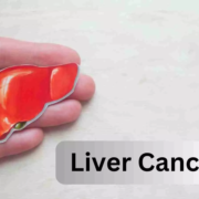
Fatty liver disease often leads to chronic liver inflammation and can even result in liver cancer. Scientists from the German Cancer Research Center (DKFZ) and the University of Tübingen have now shown in mice* that intermittent fasting on a 5:2 schedule can halt this development. The fasting regime reduces the development of liver cancer in mice with pre-existing liver inflammation. The researchers identified two proteins in liver cells that are jointly responsible for the protective effect of fasting. An approved drug can partially mimic this effect.

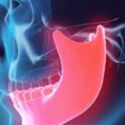
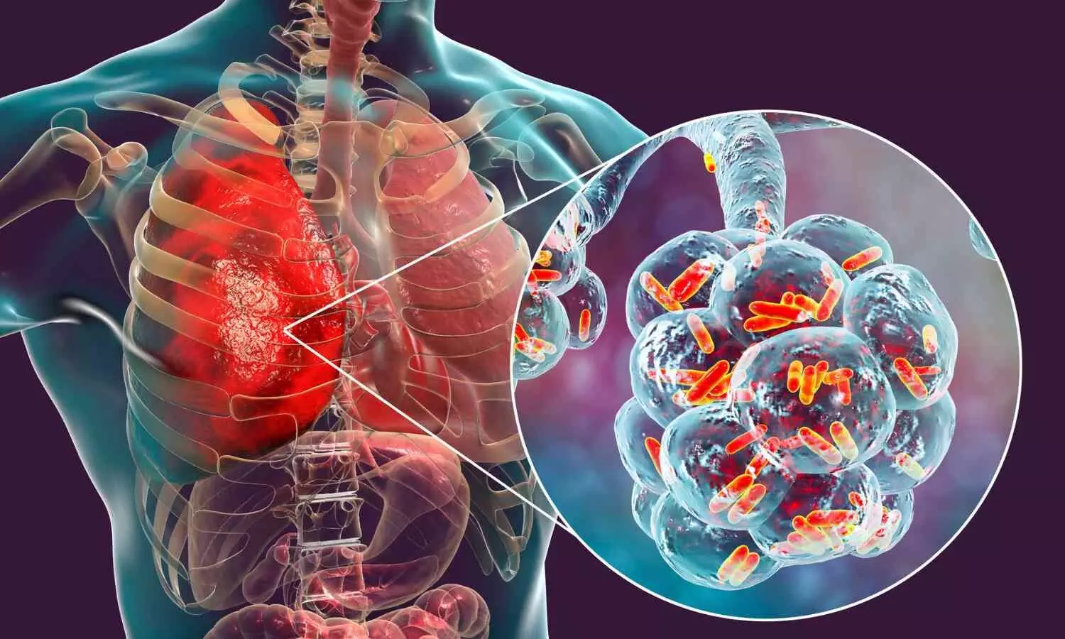
The most common chronic liver condition is non-alcoholic fatty liver disease. It can have serious consequences: If left untreated, it can lead to liver inflammation (metabolic dysfunction-associated steatohepatitis, MASH), liver cirrhosis and even liver cancer. Fatty liver disease is largely considered to be a direct consequence of obesity. It is not only people in Europe and the USA who have put on enormous amounts of weight in recent decades; obesity is also becoming increasingly widespread in emerging countries such as India and China. As a result, the number of cases of liver failure and liver cancer is rising sharply in the countries affected.

“The vicious circle of an unhealthy diet, obesity, liver inflammation and liver cancer is associated with major restrictions and suffering for those affected and also represents a considerable burden on healthcare systems,” says Mathias Heikenwälder, DKFZ and University of Tübingen. “We have therefore investigated whether simple dietary changes can specifically interrupt this fatal process.”

Intermittent fasting has already been shown in several studies to be an effective means of reducing weight and alleviating certain metabolic disorders. Heikenwälder’s team has now tested in mice whether this approach can also protect the liver from fatty degeneration and chronic inflammation.

Resistance to liver inflammation is independent of calorie intake

The animals were fed with a high-sugar and high-fat diet correspondind to the typical Western diet. One group of mice had constant access to the food. As expected, these animals gained weight and body fat and developed chronic liver inflammation.

The mice in the other group were given nothing to eat on two days a week (5:2 intermittent fasting, or 5:2 IF for short), but were allowed to eat as much as they wished on the other days. Despite the high-calorie diet, these animals did not put on weight, showed fewer signs of liver disease and had lower levels of biomarkers that indicate liver damage. In short, they were resistant to the development of MASH.

Interestingly, resistance to the development of a fatty liver was independent of the total calorie intake, as the animals immediately made up for the lost rations after the end of the fasting periods.

When experimenting with different variants of intermittent fasting, it was found that several parameters determine protection against liver inflammation: The number and duration of fasting cycles play a role, as does the start of the fasting phase. A 5:2 dietary pattern works better than 6:1; 24-hour fasting phases better than 12-hour ones. A particularly unhealthy diet requires more frequent dieting cycles.

Heikenwälder’s team now wanted to find out the molecular background of the response to fasting. To this end, the researchers compared protein composition, metabolic pathways and gene activity in the liver of fasting and non-fasting mice. Two main players responsible for the protective fasting response emerged: the transcription factor PPARα and the enzyme PCK1. The two molecular players work together to increase the breakdown of fatty acids and gluconeogenesis and inhibit the build-up of fats.

“The fasting cycles lead to profound metabolic changes, which together act as beneficial detoxification mechanisms and help to combat MASH,” says Heikenwälder, summarizing the molecular details.

The fact that these correlations are not just a mouse phenomenon was shown when tissue samples from MASH patients were examined: Here, too, the researchers found the same molecular pattern with reduced PPAR α and PCK1. Are PPAR α and PCK1 actually responsible for the beneficial effects of fasting? When both proteins were genetically switched off simultaneously in the liver cells of the mice, intermittent fasting was unable to prevent either chronic inflammation or fibrosis.

The drug pemafibrate mimics the effects of PPARα in the cell. Can the substance also mimic the protective effect of fasting? The researchers investigated this question in mice. Pemafibrate induced some of the favorable metabolic changes that were observed with 5:2 fasting. However, it was only able to partially mimic the protective effects of fasting. “This is hardly surprising, as we can only influence one of the two key players with pemafibrate. Unfortunately, a drug that mimics the effects of PCK1 is not yet available,” explains Mathias Heikenwälder.

Intermittent fasting as liver therapy

While Heikenwälder and his team initially focused on the effects of intermittent fasting on the prevention of MASH, they then investigated whether the 5:2 diet could also alleviate existing chronic liver inflammation.

To this end, the team examined mice that had developed MASH after months of being fed a high-sugar, high-fat diet. After a further four months of 5:2 intermittent fasting (on the same diet), these animals were compared with the non-fasting control group. The fasting mice had better blood values, less fatty liver and liver inflammation and above all: they developed less liver cancer and had fewer cancer foci in the liver.

“This shows us that 5:2 intermittent fasting has great potential – both in the prevention of MASH and liver cancer, as well as in the treatment of established chronic liver inflammation,” summarizes principal investigator Heikenwälder. “The promising results justify studies in patients to find out whether intermittent fasting protects against chronic liver inflammation as well as in the mouse model.”

The 5:2 fasting regimen is popular. It is considered comparatively easy to integrate into everyday life, as the fasting days can be tailored to personal needs and no specific foods are prohibited. “Nevertheless, there will always be people who can’t stick to a strict diet in the long term,” says Heikenwälder. “That’s why we want to continue to investigate which combinations of drugs we can use to fully mimic the protective effects of fasting.”

Why was it necessary to study mice for this experiments?

A high-calorie, unbalanced diet and a sedentary lifestyle are contributing to the sharp rise in the proportion of obese people in many parts of the world. Obesity also increases the risk of a whole range of secondary diseases for those affected, including cardiovascular diseases, metabolic diseases, cancer and joint problems. Understanding these correlations and, ideally, intervening in a targeted manner is an extremely important research goal.

Several organs and organ systems are jointly involved in the development of obesity-related health consequences. The liver, pancreas, brain, lymphatic organs and fatty tissue work together here, which cannot be simulated in the culture dish. The intestine can also play a role in the development of MASH. The spontaneous development of liver cancer from a fatty liver can therefore only be recapitulated and investigated if all these factors interact.

Also crucial is the influence of immune cells that migrate from the periphery of the body into the fatty liver, are activated there and migrate back into the body, where they interact with the fatty tissue. This can only be investigated in an intact organism. Experiments with mice are also necessary to understand which length of fasting periods achieves the most favorable health results in the context of the circadian (day/night) rhythm.

Reference:

Suchira Gallage, Adnan Ali, Jose Efren Barragan Avila, Nogayhan Seymen, Pierluigi Ramadori, Vera Joerke, Laimdota Zizmare, David Aicher, Indresh K. Gopalsamy, Winnie Fong, Jan Kosla, Enrico Focaccia, Xin Li, Suhail Yousuf, Tjeerd Sijmonsma, Mohammad Rahbari, Katharina S. Kommoss, Adrian Billeter, Sandra Prokosch, Ulrike Rothermel, Florian Mueller, Jenny Hetzer, Danijela Heide, Benjamin Schinkel, Tim Machauer, Bernd Pichler, Nisar P. Malek, Thomas Longerich, Susanne Roth, Adam J. Rose, Johannes Schwenck, Christoph Trautwein, Mohammad M. Karimi, Mathias Heikenwalder, A 5:2 intermittent fasting regimen ameliorates NASH and fibrosis and blunts HCC development via hepatic PPARα and PCK1, Cell Metabolism, https://doi.org/10.1016/j.cmet.2024.04.015.

Powered by WPeMatico

Cannabis, nicotine use during pregnancy increases rate of infant death fourfold, reveals JAMA study

Oregon Health & Science University researchers in a new study report that combined cannabis and nicotine use during pregnancy is associated with significantly higher risk of poor health outcomes for newborns compared with use of either substance alone.

The study, published today in JAMA Network Open, found infants born to mothers who used both substances throughout pregnancy were at greater risk of being small for gestational size, undergoing preterm delivery, or even death.

With approximately half of individuals who use cannabis in pregnancy also using tobacco or nicotine products, the findings suggest the need for more effective clinical counseling early on, during the preconception and prenatal periods, said the study’s corresponding author, Jamie Lo, M.D., M.C.R., associate professor of obstetrics and gynecology (maternal-fetal medicine) in the OHSU School of Medicine and Division of Reproductive and Developmental Sciences at OHSU’s Oregon National Primate Research Center.

“With the growing legalization of cannabis around the country, there is often a perception that cannabis is safe in pregnancy,” Lo said. “Because we know that many people who use cannabis often use tobacco or nicotine products, we wanted to better understand the potential health implications on both the pregnant individual and the infant.

“There is still a great deal of stigma around the use of substances during pregnancy,” she continued. “With limited research to support official clinical recommendations, it can be a difficult topic for both patients and providers to navigate. Our hope is that this research supports more open and productive conversations that ultimately result in a healthier pregnancy.”

Researchers evaluated hospital discharge data and vital statistics from more than 3 million pregnant patients with documented cannabis and nicotine use. Compared with people who didn’t use any substances, those using cannabis or nicotine alone had increased rates of infant and newborn death, small gestational size and preterm delivery-but the risk was even higher when both cannabis and nicotine were used together.

Most notable was the rate of infant death, which was four times higher in users of both cannabis and nicotine compared with non-users. The rate was nearly two times higher compared with users of just cannabis or nicotine alone.

The research team says these findings can help guide health care professionals with preconception and prenatal counseling, especially regarding the benefits of quitting at least one of the two substances. They also support expanded efforts to educate pregnant people about the risks of cannabis and nicotine.

“We hope that pregnant individuals can abstain from using both cannabis and nicotine products. However, we acknowledge the complexities of individual circumstances may make this goal challenging, and for some patients is simply not realistic,” said Adam Crosland, M.D., M.P.H., assistant professor of obstetrics and gynecology in the OHSU School of Medicine and lead author of the study. “We always strive to meet people where they are and support patients with evidence-based recommendations and treatment options that are both patient-centered and promote the healthiest outcomes possible.

“Our findings suggest that avoiding use of just one of these substances can decrease the pregnancy risks we see when both substances are used together, which is a critical piece of information providers can highlight when counseling patients.”

The multidisciplinary research team also included Eliot Spindel, M.D., Ph.D., and Cindy McEvoy, M.D., M.C.R., who are investigating how to mitigate the negative respiratory outcomes among children born to parents who smoke during pregnancy. McEvoy’s team found that vitamin C supplementation to pregnant women unable to quit smoking cigarettes significantly improves airway function and respiratory health in their offspring; moreover, these improved outcomes persist through age 5 and these children are in continued follow-up through adolescence.

Looking ahead, researchers will continue to investigate the effects of combined cannabis and nicotine use-including the influence of potency, frequency and timing-in order to better educate pregnant patients about the harms of use. Additionally, the team will continue to explore the viability of prenatal interventions, such a vitamin C supplementation, and other treatment options for individuals who are unable to quit smoking during their pregnancy.

Reference:

Crosland BA, Garg B, Bandoli GE, et al. Risk of Adverse Neonatal Outcomes After Combined Prenatal Cannabis and Nicotine Exposure. JAMA Netw Open. 2024;7(5):e2410151. doi:10.1001/jamanetworkopen.2024.10151.

Powered by WPeMatico

Pediatric hypertension associated with long-term cardiovascular events: JAMA

A recent study from Ontario, Canada illuminated the long-term risks associated with childhood hypertension. This comprehensive study underlines the pressing need for increased awareness and effective management of hypertension in children. The major findings of this study were published in the recent issue of Journal of American Medical Association.

Hypertension was once considered primarily an affliction of adulthood is increasingly affecting children with its estimates suggesting to 6% effect on all children and which is still on the rise. Despite this trend, the evidence that links childhood hypertension to adverse cardiovascular outcomes was lacking, which leads to underdiagnosis and undertreatment.

This population-based, retrospective, matched cohort analysis meticulously examined the data from 1996 to 2022 and encompassed all children who were aged 3 to 18 years in Ontario. The research identified children with incident hypertension using validated case definitions derived from diagnostic and physician billing claims. Following this, they matched them with controls without hypertension based on various parameters including age, sex, birth weight, maternal gestational hypertension and prior comorbidities.

Over a median follow-up period of 13.6 years, the children who were diagnosed with hypertension expressed a significantly higher risk of major adverse cardiac events (MACE) when compared to their non-hypertensive counterparts. These events included cardiovascular death, stroke, hospitalization for myocardial infarction or unstable angina, coronary intervention and congestive heart failure.

The incidence of MACE was significantly higher among the children with hypertension with a rate of 4.6 per 1000 person-years when compared to 2.2 per 1000 person-years in controls. This translates to a more than twofold increased risk of MACE among the children with hypertension which emphasizes the profound impact of hypertension on cardiovascular health even from a young age.

While prior comorbidities such as malignancy, congenital heart disease and diabetes were uncommon in both cohorts, hypertension remained a significant independent risk factor for adverse cardiovascular outcomes. The implications of these findings underline the critical importance of early detection, regular monitoring and effective management of hypertension in children to reduce the long-term risks of cardiovascular disease in adulthood. Overall, clinicians can potentially avert a substantial burden of cardiovascular morbidity and mortality in the future by addressing hypertension in childhood. 

Source:

Robinson, C. H., Hussain, J., Jeyakumar, N., Smith, G., Birken, C. S., Dart, A., Dionne, J., Garg, A., Kandasamy, S., Karam, S., Marjerrison, S., South, A. M., Thabane, L., Wahi, G., Zappitelli, M., & Chanchlani, R. (2024). Long-Term Cardiovascular Outcomes in Children and Adolescents With Hypertension. In JAMA Pediatrics. American Medical Association (AMA). https://doi.org/10.1001/jamapediatrics.2024.1543

Powered by WPeMatico

30-year US study links ultra-processed food to higher risk of early death

Higher consumption of most ultra-processed foods is linked to a slightly higher risk of death, with ready-to-eat meat, poultry, and seafood based products, sugary drinks, dairy based desserts, and highly processed breakfast foods showing the strongest associations, finds a 30-year US study in The BMJ today.

The researchers say not all ultra-processed food products should be universally restricted, but that their findings “provide support for limiting consumption of certain types of ultra-processed food for long term health.”

Ultra-processed foods include packaged baked goods and snacks, fizzy drinks, sugary cereals, and ready-to-eat or heat products. They often contain colours, emulsifiers, flavours, and other additives and are typically high in energy, added sugar, saturated fat, and salt, but lack vitamins and fibre.

Mounting evidence links ultra-processed foods to higher risks of obesity, heart disease, diabetes and bowel cancer, but few long term studies have examined links to all cause and cause specific deaths, especially due to cancer.

To address this knowledge gap, researchers tracked the long-term health of 74,563 female registered nurses from 11 US states in the Nurses’ Health Study (1984-2018) and 39,501 male health professionals from all 50 US states in the Health Professionals Follow-up Study (1986-2018) with no history of cancer, cardiovascular diseases, or diabetes at study enrolment.

Every two years participants provided information on their health and lifestyle habits, and every four years they completed a detailed food questionnaire. Overall dietary quality was also assessed using the Alternative Healthy Eating Index-2010 (AHEI) score.

During an average 34-year follow-up period, the researchers identified 48,193 deaths, including 13,557 deaths due to cancer, 11,416 deaths due to cardiovascular diseases, 3926 deaths due to respiratory diseases, and 6343 deaths due to neurodegenerative diseases.

Compared with participants in the lowest quarter of ultra-processed food intake (average 3 servings per day), those in the highest quarter (average 7 servings per day) had a 4% higher risk of total deaths and a 9% higher risk of other deaths, including an 8% higher risk of neurodegenerative deaths.

No associations were found for deaths due to cardiovascular diseases, cancer, or respiratory diseases.

In absolute numbers, the rate of death from any cause among participants in the lowest and highest quarter of ultra-processed food intake was 1472 and 1536 per 100,000 person years, respectively.

The association between ultra-processed food intake and death varied across specific food groups, with meat, poultry, and seafood based ready-to-eat products showing the strongest and most consistent associations, followed by sugar sweetened and artificially sweetened beverages, dairy based desserts, and ultra-processed breakfast food.

And the association was less pronounced after overall dietary quality was taken into account, suggesting that dietary quality has a stronger influence on long term health than ultra-processed food consumption, note the authors.

This is an observational study, so no firm conclusions can be drawn about cause and effect, and the authors point out that the ultra-processed food classification system does not capture the full complexity of food processing, leading to potential misclassification. In addition, participants were health professionals and predominantly white, limiting the generalizability of the findings.

However, this was a large study with long follow-up, using detailed, validated, and repeated measurements, and results were similar after further analyses, providing greater confidence in the conclusions.

The researchers stress that not all ultra-processed food products should be universally restricted and say oversimplification when formulating dietary recommendations should be avoided.

But they conclude: “The findings provide support for limiting consumption of certain types of ultra-processed food for long term health,” adding that “future studies are warranted to improve the classification of ultra-processed foods and confirm our findings in other populations.”

In a linked editorial, researchers in New Zealand point out that recommendations to avoid ultra-processed food may also give the impression that foods that are not ultra-processed, such as red meat, can be frequently consumed.

They argue that debate about the ultra-processed concept must not delay food policies that improve health, such as restrictions on marketing unhealthy foods to children, warning labels on nutritionally poor food products, and taxes on sugary drinks.

“Our focus should be on advocating for greater global adoption of these and more ambitious interventions and increasing safeguards to prevent policies from being influenced by multinational food companies with vested interests that do not align with public health or environmental goals,” they conclude.

Reference:

https://www.bmj.com/content/385/bmj-2023-078476

Powered by WPeMatico

Lung abnormality progression linked to acute respiratory disease in smokers, claims study

Smokers who have small abnormalities on their CT scans that grow over time have a greater likelihood of experiencing acute respiratory disease events, according to a new study published in Radiology, a journal of the Radiological Society of North America (RSNA).

Quantitative interstitial abnormalities (QIA) are subtle abnormalities on chest CTs that do not meet the diagnostic criteria for advanced pulmonary diseases but are nonetheless associated with decreased lung function and capacity, increased respiratory symptoms and death.

“QIA includes features like reticulation and ground-glass opacities as well as subtle density changes with important clinical implications,” said Bina Choi, M.D., associate physician in the Division of Pulmonary and Critical Care Medicine at Brigham and Women’s Hospital and postdoctoral fellow at the Applied Chest Imaging Laboratory, Harvard Medical School in Boston. “In some patients, QIA may be a precursor to advanced diseases such as pulmonary fibrosis or emphysema.”

Acute respiratory disease events are episodes of increased cough, phlegm or shortness of breath that last at least two days and require treatment with steroids or antibiotics. Severe acute respiratory disease episodes require an emergency room visit or hospitalization.

“We wanted to determine whether progression in QIA on chest CT is associated with acute respiratory disease events in individuals with a history of smoking,” Dr. Choi said. “While many acute respiratory disease events are likely related to airway disease and COPD, some may instead be associated with QIA especially in people without obstruction or emphysema.”

Dr. Choi’s team performed a secondary analysis of the CT scans of 3,972 participants (mean age 60.7; 2,120 women) in the COPDGene® Study, one of the largest studies ever to investigate the underlying genetic factors of COPD. The study included individuals with a 10-pack-year or greater smoking history recruited from multiple centers between November 2007 and July 2017.

QIA was measured with machine learning-based tools as a percentage of lung volume on a CT scan. QIA progression was assessed using the participants’ QIA measurements at baseline and five-year follow-up CT exams.

Statistical analyses revealed that participants in the highest quartile of QIA progression had more frequent acute respiratory disease and severe acute respiratory disease events than those in the lowest quartile.

“We found that progression in QIA is independently associated with these acute respiratory disease events both intercurrent and subsequent to progression,” she said.

Dr. Choi said the results suggest that QIA progression may represent changes in lung tissue processes that have both short- and long-term impacts on patient symptoms and the worsening of those symptoms.

“Severe acute respiratory disease events may be a sign of disease activity and a source of morbidity at the earliest stages of lung tissue injury,” she said. “Some people with QIA progression may merit more aggressive monitoring and earlier intervention.”

Reference:

Bina Choi , Alejandro A. Díaz, Ruben San José Estépar, Nicholas Enzer, Victor Castro, MeiLan K. Han, George R. Washko, Association of Acute Respiratory Disease Events with Quantitative Interstitial Abnormality Progression at CT in Individuals with a History of Smoking, Radiology, https://doi.org/10.1148/radiol.231801.

Powered by WPeMatico

Weight-loss surgery before kidney transplantation improves health and eligibility of patients with obesity and renal failure, finds study

Laparoscopic sleeve gastrectomy promotes relatively rapid weight loss, reduces obesity-related health issues and improves eligibility for kidney transplantation for patients with advanced chronic kidney disease and obesity, according to new research from Mayo Clinic.

Patients with advanced chronic kidney failure and severe obesity often are not considered for kidney transplantation, but according to a study published in Mayo Clinic Proceedings, sleeve gastrectomy can improve high-risk patients meeting the criteria for transplantation. The findings also show that the surgical weight-loss procedure reduced cardiovascular risks, including diabetes and hypertension.

“In earlier research, we found that conservative weight-loss approaches do not adequately result in significant weight loss in patients with advanced chronic kidney disease,” says Aleksandra Kukla, M.D., a Mayo Clinic transplant nephrologist and the study’s first author. “These new findings support the value of surgical gastrectomy for patients with advanced chronic kidney failure, stage 4-5D, to improve general health and access to kidney transplantation.”

The retrospective study involved 104 patients with advanced chronic kidney failure and obesity who were treated at Mayo Clinic between 2020 and 2023. Fifty-four underwent laparoscopic sleeve gastrectomy, the most frequently performed surgical weight-loss procedure in patients seeking kidney transplant, and 50 patients opted for a nonsurgical weight-loss approach.

Undergoing sleeve gastrectomy reduced the time for patients to be added to waiting lists for kidney transplantation. It also improved the likelihood of receiving a transplant, with 37% of those who underwent sleeve gastrectomy receiving a transplant within 18 months, versus 10% in the nonsurgical cohort.

The risk of postsurgical complications was low, and the rate of hospitalizations and infections for patients who underwent sleeve gastrectomy was similar to the nonsurgical cohort.

Effective therapy for obesity among patients with advanced chronic kidney disease is understudied, in part because high body mass index (BMI) is often considered beneficial in patients receiving kidney dialysis. “Treatment of obesity in this population has been limited to nonsurgical options in the past,” says Tayyab Diwan, M.D., a Mayo Clinic transplant surgeon and co-author of the study.

More research is needed on the optimal timing of the surgery for kidney transplant candidates, Dr. Diwan says.

Reference:

Aleksandra Kukla, Sukhdeep S. Sahi, Pavel Navratil, Roberto P. Benzo, Byron H. Smith, Dustin Duffy, Walter D. Park, Meera Shah, Pankaj Shah, Matthew M. Clark, David C. Fipps, Aleksandar Denic, Carrie A. Schinstock, Patrick G. Dean, Mark D. Stegall, Yogish C. Kudva, Tayyab S. Diwan, Weight Loss Surgery Increases Kidney Transplant Rates in Patients With Renal Failure and Obesity, Mayo Clinic Proceedings, https://doi.org/10.1016/j.mayocp.2024.01.017.

Powered by WPeMatico