Anti-aging drug Rapamycin extends lifespan as effectively as eating less: Study

The anti-aging drug Rapamycin has the same life-extending effect as eating less, according to new research from the University of East Anglia and University of Glasgow.

Dietary restriction has long been considered one of the most reliable methods for increasing lifespan across species.But if fasting for hours sounds unpleasant, science may suggest another route to achieving a longer and healthier life.A new study published today reveals compelling evidence that Rapamycin, a compound originally developed as an immunosuppressant, offers comparable life-extending benefits in eight species of vertebrates, not including humans.Co-lead researcher Dr Zahida Sultanova, from UEA’s School of Biological Sciences, said: “Dietary restriction – for example through intermittent fasting or reduced calorie intake – has been the gold standard for living longer. But it’s difficult for most of us to maintain long-term.“We wanted to know if popular anti-aging drugs like Rapamycin or Metformin could offer similar effects without the need to cut calories.”The research team looked at data from 167 studies of lifespan across eight vertebrate species including fish, mice, rats and primates – in this, the largest study of its kind.

They investigated the effect of dietary restriction on longevity – as well as that of Rapamycin and Metformin, both of which have been touted as life-extending drugs.

The team found that Rapamycin extends lifespan almost as consistently as eating less, while the Type 2 diabetes medicine, Metformin, does not.

Key findings:

  • Dietary restriction – from intermittent fasting to cutting calories – consistently extended lifespan across all vertebrate species analysed in this study.
  • Rapamycin increased lifespan to the same extent as dietary restriction.
  • Metformin showed no clear longevity benefit although it is widely used for type 2 diabetes.
  • Lifespan gains were the same for males and females, and did not depend on the type of diet restriction.

As scientists continue the search for interventions that can improve our health and help us live longer, Rapamycin may stand out as one of the most promising tools – potentially sidestepping the challenges of long-term caloric restriction while offering similar benefits.

Co-lead researcher Dr Edward Ivimey-Cook, from the University of Glasgow, said: “These findings don’t suggest we should all start taking Rapamycin. But they do strengthen the case for its further study in aging research and raise important questions about how we approach longevity therapeutics.”Dr Sultanova added: “Our findings show that drug repurposing is a promising approach to improving people’s health and lifespan”.Both Rapamycin and Metformin are currently being used in human trials, with results still pending.

The authors note that Rapamycin may have negative effects on the immune system, and further research into safety in humans is necessary, although recent work indicates that low-dose Rapamycin has no serious adverse effects in healthy people.

This research was funded by the Natural Environment Research Council (NERC) and the Leverhulme Trust.‘Rapamycin, not metformin, mirrors dietary restriction-driven lifespan extension in vertebrates: a meta-analysis’ is published in the journal Aging Cell.

Reference:

Zahida Sultanova, Aging Cell, Rapamycin, not metformin, mirrors dietary restriction-driven lifespan extension in vertebrates: a meta-analysis.

Powered by WPeMatico

Zimislecel Therapy Achieves Insulin Independence in Majority of Type 1 Diabetes Patients in Phase 2 trial

In phase 2 of the FORWARD study, 83% of participants with type 1 diabetes achieved insulin independence a year after receiving zimislecel (formerly VX-880), an islet cell therapy developed by Vertex Pharmaceuticals. 10 out of 12 patients who received the full dose no longer required exogenous insulin, demonstrating promising potential for long-term diabetes management. These findings were published in The New England Journal of Medicine.

Zimislecel is an allogeneic, stem cell–derived islet-cell therapy designed to replace the insulin-producing beta cells destroyed by autoimmune activity in type 1 diabetes. This study which assessed the efficacy Zimislecel of enrolled 14 adults with long-standing type 1 diabetes, all of whom had undetectable C-peptide levels at baseline, indicating absent endogenous insulin production.

The participants were enrolled across three parts of the trial. In part A, two individuals received a half dose of zimislecel (0.4×10⁹ cells), with the possibility of a second dose within two years. In parts B and C, 12 patients received the full dose (0.8×10⁹ cells) via a single infusion into the portal vein. All participants were treated with glucocorticoid-free immunosuppressive regimens to prevent rejection of the implanted cells.

All 12 patients in parts B and C, who received the full zimislecel dose, experienced no severe hypoglycemic events between days 90 and 365 post-infusion. Moreover, they all achieved tight glucose control, reflected in glycated hemoglobin (HbA1c) levels under 7%. Most impressively, 10 of the 12 participants (83%) were insulin independent at day 365, meaning they no longer required external insulin to manage their blood sugar levels.

C-peptide was detectable in all participants post-treatment which suggests successful engraftment and functional islet cell restoration. This transformation occurred despite their complete lack of C-peptide at the beginning of the study. Neutropenia was the most common serious adverse event, occurring in 3 patients. Tragically, two deaths were reported: one from cryptococcal meningitis and another from progression of pre-existing severe neurocognitive impairment. 

The primary goal in part C was to ensure patients avoided severe hypoglycemic episodes while achieving meaningful improvements in blood sugar control. Secondary endpoints included safety and independence from insulin. Each participant’s response was assessed using a 4-hour mixed-meal tolerance test to evaluate natural insulin production via C-peptide detection.

Overall, these preliminary results suggest zimislecel may represent a major advance in regenerative treatment for type 1 diabetes, restoring natural insulin production in most patients without the need for lifelong insulin therapy. Further studies are planned to confirm these findings and better understand the safety profile of this therapy.

Reference:

Reichman, T. W., Markmann, J. F., Odorico, J., Witkowski, P., Fung, J. J., Wijkstrom, M., Kandeel, F., de Koning, E. J. P., Peters, A. L., Mathieu, C., Kean, L. S., Bruinsma, B. G., Wang, C., Mascia, M., Sanna, B., Marigowda, G., Pagliuca, F., Melton, D., Ricordi, C., & Rickels, M. R. (2025). Stem cell–derived, fully differentiated islets for type 1 diabetes. The New England Journal of Medicine. https://doi.org/10.1056/nejmoa2506549

Powered by WPeMatico

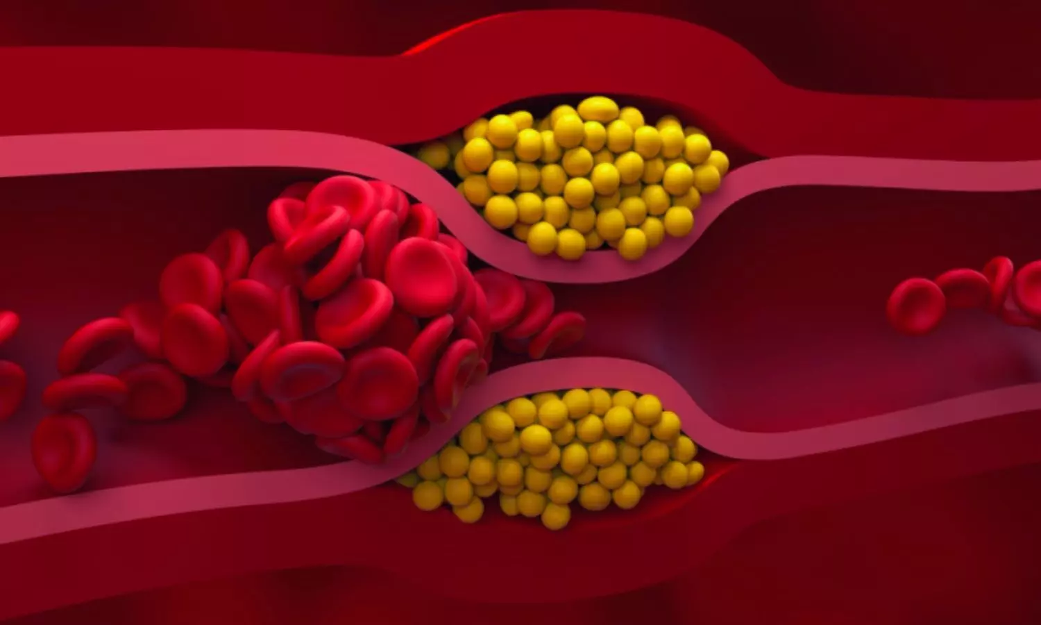
High Triglycerides Drive Residual Cardiovascular Risk Despite Optimal Lipid-Lowering Therapy: Study Shows

Netherlands: A new study published in Atherosclerosis reveals that elevated triglyceride (TG) levels are strongly associated with an increased risk of recurrent cardiovascular events and mortality in patients with established cardiovascular disease (CVD), regardless of how well other lipid targets are managed. The study, led by Pauline C.E. Schuitema and colleagues from the Department of Vascular Medicine at University Medical Center Utrecht, underscores the growing importance of addressing triglycerides as part of secondary prevention strategies.

“Elevated triglyceride levels strongly predicted higher residual risk in patients with established CVD. Each 1-unit increase in log-TG was linked to greater risk of myocardial infarction (HR 1.34), recurrent cardiovascular events (HR 1.17), cardiovascular mortality (HR 1.23), and a 12% rise in all-cause mortality—independent of LDL-C, non-HDL-C target achievement, HDL-C levels, or lipid-lowering therapy intensity,” the researchers reported.

While low-density lipoprotein cholesterol (LDL-C) management is a central pillar of CVD treatment, many patients continue to experience residual cardiovascular risk even after reaching guideline-recommended LDL-C levels. The research team investigated whether elevated triglycerides contribute to this persistent risk and whether this risk is influenced by LDL-C and non-HDL-C target achievement, high-density lipoprotein cholesterol (HDL-C) levels, or the intensity of lipid-lowering therapy (LLT).

The prospective cohort study included 9,436 individuals with established CVD. Researchers used Cox regression models to evaluate the relationship between triglyceride levels and the incidence of cardiovascular events and mortality over a median follow-up period of 9 years (interquartile range: 4.5 to 14.1 years).

The study led to the following findings:

  • Each one-unit rise in log-transformed triglyceride levels was associated with a 17% higher risk of recurrent cardiovascular events.
  • A 34% higher risk of myocardial infarction was linked to elevated triglyceride levels.
  • Triglyceride elevation corresponded to a 23% increase in cardiovascular mortality.
  • All-cause mortality increased by 12% with higher triglyceride levels.
  • These risks remained consistent regardless of LDL-C or non-HDL-C target achievement, HDL-C levels, or lipid-lowering therapy intensity.
  • During the study, there were 2,075 recurrent cardiovascular events, 736 myocardial infarctions, 586 strokes, 1,231 cardiovascular deaths, and 2,729 all-cause deaths.
  • No significant association was found between elevated triglycerides and stroke risk alone.

According to the authors, these findings emphasize the importance of monitoring and managing triglyceride levels in patients with CVD, alongside traditional lipid targets like LDL-C. The research highlights the potential benefit of incorporating targeted interventions—such as lifestyle changes and dietary adjustments—into secondary prevention plans to address the residual cardiovascular risk associated with elevated TGs.

The study concludes that triglyceride management should be prioritized as part of comprehensive cardiovascular care to help mitigate ongoing risk in patients already receiving intensive lipid-lowering therapy.

Reference:

Schuitema, P. C., Visseren, F. L., Nordestgaard, B. G., Teraa, M., Van der Meer, M. G., Ruigrok, Y. M., Onland-Moret, N. C., & Koopal, C. (2025). Elevated triglycerides are related to higher residual cardiovascular disease and mortality risk independent of lipid targets and intensity of lipid-lowering therapy in patients with established cardiovascular disease. Atherosclerosis, 120411. https://doi.org/10.1016/j.atherosclerosis.2025.120411

Powered by WPeMatico

Finerenone Benefits Heart Failure Patients Regardless of Frailty Status: JAMA

UK: A new analysis of the FINEARTS-HF clinical trial reveals that finerenone, a nonsteroidal mineralocorticoid receptor antagonist, offers significant benefits for patients with heart failure (HF) and mildly reduced or preserved ejection fraction, regardless of their level of frailty. The study, published in JAMA Cardiology, was led by Dr. Jawad H. Butt and colleagues from the British Heart Foundation Cardiovascular Research Centre at the University of Glasgow.

Frailty is often a concern when introducing novel therapies in HF patients, as it is typically linked with poorer outcomes and increased vulnerability to adverse events. This research evaluated whether frailty influences the safety and effectiveness of finerenone in individuals with HFmrEF or HFpEF.

The prespecified secondary analysis drew on data from the phase 3 FINEARTS-HF randomized clinical trial, conducted across 653 sites in 37 countries. The study enrolled patients with NYHA class II-IV HF, left ventricular ejection fraction of 40% or higher, structural heart disease, and elevated natriuretic peptide levels. Participants were randomized to receive either once-daily finerenone or placebo in addition to standard therapy. The primary endpoint was a composite of cardiovascular death and total worsening HF events. Frailty was assessed using the Rockwood cumulative deficit model.

The study led to the following findings:

  • Among 6,001 patients enrolled, frailty data were available for 5,952 participants (mean age 72 years; 54.4% male).
  • 26.7% of patients were classified as not frail, 36.0% as moderately frail, and 37.3% as most frail.
  • Higher frailty levels were associated with worse clinical outcomes, including increased risks of cardiovascular death and heart failure events.
  • Finerenone reduced the risk of cardiovascular death and worsening heart failure events across all frailty groups.
  • Finerenone improved symptom scores in patients regardless of frailty status.
  • The treatment effects of finerenone did not significantly vary by level of frailty.
  • The incidence of common adverse events—including hypotension, kidney dysfunction, and electrolyte imbalances—was similar across all frailty groups.

These findings suggest that finerenone maintains a favorable benefit-risk profile even in frail patients, a population often overlooked in HF treatment due to safety concerns. The results advocate for broader consideration of finerenone use in clinical practice, particularly for frail patients who may still benefit from the therapy.

While the study is limited by the underrepresentation of the most frail patients, typically excluded from clinical trials, and the absence of certain frailty measures, such as muscle strength, it provides compelling evidence for the utility of finerenone in a real-world HF population.

The authors concluded, “finerenone appears to be an effective and safe option for managing HFmrEF and HFpEF, irrespective of a patient’s frailty status. These findings should encourage clinicians to consider its use even in frail individuals, helping to optimize care for this vulnerable patient group.”

Reference:

Butt JH, Jhund PS, Henderson AD, et al. Finerenone According to Frailty in Heart Failure: A Prespecified Analysis of the FINEARTS-HF Randomized Clinical Trial. JAMA Cardiol. Published online June 18, 2025. doi:10.1001/jamacardio.2025.1775

Powered by WPeMatico

Automated Insulin Delivery Boosts Glycaemic Control in Type 1 Diabetes: Real-World Australian Study

Australia: A recent multicentre audit conducted in Queensland, Australia, has provided compelling real-world evidence supporting the use of automated insulin delivery (AID) systems in managing type 1 diabetes mellitus (T1DM). The study, published in Diabetes, Obesity and Metabolism, evaluated the impact of AID therapy on glycaemic control in both metropolitan and regional healthcare settings, revealing significant improvements in blood glucose metrics.

The researchers reported, “The meta-analysis of 16 trials found that fully AID improved time-in-range (TIR) by 10% compared to conventional insulin therapy in type 1 diabetes, with greater benefits seen in younger individuals and those with shorter disease duration. However, fully AID was less effective than hybrid systems (−3.05% vs 17.44% TIR improvement).”

The research involved a retrospective analysis of 158 adults with T1DM who were initiated on AID therapy across three sites—Logan (metropolitan), Mackay, and Townsville (regional). AID systems integrate continuous glucose monitoring with automated insulin dosing, aiming to maintain blood glucose levels within a target range. Although clinical trials have shown promise, the study sought to assess how the technology performs in routine care, especially in less urban areas where access to specialized diabetes services may be limited.

The study led to the following findings:

  • Average time-in-range (TIR) improved from 53.4% to 70.0% after up to 12 months of AID therapy.
  • Time spent in hyperglycaemia (>13.9 mmol/L) decreased from 18.7% to 8.4%.
  • Mean HbA1c levels dropped from 8.62% to 7.34%.
  • 64.1% of participants achieved HbA1c levels below 7.5% at follow-up.
  • 42.7% of participants reached the recommended HbA1c target of below 7%.
  • There were greater HbA1c reductions in patients with higher baseline HbA1c values.
  • Improvements in glycaemic control were consistent across different AID systems and geographic locations.

The study findings echo previous meta-analyses, which have shown that fully automated insulin delivery systems improve TIR by about 10% compared to traditional insulin methods. Interestingly, the recent audit also supports the idea that younger individuals and those earlier in their disease course may gain more pronounced benefits from AID.

“The real-world audit demonstrates that AID systems offer a significant advancement in diabetes management, especially in bridging the urban-rural divide in care delivery. As these systems continue to evolve, ongoing research is essential to maximize their accessibility and long-term benefit for all patients living with T1DM,” the authors concluded.

Reference:

Konantambigi A, Wang W, Boggild D, Rana A, Syphers L, Presley C, Cummins C, Malabu U, Hawke K, Shenoy V, Puri G, Deshmukh H. Real-world evaluation of automated insulin delivery therapy in type 1 diabetes: A multicentre study across regional and metropolitan Queensland, Australia. Diabetes Obes Metab. 2025 May 28. doi: 10.1111/dom.16485. Epub ahead of print. PMID: 40432403.

Powered by WPeMatico
New research shows excessive oleic acid, found in olive oil, drives fat cell growth

Eating a high-fat diet containing a large amount of oleic acid – a type of fatty acid commonly found in olive oil – could drive obesity more than other types of dietary fats, according to a study published in the journal Cell Reports.

The study found that oleic acid, a monounsaturated fat associated with obesity, causes the body to make more fat cells. By boosting a signaling protein called AKT2 and reducing the activity of a regulating protein called LXR, high levels of oleic acid resulted in faster growth of the precursor cells that form new fat cells.

“We know that the types of fat that people eat have changed during the obesity epidemic. We wanted to know whether simply overeating a diet rich in fat causes obesity, or whether the composition of these fatty acids that make up the oils in the diet is important. Do specific fat molecules trigger responses in the cells?” said Michael Rudolph, Ph.D., assistant professor of biochemistry and physiology at the University of Oklahoma College of Medicine and member of OU Health Harold Hamm Diabetes Center.

Rudolph and his team, including Matthew Rodeheffer, Ph.D., of Yale University School of Medicine and other collaborators at Yale and New York University School of Medicine, fed mice a variety of specialized diets enriched in specific individual fatty acids, including those found in coconut oil, peanut oil, milk, lard and soybean oil. Oleic acid was the only one that caused the precursor cells that give rise to fat cells to proliferate more than other fatty acids.

“You can think of the fat cells as an army,” Rudolph said. “When you give oleic acid, it initially increases the number of ‘fat cell soldiers’ in the army, which creates a larger capacity to store excess dietary nutrients. Over time, if the excess nutrients overtake the number of fat cells, obesity can occur, which can then lead to cardiovascular disease or diabetes if not controlled.”

Unfortunately, it’s not quite so easy to isolate different fatty acids in a human diet. People generally consume a complex mixture if they have cream in their coffee, a salad for lunch and meat and pasta for dinner. However, Rudolph said, there are increasing levels of oleic acid in the food supply, particularly when access to food variety is limited and fast food is an affordable option.

“I think the take-home message is moderation and to consume fats from a variety of different sources,” he said. “Relatively balanced levels of oleic acid seem to be beneficial, but higher and prolonged levels may be detrimental. If someone is at risk for heart disease, high levels of oleic acid may not be a good idea.”

Reference:

Wing, Allison et al., Dietary oleic acid drives obesogenic adipogenesis via modulation of LXRα signaling, Cell Reports, DOI: 10.1016/j.celrep.2025.115527 

Powered by WPeMatico

Study Revolutionizes IHCA Management: The Promise of Triple Therapy with Adrenaline, Vasopressin, and Corticosteroids

A recent pilot study called “Vasopressin and steroids in addition to adrenaline in cardiac arrest (VAST-A)” aimed to assess the feasibility and safety of administering a combination therapy involving adrenaline, vasopressin, and corticosteroids in in-hospital cardiac arrest (IHCA) patients compared to standard care. The study ran from December 2022 to June 2024 across three Swedish hospitals and enrolled 39 patients (median age 77, 64% male) out of 183 screened IHCA cases. The primary outcomes focused on feasibility, protocol adherence, safety, and return of spontaneous circulation (ROSC).

Research Background

Previous research suggested benefits of combining vasopressin and corticosteroids with adrenaline in cardiac arrest, showing improved ROSC and survival rates. The study prepared for a larger investigation aiming to evaluate the effect of this combination on 30-day survival in IHCA patients. The pilot study highlighted challenges in randomization and drug administration timing, with protocol deviations occurring at the scene and in the ICU, indicating the need for protocol adjustments. While no major adverse events were reported, there was a tendency for delays in drug administration in the intervention group, emphasizing the importance of timely interventions during cardiopulmonary resuscitation (CPR).

Safety Analysis

The safety analysis revealed no premature administration of study drugs before adrenaline. Protocol deviations were seen in 10% of cases, particularly in the ICU, impacting study drug administration post-arrest. The median time to adrenaline was 7:00 minutes in the intervention group and 5:00 minutes in the standard care group, with similar trends observed for study drug administration. The rate of ROSC was 38% in the intervention group and 17% in the standard care group, although statistical significance was not reached.

Study Limitations

The pilot study identified limitations such as a lower-than-expected enrollment rate, protocol deviations, and the need for streamlined drug administration processes. These findings underline the importance of optimizing the study protocol before launching the main VAST-A study nationwide in 2025. Adjustments in randomization procedures, drug preparation, and administration are crucial to enhance the feasibility and safety of administering a combination regimen in IHCA patients. In conclusion, the pilot study demonstrated the safety of the proposed combination therapy and highlighted the necessity of refining protocols for improved patient recruitment and streamlined drug delivery in preparation for the larger VAST-A main study. This comprehensive assessment laid the groundwork for future investigations into the efficacy of combining vasopressin and corticosteroids with adrenaline in IHCA scenarios.

Key Points

– The pilot study named “Vasopressin and steroids in addition to adrenaline in cardiac arrest (VAST-A)” aimed to evaluate the feasibility and safety of administering a combined therapy involving adrenaline, vasopressin, and corticosteroids in in-hospital cardiac arrest (IHCA) patients, compared to standard care. It took place across three Swedish hospitals from December 2022 to June 2024 and included 39 patients with a median age of 77, of which 64% were male.

– Previous research indicated potential benefits of combining vasopressin and corticosteroids with adrenaline in cardiac arrest, leading to improved return of spontaneous circulation (ROSC) and survival rates, which set the stage for a larger investigation into the impact of this combination on 30-day survival in IHCA patients.

– The safety analysis of the pilot study revealed no instances of premature administration of study drugs before adrenaline, although protocol deviations were observed in 10% of cases, particularly affecting drug administration post-arrest in the ICU. The median time to adrenaline was 7 minutes in the intervention group and 5 minutes in the standard care group, with similar trends noticed for study drug administration. The rate of ROSC was 38% in the intervention group and 17% in the standard care group, although statistical significance was not achieved.

– Limitations of the pilot study included a lower-than-expected enrollment rate, protocol deviations, and the necessity for optimized drug administration processes. These identified shortcomings emphasize the crucial need for refinements in randomization procedures, drug preparation, and administration for enhanced feasibility and safety before launching the main VAST-A study nationwide in 2025.

– The pilot study established the safety of the proposed combined therapy and underscored the importance of protocol enhancements to facilitate improved patient recruitment and efficient drug delivery for the upcoming larger VAST-A main study. This preliminary assessment serves as a foundation for future research investigating the efficacy of combining vasopressin and corticosteroids with adrenaline in IHCA scenarios.

Reference –

Malin Albert et al. (2025). Vasopressin And Steroids In Addition To Adrenaline In Cardiac Arrest (VAST-A) – A Randomised Pilot Study.. *Resuscitation*, 110593 . https://doi.org/10.1016/j.resuscitation.2025.110593.

Powered by WPeMatico

Bevacizumab and erlotinib combo effective in papillary renal-cell carcinoma patients: NEJM

A new study published in The New England Journal of Medicine showed that bevacizumab and erlotinib together demonstrated efficacy in individuals with spontaneous or Hereditary leiomyomatosis and renal-cell cancer (HLRCC)-associated papillary renal-cell carcinoma.

Germline mutations in fumarate hydratase (FH) cause the genetic disease known as hereditary leiomyomatosis and renal cell cancer syndrome. Skin leiomyomas, early-onset numerous uterine leiomyomas, and an aggressive form of type 2 papillary renal cell carcinoma (RCC) are the hallmarks of this autosomal dominant disorder. The WHO has modified its 2016 genitourinary cancer classification, including RCC in HLRCC as a new entity, HLRCC-associated RCC, even though RCC developing in HLRCC syndrome had previously been classified as type 2 papillary RCC.

The patients with severe HLRCC-associated papillary renal-cell carcinoma have no known therapeutic treatment, and the majority of them pass away as their condition worsens. Thus, this research evaluated the effectiveness of using bevacizumab and erlotinib in combination to treat HLRCC.

This research assessed the effectiveness of erlotinib (150 mg once daily) and bevacizumab (10 mg per kilogram of body weight every two weeks) in patients with advanced HLRCC-associated or spontaneous papillary renal-cell carcinoma. Overall response was the main outcome, while progression-free and overall survival were the secondary outcomes.

40 patients with spontaneous papillary renal-cell carcinoma and 40 patients with HLRCC-associated papillary renal-cell carcinoma were included in the study. Nearly, 31 patients (72%; 95% CI, 57 to 83) with HLRCC-associated papillary renal-cell carcinoma experienced a confirmed response; the median overall survival was 44.6 months (95% CI, 32.7 to could not be estimated) and the median progression-free survival was 21.1 months (95% CI, 15.6 to 26.6).

With a median progression-free survival of 8.9 months (95% CI, 5.5 to 18.3) and a median overall survival of 18.2 months (95% CI, 12.6 to 29.3), 14 patients (35%; 95% CI, 22 to 51) with sporadic papillary renal-cell carcinoma experienced a verified response.

The most frequent side effects of therapy were proteinuria (78%), diarrhea (89%), and acneiform rash (93%). And the most frequent adverse events of grade 3 or above that were associated to therapy were proteinuria (17%) and hypertension (34%). Overall, a combination of bevacizumab and erlotinib demonstrated anticancer efficacy in patients with HLRCC-associated or spontaneous papillary renal-cell carcinoma. 

Reference:

Srinivasan, R., Gurram, S., Singer, E. A., Sidana, A., Al Harthy, M., Ball, M. W., Friend, J. C., Mac, L., Purcell, E., Vocke, C. D., Ricketts, C. J., Kong, H. H., Cowen, E. W., Malayeri, A. A., Shih, J. H., Merino, M. J., & Linehan, W. M. (2025). Bevacizumab and erlotinib in hereditary and sporadic papillary kidney cancer. The New England Journal of Medicine, 392(23), 2346–2356. https://doi.org/10.1056/NEJMoa2200900

Powered by WPeMatico

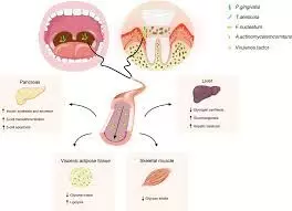
TyG Index Linked to Increased Risk of Periodontitis, suggests research

Researchers have found in an observational analysis that there was a significant association between the triglyceride-glucose (TyG) index and the risk of developing periodontitis. However further research was needed to explore the underlying mechanisms of this link, they suggested.

The relationship between the Triglyceride-Glucose (TyG) index and periodontitis remains unclear. This study aims to elucidate this relationship using data from two large population-based surveys. Datasets from NHANES (2009-2014) and KNHANES (2007-2018, except for 2011) were utilized. We applied multivariate logistic regression, stratified analysis, restricted cubic splines (RCS), and subgroup analyses to examine the correlation between the TyG index and periodontitis risk. The predictive value of the TyG index was assessed using receiver operating characteristic (ROC) curves. Mediation analyses investigated variables mediating this relationship. Results: The NHANES and KNHANES cohorts included 2,511 and 16,239 participants with periodontitis, respectively. After adjusting for covariates, the TyG index was significantly associated with periodontitis risk (NHANES: OR 1.19, 95%CI: 1.07-1.34; Q2 vs. Q1, OR 1.20, 95% CI: 1.02-1.42; Q4 vs. Q1, OR 1.23, 95%CI: 1.02-1.49. KNHANES: OR 1.09, 95% CI: 1.05-1.13; Q4 vs. Q1, OR 1.09, 95%CI: 1.02-1.17, P for trend = 0.025). RCS analyses revealed a nonlinear relationship. ROC curves indicated that the predictive values of the TyG index were 8.24 (NHANES) and 8.69 (KNHANES). Mediation analysis showed that inflammatory (alkaline phosphatase and white blood cell) and metabolic factors (vitamin D and high-density lipoprotein cholesterol) partially mediated this association. The observational analysis reveals a significant association between the TyG index and the risk of periodontitis. Further studies are needed to clarify the underlying mechanisms.

Reference:

Huang J, Zhang D, Li H, Zhang Y, Long T, Guo X, Cui H, Wei Z, Zhao J, Li M, Wang P. Triglyceride-glucose index and periodontitis: evidence from two population-based surveys. Front Endocrinol (Lausanne). 2025 May 19;16:1558692. doi: 10.3389/fendo.2025.1558692. PMID: 40458175; PMCID: PMC12127200.

Keywords:

TyG, Index , Linked, Increased, Risk, Periodontitis, suggests, research, triglyceride-glucose index, periodontitis, NHANES, KNHANES, insulin resistance, Huang J, Zhang D, Li H, Zhang Y, Long T, Guo X, Cui H, Wei Z, Zhao J, Li M, Wang P.

Powered by WPeMatico

Simple blood test predicts cognitive decline in Alzheimer’s patients, new study shows

Insulin resistance detected by routine triglyceride-glucose (TyG) index can flag people with early Alzheimer’s who are four times more likely to present rapid cognitive decline, according to new research presented at the European Academy of Neurology (EAN) Congress 2025.

Neurologists at the University of Brescia reviewed records for 315 non-diabetic patients with cognitive deficits, including 200 with biologically confirmed Alzheimer’s disease. All subjects underwent an assessment of insulin resistance using the TyG index and a clinical follow-up of 3 years. When patients were divided according to TyG index, those in the highest third of the Mild Cognitive Impairment AD subgroup deteriorated far more quickly than their lower-TyG peers, losing >2.5 points on the Mini Mental State Examination per year (hazard ratio 4.08, 95% CI 1.06–15.73). No such link appeared in the non-AD cohort.

“Once mild cognitive impairment is diagnosed, families always ask how fast it will progress”, said lead investigator Dr Bianca Gumina. “Our data show that a simple metabolic marker available in every hospital laboratory can help identify more vulnerable subjects who may be suitable candidates for targeted therapy or specific intervention strategies.”

While insulin resistance has been linked to the onset of Alzheimer’s disease, its role in how quickly the condition progresses has received less attention. This study aimed to fill that gap by focusing on its impact during the prodromal mild cognitive impairment (MCI) stage, when patients follow highly variable trajectories. The researchers used the TyG index, which offers a low-cost, routinely available surrogate for insulin resistance, to explore whether metabolic dysfunction could help predict the pace of cognitive decline after diagnosis.

In AD specifically, insulin resistance is believed to impair neuronal glucose uptake, promote amyloid accumulation, disrupt the blood–brain barrier, and fuel inflammation – pathways that are less relevant or differently regulated in other neurodegenerative diseases.

“We were surprised to see the effect only in the Alzheimer’s spectrum and not in other neurodegenerative diseases”, Dr Gumina noted. “It suggests a disease-specific vulnerability to metabolic stress during the prodromal window, when interventions may still change the trajectory.”

The researchers at University of Brescia, led by Professor Padovani and Professor Pilotto, found that high TyG was also associated with blood–brain barrier disruption and cardiovascular risk factors, yet it showed no interaction with the APOE ε4 genotype, indicating that metabolic and genetic risks may act through distinct pathways.

Identifying high-TyG patients could refine enrolment for anti-amyloid or anti-tau trials and prompt earlier lifestyle or pharmacological measures to improve insulin sensitivity. The researchers are currently investigating whether TyG levels also track with neuroimaging biomarkers to aid earlier detection and stratification.

“If targeting metabolism can delay progression, we will have a readily modifiable target that works alongside emerging disease-modifying drugs”, concluded Dr Gumina.

Reference:

Simple blood test predicts cognitive decline in Alzheimer’s patients, new study shows, Beyond, Meeting: European Academy of Neurology (EAN) Congress 2025.

Powered by WPeMatico