Age-Related Macular Degeneration Increases Rheumatoid Arthritis Risk, Study Finds

Researchers report that the prevalence of rheumatoid arthritis (RA) is greatly higher in patients with age-related macular degeneration (AMD), regardless of the presence of visual disability. A recent study was published in the journal Scientific Reports by Jee Moon Yoon and colleagues..

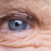
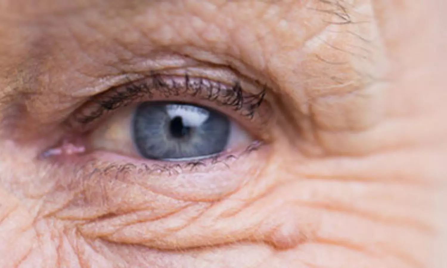
Age-related macular degeneration is a leading cause of blindness among older adults. Rheumatoid arthritis is an autoimmune disease, characteristically involving the joints. They investigated this in a risk study of RA in individuals with AMD, with and without visual disability, using data on over 2 million South Koreans.

This was a large cohort study, with 3,537,293 people who underwent health checkup in 2009 and were followed for 10 years until 2019. Patients were stratified into three groups: patients suffering from AMD without VD, patients with AMD with VD, and controls without AMD. Loss of vision or defects in the visual field according to certified criteria by the Ministry of Health and Welfare of Korea was used as the condition for diagnosing visual disability. The incidence of RA diagnoses was tracked during the study period, and the analysis used multivariable-adjusted Cox regression to estimate RA risk in the exposed relative to the control groups. Lifestyle factors and other possible confounders were controlled for in the hazard ratios (HRs).

Results

  • A total of 43,772 people (1.24% of the study population) were diagnosed with RA during the follow-up period.

  • The risk of RA was higher in the AMD group than in controls regardless of visual disability status (aHR 1.11; 95% CI, 1.02–1.21).

  • The additional analysis in the AMD group shows that patients with AMD without visual disability had a higher risk of RA (aHR 1.13; 95% CI, 1.03–1.21), while those with AMD and visual disability had a non-significant lower risk of RA (aHR 0.90; 95% CI, 0.64–1.27).

  • There is also a significantly associated risk of RA with AMD even after accounting for lifestyle factors such as smoking and alcohol consumption, and comorbid conditions such as hypertension and diabetes.

This study found that there is a modestly increased risk of rheumatoid arthritis (RA) with age-related macular degeneration (AMD). Even after adjusting for lifestyle and comorbid factors, this association remained, which potentially brings to the fore a common pathway existing between the two diseases. This discrepancy could reflect differences in mechanisms between the two conditions operated with or without visual impairment or the effects of chronic inflammatory processes in both diseases.

Reference:

Yoon, J. M., Eun, Y., Han, K., Kim, B. S., Jung, W., Kim, H., Shin, D. W., & Lim, D. H. (2024). Association of rheumatoid arthritis with age-related macular degeneration in nationwide longitudinal cohort study. Scientific Reports, 14(1), 1–8. https://doi.org/10.1038/s41598-024-71524-x

Powered by WPeMatico

Flow within radial artery remains unchanged after cannulation among patients undergoing abdominal surgery, finds study

The radial artery is commonly cannulated during the perioperative period and in critical care situations. Despite being invasive, this procedure offers real-time monitoring of arterial blood pressure, enabling clinicians and anesthesiologists to promptly assess and treat patients, as well as to obtain arterial blood samples for various diagnostic purposes such as blood chemistry, blood gas analysis, and coagulation management. Recent study evaluated the impact of arterial cannulation on flow in the radial artery and the ulnar artery in patients undergoing abdominal surgery. The primary outcome was to measure the resistive indices (RIs) before cannula insertion and 6 hours after decannulation, while the secondary outcomes included measuring the artery size by anteroposterior (AP) diameter at the insertion site and at a point 4 cm proximal to the insertion site in both the radial and ulnar arteries. The study included 96 patients and used peak systolic velocity (PSV) and end‑diastolic velocity (EDV) to derive RI using a linear ultrasound transducer.

Study Results

The results showed that the flow in the radial artery did not change following cannulation, and the resistive index (RI) and AP diameter at the insertion site and proximal to the insertion site were similar before and after cannulation. However, the peak systolic velocity (PSV) and anteroposterior (AP) diameter in the ulnar artery increased significantly after decannulation, indicating a compensatory increase in flow following radial artery decannulation. Subgroup analysis revealed that patients with cannulas retained for less than 24 hours showed increased end‑diastolic velocity (EDV) of the radial artery following decannulation, while those with cannulas retained for more than 24 hours showed an increase in PSV and AP diameter of the ulnar artery after decannulation. The study also considered other research findings and discussed the impact of different techniques of radial arterial cannulation on complications. The study acknowledged limitations, such as the inability to document decreased pulsatility of the radial pulse on ultrasound Doppler due to study design constraints and the lack of follow-up beyond 6 hours of decannulation. In conclusion, the study found that resistive indices and artery size in the radial artery did not change significantly following cannulation, while there was a compensatory increase in flow in the ulnar artery after decannulation. The study emphasized the need for further research to evaluate flow patterns at decannulation and extend the measurement period beyond immediate decannulation. Overall, the study provided insight into the impact of arterial cannulation on arterial flow in patients undergoing abdominal surgery and highlighted the need for further research in this area.

Key Points

– The study aimed to assess the impact of arterial cannulation on flow in the radial and ulnar arteries in patients undergoing abdominal surgery. It included 96 patients and measured resistive indices (RIs) before cannula insertion and 6 hours after decannulation, as well as artery size by anteroposterior (AP) diameter at the insertion site and proximal to it in both arteries, using peak systolic velocity (PSV) and end‑diastolic velocity (EDV) derived from a linear ultrasound transducer.

– The analysis of the results revealed that the flow in the radial artery did not change following cannulation, and the resistive index (RI) and AP diameter at the insertion site and proximal to it were similar before and after cannulation. However, there was a significant increase in peak systolic velocity (PSV) and anteroposterior (AP) diameter in the ulnar artery after decannulation, suggesting a compensatory increase in flow following radial artery decannulation.

– Subgroup analysis indicated that patients with cannulas retained for less than 24 hours showed increased end‑diastolic velocity (EDV) of the radial artery following decannulation, while those with cannulas retained for more than 24 hours showed an increase in PSV and AP diameter of the ulnar artery after decannulation.

– The study also acknowledged limitations, including the inability to document decreased pulsatility of the radial pulse on ultrasound Doppler due to study design constraints and the lack of follow-up beyond 6 hours of decannulation. – In conclusion, the study found that resistive indices and artery size in the radial artery did not change significantly following cannulation, while there was a compensatory increase in flow in the ulnar artery after decannulation. The study emphasized the need for further research to evaluate flow patterns at decannulation and extend the measurement period beyond immediate decannulation.

– Overall, the study provided valuable insight into the impact of arterial cannulation on arterial flow in patients undergoing abdominal surgery and highlighted the need for more comprehensive research in this area.

Reference –

Kumar L, Subramaniam R, Kartha PA, Thomas DE, Patel S, Kartha N. Does the resistive index of the radial artery change after arterial cannulation in patients undergoing abdominal surgery? A prospective observational study. Indian J Anaesth 2024;68:909‑14

Powered by WPeMatico

Using transparent capsules in dry powder inhalers could significantly improve medication delivery, reports study

Background and Goal: 

This study examined whether patients with non-reversible chronic airway disease using a transparent capsule in single-dose dry powder inhalers affects the amount of medication delivered. The goal was to determine if patients who use transparent capsules that allow them to see if the medication has been fully inhaled have better inhalation results compared to those using opaque capsules.

Study Approach:

Researchers conducted an observational cross-sectional study between October 2020 and October 2022 at a tertiary university hospital. The study involved 91 patients using long-acting bronchodilators with single-dose dry powder inhalers for chronic airway disease. Patients used their inhalers under supervision, and the weight of the capsule was measured before and after inhalation to assess how much of the medication was delivered.

Main Results:

• Patients using transparent capsules had a significant decrease in capsule weight after inhalation, with a 30.1% reduction compared to an 8.6% reduction for opaque capsules, indicating more medication was delivered.

• The change in capsule weight was consistent regardless of the severity of the patient’s airway disease.

Why It Matters:

The findings suggest that using transparent capsules in dry powder inhalers could improve medication delivery by providing patients with visual feedback on their inhalation technique. This could lead to better management of chronic airway diseases by ensuring that patients receive their full dose of medication.

Reference:

Rocío Reinoso Arija, Rosario Ruiz-Serrano de la Espada, Mª Dolores Núñez Ollero, Laura Carrasco Hernandez, Esther Quintana Gallego and Jose Luis López-Campos, Evaluation of the Importance of Capsule Transparency in Dry Powder Inhalation Devices, The Annals of Family Medicine September 2024, 22 (5) 417-420; DOI: https://doi.org/10.1370/afm.3147.

Powered by WPeMatico

Co-Use of Amphetamines and Alcohol Linked to Serious Liver and Kidney Damage: Study Reveals

Saudi Arabia: A recent study published in Scientific Reports has highlighted the dangerous effects of co-abusing amphetamines and alcohol, revealing that this combination significantly increases toxicity in both the kidneys and liver. The findings suggest a concerning synergy between these substances, leading to greater harm than when either is used alone.

Researchers have long understood the individual risks associated with amphetamines and alcohol. The prevalence of alcohol use disorder was 75% higher in patients dependent on amphetamines. Amphetamines, often prescribed for conditions like ADHD, can lead to increased heart rate, elevated blood pressure, and anxiety. Meanwhile, alcohol consumption is well-documented for its negative effects on the liver, contributing to conditions such as fatty liver disease and cirrhosis. However, the interaction between the two substances has not been explored thoroughly until now.

To explore the same, Ashraf M. Emara, Department of Pharmacology and Toxicology, College of Pharmacy, Qassim University, Al Qassim, Buraydah, Saudi Arabia, and colleagues aimed to evaluate the toxic effects of co-abusing amphetamines and alcohol on the liver and kidneys.

For this purpose, the researchers conducted a cross-sectional study at Amal Hospital for Mental Health in the Qassim region of Saudi Arabia, involving 100 participants. Seventy-five of these were patients hospitalized for substance abuse treatment, while 25 were healthy volunteers with no history of abuse. An experienced psychiatrist interviewed the patients and assessed them using DSM-5 criteria. The data from the healthy participants served as a control group, which was matched with the abuse group based on age and lifestyle.

Participants were divided into four groups: Group I included 25 controls; Group II consisted of 25 amphetamine (AMP) users; Group III included 25 alcohol users; and Group IV comprised 25 individuals who abused both amphetamines and alcohol. Researchers collected socio-demographic data, conducted complete medical examinations, and measured Body Mass Index (BMI). Blood and urine samples were taken from all participants for analytical tests to determine levels of alcohol and amphetamines, as well as to assess kidney and liver function.

The research produced the following results:

  • The mean BMI values in groups II, III, and IV did not significantly differ from the control group. However, serum levels of albumin and alkaline phosphatase significantly decreased in all substance-abusing groups. In contrast, levels of alanine transaminase (ALT), aspartate transaminase (AST), and osteopontin increased significantly across all abuser groups.
  • Fasting blood sugar levels rose significantly in alcohol users, while there were no significant changes in the amphetamine and combined groups.
  • The mean urea levels remained stable in both amphetamine and alcohol users but showed a significant increase in the combined drug abuser group.
  • Serum creatinine levels significantly increased in all abuser groups, with Cystatin C levels also rising significantly.
  • Most biochemical parameters exhibited more than a twofold alteration in the combined group compared to the amphetamine or alcohol groups.

This study found that addiction to alcohol, amphetamines, or a combination of both is linked to liver and kidney damage. Co-abusing amphetamines and alcohol significantly increases toxicity to these organs. The combined use creates a stronger toxic effect, highlighting the need for treatment strategies that address this synergy.

“Recognizing these risks can help individuals make informed choices and seek appropriate support and treatment,” the researchers concluded.

Reference:

Alharbi, S., Aldubayan, M. A., Alhowail, A. H., Almogbel, Y. S., & Emara, A. M. (2024). Co-abuse of amphetamine and alcohol harms the kidney and liver. Scientific Reports, 14(1), 1-17. https://doi.org/10.1038/s41598-024-74459-5

Powered by WPeMatico

Airway ultrasound superior to traditional clinical parameters for predicting difficult laryngoscopy, suggests study

Assessment of the airway is crucial for managing airways effectively. Ultrasound has increasingly become a viable alternative for evaluating airways. Recent research paper aims to assess the accuracy of various clinical and ultrasonographic parameters in predicting difficult laryngoscopy in the emergency department. The primary objective is to determine the accuracy of ultrasonography parameters compared to Cormack-Lehane grading in predicting difficult laryngoscopy in adults. The secondary objective is to assess the accuracy of clinical parameters compared to Cormack-Lehane grading in predicting difficult laryngoscopy in adults.

Study Design and Procedure

The study will be a prospective cross-sectional study involving 62 adult patients over 18 years who come to the emergency department and require endotracheal intubation. An emergency medicine physician will perform an ultrasonographic examination of the upper airway, measuring the skin-to-epiglottis distance and tongue thickness using a GE Healthcare ultrasound. Clinical airway assessment will also be conducted using the LEMON criteria. A trained emergency medicine physician, unaware of the clinical and ultrasound airway parameters, will perform a laryngoscopy and record the Cormack-Lehane grading. The study is expected to provide insights into the comparative effectiveness of airway ultrasound versus conventional clinical parameters in predicting difficult laryngoscopy within an emergency department setting. Airway ultrasound has demonstrated significant potential due to its real-time imaging capabilities and ability to visualize anatomical structures that may not be readily accessible through clinical examination alone. Ultrasound parameters such as the distance from the skin to the epiglottis and the anterior neck soft tissue thickness at the level of the hyoid bone and vocal cords have been shown to be effective in predicting difficult laryngoscopy.

Hypothesis and Conclusion

The researchers hypothesize that airway ultrasound may offer superior predictive value compared to traditional clinical parameters. The integration of ultrasound into airway assessment protocols could revolutionize the approach to managing airways in the emergency department, ensuring that patients receive the best possible care even in the most challenging situations. The conclusion of the study will be drawn regarding the correlation between ultrasound parameters, Cormack-Lehane grading, and clinical parameters in predicting difficult laryngoscopy.

Key Points

1. The primary objective is to determine the accuracy of ultrasonography parameters compared to Cormack-Lehane grading in predicting difficult laryngoscopy in adults.

2. The secondary objective is to assess the accuracy of clinical parameters compared to Cormack-Lehane grading in predicting difficult laryngoscopy in adults.

3. The study is a prospective cross-sectional study involving 62 adult patients over 18 years who come to the emergency department and require endotracheal intubation.

4. An emergency medicine physician will perform an ultrasonographic examination of the upper airway, measuring the skin-to-epiglottis distance and tongue thickness, and a clinical airway assessment using the LEMON criteria.

5. The researchers hypothesize that airway ultrasound may offer superior predictive value compared to traditional clinical parameters in predicting difficult laryngoscopy.

6. The conclusion of the study will be drawn regarding the correlation between ultrasound parameters, Cormack-Lehane grading, and clinical parameters in predicting difficult laryngoscopy.

Reference –

Kadadi S, Gadkari C, Pundkar A (September 16, 2024) A Comparison of Airway Ultrasound and Clinical Parameters for Predicting Difficult Laryngoscopy in the Emergency Department: A Study Protocol. Cureus 16(9): e69543. DOI 10.7759/cureus.69543

Powered by WPeMatico

CENTAC Invites Applications for Special Stray Round Admissions to MDS Courses, Releases Seat Matrix

Puducherry- The Centralised Admission Committee (CENTAC) had invited fresh applications for the special stray for admission to PG Dental (MDS) courses. Applications were invited for PG Dental (MDS) vacancies under the Management Quota due to the low cut-off.

As per the official notice in this regard, candidates can submit their application form along with course preferences and payment of Registration fees of Rs.50,000/- after referring to vacancy positions published on the official website of CENTAC for the special stray for admission to PG Dental (MDS) courses using the dashboard login credentials.

The last date for submission of fresh Application is 16th October 2024, 5.00 pm and the last date for submission of willingness, Payment of Registration fee and submission of course Preferences is 18th October 2024, 5.00 pm.

Meanwhile, the candidates are requested to transfer the registration fee to the CENTAC account through NET BANKING or by generating NEFT / RTGS (preferably RTGS) challan using the payment link in their dashboard login credentials and proceed for payment.

ELIGIBILITY FOR REGISTRATION

All candidates EXCEPT

1 Candidates who are in admitted status after Stray Round in All India Quota or State Quota.

2 Candidates who have been allotted any seat in Round 3 or in any Stray round in All India Quota / State quota and either joined or not joined.

EXISTING / APPLIED CANDIDATES OF CENTAC OPTING FOR MANAGEMENT QUOTA

Candidates who have already applied to CENTAC for PG NEET – MDS admission for Government quota only including candidates in the ineligible list may now submit willingness for Management quota after payment of balance application fees for admission to the MDS – Management vacant seats.

SUBMISSION OF WILLINGNESS, PAYMENT, DECLARATION AND COURSE PREFERENCES

Both fresh and existing candidates may participate after submitting willingness, paying the Registration Fee and filling out Course preferences before 18 October 2024, 5.00 PM. If the course preferences list is empty, the candidate will not be considered for allotment.

The fresh applications are invited from the candidates including candidates who become eligible due to the lowering of the cut-off as detailed below for the vacancies published on the official website of CENTAC for admission in PG Dental (MDS) courses.

CUTT-OFF

S.NO

CATEGORY

MINIMUM

QUALIFYING

CRITERIA AS PER

INFORMATION

BULLETIN

SCORE AS

PER

INFORMATION

BULLETIN (OUT

OF 960)

REVISED MINIMUM

QUALIFYING CRITERIA

REVISED

CUT-OFF

SCORE

(OUT OF

960)

1

General/EWS

50th Percentile

263

28.308

196

2

General

PwBD

45th Percentile

246

23.308

180

3

SC/ST/OBC

(Including

PwBD of

SC/ST/OBC)

40th Percentile

230

18.308

164

REFUND/FORFEITURE OF FEES PAID FOR SPECIAL STRAY ROUND

1 In case the candidate is not allotted any seat, the Registration fee paid, will be refunded to the same account of the candidate from which payment had been made.

2 Otherwise, if the seat is allotted and the candidate does not join the course, the Registration fee amount will be forfeited and such candidate will be debarred from taking the NEET exam next year.

3 If a candidate is allotted a seat and joins the alloted seat the registration fee will be adjusted against the Tuition fee.

4 Therefore, it is advised that the candidate makes a thoughtful decision before submitting course preferences.

Along with this, CENTAC has also released PG NEET Dental (M.D.S.) courses -seat matrix After Stray Round. As per the seat matrix, a total of 5 Seats are vacant after a stray round for Management Quota.

Below is the detailed seat matrix-

SEAT MATRIX

S.NO

COURSE NAME

VACANT SEATS

1

CONSERVATIVE DENTISTRY & ENDODONTICS.

1

2

ORTHODONTICS AND DENTOFACIAL ORTHOPAEDICS.

1

3

PAEDODONTICS AND PREVENTIVE DENTISTRY.

1

4

PERIODONTOLOGY / PERIODONTICS.

1

5

PROSTHODONTICS / PROSTHODONTICS AND CROWN & BRIDGE.

1

To view the notice, click the link below

To view the seat matrix, click the link below

Powered by WPeMatico

WBCERC directs Apollo Gleneagles Hospital to refund overcharged amount

Holding that the hospital overcharged the patient for a hernia operation, the West Bengal Clinical Establishment Regulatory Commission (WBCERC) has issued a directive to the Apollo Gleneagles Hospital.

In its order, the regulatory panel directed the hospital to set up a fixed deposit of Rs. 13,488 in the name of a patient which can later used by the concerned patient for future treatments.

For more details, click the link below

Ray back overcharged Amount! Regulatory panel orders Apollo Gleneagles Hospital

Powered by WPeMatico

Premature newborn’s hand amputated: Hospital, Doctor slapped Rs 45 lakh compensation for negligence

The District Consumer Disputes Redressal Commission, Amritsar recently directed a private hospital and its doctor to pay Rs 45 lakh compensation and Rs 1 Lakh legal cost to a man for alleged medical negligence while providing treatment to his newborn child. 

After being born prematurely, the child was admitted to the hospital as he was suffering from a low oxygen level. However, during treatment, gangrene formed in his right hand leading to the amputation of the hand at PGI Chandigarh.

For more details, click the link below

Premature newborn’s hand amputated due to negligence! Hospital, Doctor slapped Rs 45 Lakh Compensation

Powered by WPeMatico

1 in 4 adults suspect undiagnosed ADHD, survey finds

Attention deficit/hyperactivity disorder—also known as ADHD—is typically thought of as a childhood condition. But more adults are realizing that their struggles with attention, focus and restlessness could in fact be undiagnosed ADHD, thanks in large part to trending social media videos racking up millions of views.

Powered by WPeMatico

Key signs of major trauma could speed up treatment of severely injured children brought to emergency departments

Severely injured children who are brought to an emergency medical department by their parents or caregivers are often not seen as quickly as those who arrive at hospital via ambulance, according to findings presented at the European Emergency Medicine Congress.

Powered by WPeMatico