Surrogacy associated with higher risk of severe pregnancy outcomes, claims research

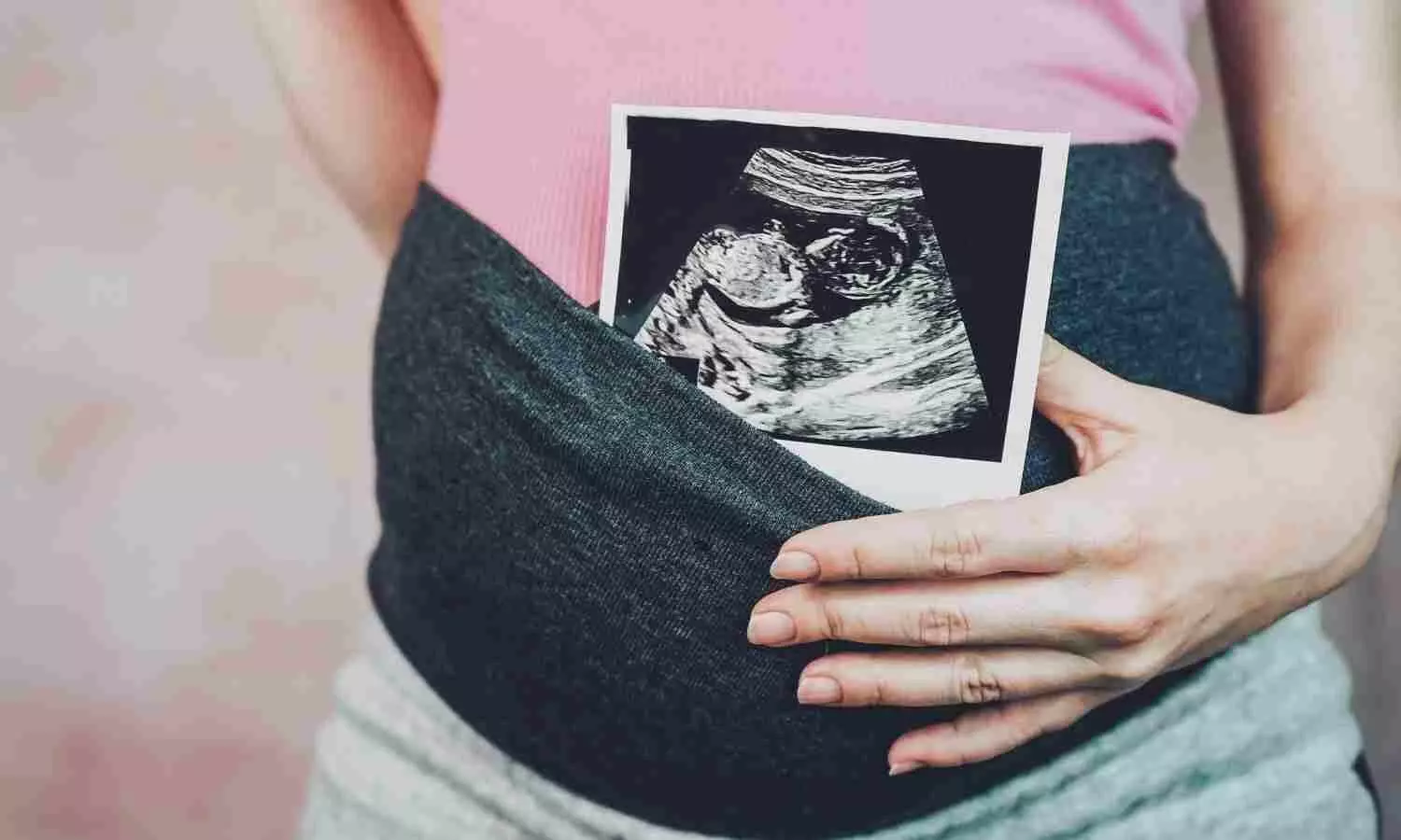
People who are gestational carriers (or “surrogates”) may have a higher risk of severe complications during pregnancy and early postpartum, hypertension in pregnancy, and postpartum hemorrhage, compared to people who conceive without assistance or with IVF, according to new research from ICES and Queen’s University.

Gestational carriers carry and give birth to children for couples or individuals who are otherwise unable to carry a pregnancy. Little is known about whether gestational carriers and babies are at a higher risk for severe health outcomes, both during pregnancy and following birth.

This is one of the first large population-based studies to analyze linked health datasets comparing health outcomes for three different types of conception: unassisted, in vitro fertilization (IVF), and gestational carriage.

“The study was prompted by an increased in the use of gestational carriers worldwide and a lack of information about the impact of this reproductive modality on pregnancy outcomes, for the gestational carrier and the offspring,” says lead author Dr. Maria Velez, an adjunct scientist at ICES and at the time of this study, an associate professor in the department of Obstetrics and Gynaecology at Queen’s University. Velez is currently an associate professor and a clinician scientist at McGill University and the Research Institute of the McGill University Health Centre (RI-MUHC)

The study, published in the journal Annals of Internal Medicine, included 863,017 singleton births at more than 20 weeks’ gestation in Ontario, Canada, between 2012 and 2021. The groups included 846,124 (97.6%) who were conceived without assistance, 16,087 (1.8%) by IVF, and 806 (0.1%) using gestational carriers.

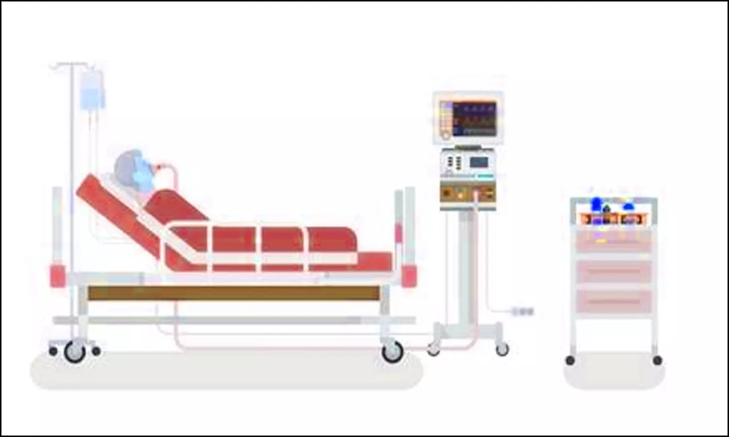
The researchers analyzed severe maternal morbidity (SMM) and severe neonatal morbidity (SNM), which combine many different health indicators for both birthing people and babies. They also assessed hypertensive disorders (such as pre-eclampsia), cesarean delivery, preterm birth, and postpartum hemorrhage.

Key Findings

• The risk of SMM was 2% for the unassisted group, 4% for the IVF group and 8% for the gestational carriage group.

• The gestational carriage group also had a higher risk for hypertensive disorders and postpartum hemorrhage when analyzed separately from SMM.

• Although gestational carriage was associated with preterm birth (less than 37 weeks’ gestation), there was less clear evidence of a higher risk of SNM.

One limitation of the study was that there was a lack of information about why gestational carriage was chosen by the intended parents, egg and sperm donor sources, the type of IVF used, and reasons why people chose to become gestational carriers. Future research could help to assess whether any of these factors would impact the health outcomes of the pregnant person or the baby.

“Clinicians involved in the care of individuals and couples who need a gestational carrier to build their family should counsel their patients and the gestational carriers about the potential risk during pregnancy and early postpartum,” says Velez.

“There are guidelines about the eligibility criteria to minimize the risk of pregnancy complications among gestational carriers,” she adds. “However, these guidelines are not always strictly followed.”

Reference:

Maria P. Velez, Marina Ivanova, Jonas Shellenberger, et al. Severe Maternal and Neonatal Morbidity Among Gestational Carriers: A Cohort Study. Ann Intern Med. [Epub 24 September 2024]. doi:10.7326/M24-0417.

Powered by WPeMatico

Single-dose gene therapy potentially life-changing for adults with hemophilia B: Study

 Adults with hemophilia B saw their number of bleeding episodes drop by an average of 71 percent after a single infusion of gene therapy, according to the results of an international Phase III clinical trial published today in the New England Journal of Medicine by researchers from the University of Pennsylvania Perelman School of Medicine and a multicenter group of investigators.

Hemophilia is a genetic disorder that limits the blood’s ability to clot and affects around 30,000 people in the United States, mostly males. Left untreated, it can cause spontaneous bleeding, particularly internal bleeding into the joints, which, over time, can cause painful joint damage and mobility issues. Hemophilia B is caused by a lack of clotting factor IX. The gene therapy enables the liver to create factor IX, which allows the blood to clot and protects patients from frequent bleeds.

“What we saw from patients in this study was that within a few days of receiving the gene therapy infusion, it took root, and their bodies started making factor IX for the first time in their lives,” said study investigator and lead author Adam Cuker, MD, MS, section chief for Hematology, and clinical director of the Penn Blood Disorders Center and the Penn Comprehensive Hemophilia Program. “We always want to be careful about using the word ‘cure’ especially until we have longer follow-up data, but for many of these patients, it’s been life changing.”

After at least one year of follow-up, participants in the study had an average 71 percent reduction in bleed rate after receiving the gene therapy, compared to the year prior, when they were treated with prophylactic infusions of factor IX, the standard treatment for the disease. More than half of the 45 patients in the study did not have any bleeds after receiving gene therapy.

FDA-approved gene therapies available at Penn Medicine

Based on the results of this study, the FDA approved the gene therapy (fidanacogene elaparvovec) in April 2024. Cuker was the site lead for the clinical trial at Penn Medicine, which was one of the top-enrolling sites for the study. It represents the second form of gene therapy approved to treat hemophilia B. The first such therapy (etranacogene dezaparvovec-drlb) was approved in November 2022, and Penn Medicine is one of several medical centers in the United States where this treatment is available to patients.

Gene therapies have very specific guidelines that determine eligibility and require specialized knowledge to carry out patient screening and selection, education about treatment risks and benefits, and post-therapy monitoring. Penn Medicine offers access to numerous clinical trials for gene therapy and expertise in administering FDA-approved gene therapies.

In the current study, the most common adverse effect was related to an immune system attack on liver cells that were targeted by the gene therapy, which can render the gene therapy ineffective, if not quickly treated. In the study, affected patients were treated with steroids to limit this immune reaction. Patients in the study will continue to be followed for at least five years to monitor potential long-term side effects.

Life-changing impact

For patients with hemophilia B, the current standard of care of ongoing prophylactic infusions of factor IX is generally effective, but burdensome. Depending on the specific product, a patient may require regular infusions anywhere from once every two weeks up to several times a week. Most patients learn how to put their own IV in to be able to complete their infusions at home. The goal of this prophylactic treatment is to regularly give the body enough factor IX to prevent bleeds, though they still occur. By contrast, the new gene therapy only requires a single dose, and most patients in the study did not need to resume prophylactic factor IX treatments.

“We hear from people born with hemophilia that-even if their disease is well-managed-there’s this burden that’s always in the back of their mind. The frequent infusions, the cost of treatment, the need to plan for infusions when traveling, what happens if they do experience a bleed, and so on, is always there,” Cuker said. “Now that we have patients who were treated on this study and are essentially cured of their hemophilia, they’re telling us about realizing a new, ‘hemophilia-free state of mind.’ As a physician, it’s amazing to see my patients so happy with their new reality.”

Reference:

 Adam Cuker, Laurent Frenzel, Jiaan-Der Wang, Jan Astermark, Monica H. Cerqueira, Alfonso Iorio, Gene Therapy with Fidanacogene Elaparvovec in Adults with Hemophilia B, New England Journal of Medicine, DOI: 10.1056/NEJMoa2302982.

Powered by WPeMatico

Psoriasis independent risk factor for developing neovascular AMD in Diabetes patients, suggests research

Psoriasis is an independent risk factor for developing neovascular AMD in Diabetes patients, suggests research published in the American Journal of Ophthalmology.

The objective of this study is to evaluate the relationship of psoriasis and neovascular Age-related Macular Degeneration (AMD) in diabetic mellitus (DM) population. Records of patients who had been diagnosed with type 2 diabetes mellitus over 40 years of age from January 2009 to December 2012 were analyzed. The incidence of neovascular AMD was observed from the index year to December 2018 in all subjects. We compared the incidence rate of neovascular AMD among the psoriasis group and control group. Covariates include age, sex, BMI, income level, smoking status, drinking status, regular exercise habits, hypertension, dyslipidemia, end-stage renal disease, diabetic retinopathy, glucose level, the prescription of more than three oral hypoglycemic agents, and a history of diabetes mellitus exceeding five years. Results: Of 2,245,358 type 2 DM patients, 20,853 patients were classified in the psoriasis group, and the other 2,224,505 individuals in the control group. A total of 105 neovascular AMD cases occurred in the psoriasis group and 7,459 cases in the control group. According to multivariable Cox proportional hazard models, individuals with psoriasis had a significantly higher risk for neovascular AMD compared to controls (HR=1.329, 95% confidence interval: 1.096-1.612) after adjustments for covariates. This study demonstrated that psoriasis was an independent risk factor for developing neovascular AMD in DM patients. Therefore, physicians should be alert to the development of neovascular AMD in DM patients who also have psoriasis.

Reference:

Lee MY, Han K, Min KH, Yu DS, Lee YB. Psoriasis as a Predictor of Neovascular Age-Related Macular Degeneration in Type 2 Diabetes Mellitus: A Nationwide Cohort Study. Am J Ophthalmol. 2024 Aug 30:S0002-9394(24)00395-7. doi: 10.1016/j.ajo.2024.08.033. Epub ahead of print. PMID: 39218384.

Keywords:

Psoriasis, independent, risk, factor, developing, neovascular, AMD, Diabetes, patients, research, Age-related Macular Degeneration, Diabetes mellitus, Hazard Ratio, Psoriasis, American Journal of Ophthalmology, Lee MY, Han K, Min KH, Yu DS, Lee YB

Powered by WPeMatico

Chronic kidney disease linked with adverse postoperative outcomes across various surgical procedures, claims study

Chronic kidney disease linked with adverse postoperative outcomes across various surgical procedures, claims study published in the BMC Nephrology.

Chronic kidney disease (CKD) is associated with higher incidence of major surgery. No studies have evaluated the association between preoperative kidney function and postoperative outcomes across a wide spectrum of procedures. They aimed to evaluate the association between CKD and 30-day postoperative outcomes across surgical specialties. They selected adult patients undergoing surgery across eight specialties. The primary study endpoint was major complications, defined as death, unplanned reoperation, cardiac complication, or stroke within 30 days following surgery. Secondary outcomes included Clavien-Dindo high-grade complications, as well as cardiac, pulmonary, infectious, and thromboembolic complications. Multivariable regression was performed to evaluate the association between CKD and 30-day postoperative complications, adjusted for baseline characteristics, surgical specialty, and operative time. Results: In total, 1,912,682 patients were included. The odds of major complications (adjusted odds ratio [aOR] 2.14 [95% confidence interval (CI): 2.07, 2.21]), death (aOR 3.03 [95% CI: 2.88, 3.19]), unplanned reoperation (aOR 1.57 [95% CI: 1.51, 1.64]), cardiac complication (aOR 3.51 [95% CI: 3.25, 3.80]), and stroke (aOR 1.89 [95% CI: 1.64, 2.17]) were greater for patients with CKD stage 5 vs. stage 1. A similar pattern was observed for the secondary endpoints. This population-based study demonstrates the negative impact of CKD on operative outcomes across a diverse range of procedures and patients.

Reference:

Riveros, C., Ranganathan, S., Shah, Y.B. et al. Association of chronic kidney disease with postoperative outcomes: a national surgical quality improvement program (NSQIP) multi-specialty surgical cohort analysis. BMC Nephrol 25, 305 (2024). https://doi.org/10.1186/s12882-024-03753-1

Keywords:

Chronic, kidney, disease, linked, adverse, postoperative, outcomes, across, various, surgical procedures, claims, study, Riveros, C., Ranganathan, S., Shah, Y.B, BMC Nephrology

Powered by WPeMatico

Elevated Biomarkers Predict Long-Term Mortality Risks in Patients with Suspected Pulmonary Embolism: Study

Finland: Recent research, published in the European Heart Journal Open has highlighted the critical role of several biomarkers—D-dimer, cardiac troponin T, C-reactive protein (CRP), and NT-proBNP—in predicting long-term mortality among patients evaluated for suspected pulmonary embolism (PE). The finding is particularly significant, as it highlights the high mortality risk faced by patients who present with clinical suspicion of PE, even when the diagnosis is ultimately ruled out.

The researchers observed high mortality rates both in patients with and without PE. They suggested that emergency departments give greater attention to these high-risk patients.

Patients with suspected PE often exhibit various symptoms, prompting clinicians to conduct thorough evaluations. However, the study revealed that many of these patients, particularly those who do not ultimately have PE, experience high long-term mortality rates. As such, identifying high-risk individuals early in their clinical journey is crucial.

Pulmonary embolism is a prevalent and potentially life-threatening condition that necessitates immediate diagnostic evaluation. While biomarkers are widely utilized in clinical settings, their role in predicting the long-term prognosis of patients assessed for suspected PE remains largely unclear. Considering this, Juha Kauppi, Turku University Hospital, Emergency Clinic, Turku, Finland, and colleagues sought to evaluate the predictive performance of NT-proBNP, C-reactive protein (CRP), D-dimer (FIDD), and cardiac troponin T (cTnT) in patients who underwent computed tomography pulmonary angiography (CTPA) due to clinical suspicion of PE.

The analysis included 1,001 patients, of whom 222 were diagnosed with pulmonary embolism at the time of imaging. The average age of patients with PE was 65.0 ± 17.1 years, compared to 64.5 ± 17.7 years for those without PE. The median follow-up duration was 3.9 years.

The study led to the following findings:

  • Mortality was relatively high both among patients with and without documented PE (24.8% versus 31.7%).
  • In patients with PE, only elevated NT-proBNP>1000 ng/L and CRP>50 mg/L levels at hospital admission were associated with higher mortality in an adjusted Cox regression model. Still, ROC analysis showed no improved prediction compared to clinical variables.
  • Among patients without PE, elevated NT-proBNP>1000 ng/L, CRP>10 mg/L, cTnT>50 ng/L and FIDD>1.0 mg/L all predicted mortality.
  • In a ROC analysis among patients without PE, models including NT-proBNP, cTnT, or CRP provided improved predictive performance.

The findings revealed that patients assessed for clinical suspicion of pulmonary embolism face significant long-term mortality risks. Commonly used biomarkers offer valuable prognostic information for those who do not have PE.

“Considering the relatively young age of many affected individuals, it is crucial to identify high-risk patients, conduct differential diagnoses for other potentially life-threatening conditions, and implement appropriate management strategies,” the researchers concluded.

Reference:

Kauppi, J., Airaksinen, K. E., Lehto, J., Pouru, J., Saha, J., Purola, P., Jaakkola, S., Lehtonen, J., Vasankari, T., Juonala, M., & Kiviniemi, T. Performance of D-dimer, cardiac troponin T, C-reactive protein and NT-proBNP in prediction of long-term mortality in patients with suspected pulmonary embolism. European Heart Journal Open. https://doi.org/10.1093/ehjopen/oeae079

Powered by WPeMatico

Hydroxychloroquine Fails to Lower Pre-eclampsia Risk in Women with Lupus, Study Finds

USA: A recent study examining the effects of hydroxychloroquine (HCQ) on pre-eclampsia in a diverse cohort of women with systemic lupus erythematosus (SLE) has provided critical insights, although it has not demonstrated a reduced risk of pre-eclampsia or eclampsia linked to the use of this medication.

“Although this study did not demonstrate a reduced risk of pre-eclampsia or eclampsia associated with HCQ, the presence of residual confounding factors may be masking the effect, even within an integrated healthcare delivery system that provides comprehensive clinical data,” the researchers reported in Arthritis Care & Research. 

Hydroxychloroquine is commonly prescribed for SLE, a chronic autoimmune condition that disproportionately affects women and is associated with various complications during pregnancy. Pregnant women with SLE are already at an elevated risk for developing pre-eclampsia, a serious condition characterized by high blood pressure and signs of damage to other organ systems, often requiring careful management. Given the potential benefits of HCQ in reducing flares of lupus and its anti-inflammatory properties, Julia F. Simard, Stanford University School of Medicine, Stanford, California, and colleagues investigated whether HCQ treatment in early pregnancy reduced the risk of pre-eclampsia or eclampsia using a cohort of pregnant patients with prevalent SLE at Kaiser Permanente Northern California (KPNC).

For this purpose, the researchers evaluated pregnant patients with SLE from 2011 to 2020, focusing on HCQ treatment from three months before pregnancy through the first trimester. HCQ exposure was assessed in various ways to account for treatment adherence and duration.

Propensity scores were utilized to address multiple confounding factors, while modified Poisson models estimated risk ratios (RRs) to explore the association between HCQ treatment and the incidence of pre-eclampsia or eclampsia. The researchers also conducted stratified analyses to examine how pregestational hypertension, history of nephritis, and antiphospholipid antibody (aPL) status might modify the effects.

The researchers reported the following findings:

  • There were 399 pregnancies among 324 patients with SLE at KPNC between 2011 and 2020.
  • Considering multiple exposure definitions, the researchers consistently found a null association between HCQ and pre-eclampsia or eclampsia.
  • The RRs were consistently lower among nullipara patients, and RRs were consistently protective but not statistically significant among the high-risk subgroup of patients with a history of nephritis, aPL positivity, or pregestational hypertension (for both nullipara and multipara patients).

The authors conclude that their findings align with those of other studies, which have also reported null results or protective relative risks regarding hydroxychloroquine and its impact on pre-eclampsia and eclampsia. Despite a substantial initial population, the study was underpowered to fully evaluate several prespecified research questions concerning effect modification and stratified analyses.

“While results from these analyses are presented for transparency, interpret them with caution,” the researchers wrote.

Although the study did not demonstrate a reduced risk of HCQ on pre-eclampsia or eclampsia, the authors acknowledge that residual confounding could be influencing the observed outcomes, even within a comprehensive healthcare setting with detailed clinical data.

Reference:

Simard, J. F., Liu, E. F., Rector, A., Cantu, M., Chakravarty, E., Druzin, M., Kuo, D. Z., Shaw, G. M., Weisman, M., & Hedderson, M. (2024). Hydroxychloroquine and Pre-eclampsia in a Diverse Cohort of Women With Systemic Lupus Erythematosus. Arthritis Care & Research, 76(10), 1390-1395. https://doi.org/10.1002/acr.25386

Powered by WPeMatico

IV golimumab for juvenile idiopathic arthritis in shows effective risk-benefit profile: S

A new study published in the Journal of Rheumatology unveiled that intravenous golimumab for juvenile idiopathic arthritis in a long-term extension trial had an acceptable risk-benefit profile.

Juvenile idiopathic arthritis (JIA) is the most prevalent rheumatologic disorder in children and is classified into three subtypes, namely, polyarticular, oligoarticular, and systemic onset. The patients with polyarticular JIA have a variety of etiologic risk factors, distinct disease courses, and treatment obstacles. The children with polyarticular JIA often have a more refractory course when compared to children whose joints are not as damaged. They have a longer course of active illness, which puts them at higher risk of joint injury and worse functional results as well as a lower quality of life.

This study by Hermine Brunner and the team evaluated the pharmacokinetics (PK), clinical effect, immunogenicity, and safety of intravenous (IV) golimumab in children with active polyarticular-course juvenile idiopathic arthritis (pcJIA) who took part in the open-label, long-term extension (LTE) of GO-VIVA (a medication to treat active polyarticular-course juvenile idiopathic arthritis) through week 252.

The participants in GO-VIVA who kept receiving IV golimumab (80 mg/m2 every 8 weeks) following week 52 were included in this trial. Clinical response was evaluated through week 116, while PK and safety were evaluated through weeks 244 (final dosage) and 252, respectively. Clinical outcomes measured were the clinical juvenile arthritis disease activity score in 10 joints (cJADAS10) and JIA–American College of Rheumatology (ACR) responses. Non-responder imputation was employed for binary outcomes, whereas observed data was utilized for other descriptive analyses.

69 individuals who entered the LTE and finished the week 252 visit comprised 112/127 (88.2%). From week 52 to week 244, golimumab concentrations at the median steady-state trough were mostly maintained. Also, the frequencies of antigolimumab antibodies were stable between weeks 52 and 244.

Up to week 116, when the median cJADAS10 was 1.6 and 56.7% (72/127) of individuals attained cJADAS10 < 5 (low disease activity), week 52 JIA-ACR 30/50/70/90 response rates were mostly sustained. Through week 252, the rates of major adverse events and severe infections were 7.7 and 3.9, per 100 patient-years, respectively. Overall, the data of this study demonstrate that IV golimumab therapy gives a long-term clinical outcome through week 116, with a satisfactory risk-benefit profile.

Source:

Brunner, H. I., Pacheco-Tena, C., Louw, I., Vega-Cornejo, G., Alexeeva, E., Appenzeller, S., Chasnyk, V., Griffin, T., Navarrete Suarez, C., Knupp-Oliveira, S., Zeft, A., Butbul Aviel, Y., De Ranieri, D., Gottlieb, B. S., Levy, D. M., Rabinovich, C. E., Artur Silva, C., Spivakovsky, Y., Uziel, Y., … Ruperto, N. (2024). Intravenous Golimumab in Children With Polyarticular-Course Juvenile Idiopathic Arthritis: Long-Term Extension of an Open-Label, Phase III Study. In The Journal of Rheumatology (p. jrheum.2024-0298). The Journal of Rheumatology. https://doi.org/10.3899/jrheum.2024-0298

Powered by WPeMatico

Tooth extraction not tied to Spontaneous Bacterial Peritonitis among cirrhosis patients: Study

Tooth extraction not tied to Spontaneous Bacterial Peritonitis among cirrhosis patients suggests a study published in the Oral Surgery, Oral Medicine, Oral Pathology and Oral Radiology.

A study was done to identify the association between tooth extraction and occurrence of spontaneous bacterial peritonitis (SBP) and to assess delayed soft tissue healing of extraction sockets in cirrhotic patients. 119 participants awaiting liver transplantation who required tooth extraction were included. Seven days before the surgery, the patients underwent panoramic radiography and laboratory exams. Soft tissue healing was evaluated seven days after the tooth extraction and medical records were checked after 21 days for development of SBP. The relationship between predictive factors and outcomes was assessed by using multiple binomial logistic regression. Results: 195 teeth were extracted, resulting in 146 alveolar wounds, in which the majority (47%) consisted of alveolar sockets of multirooted teeth. One participant was diagnosed with SBP (Escherichia coli) and another diagnosed with bacterascites (Streptococcus viridans group), occurring 11 and 6 days after tooth extraction. Poor soft tissue healing was observed in 20 (13.7%) patients, which was correlated to two risk factors, that is, jaundice (P=0.007, adjusted OR = 4.91; 95%CI = 1.56-15.47) and moderate neutropenia (P=0.048, adjusted OR = 13.99; 95%CI = 1.02-192.07). No association was found between tooth extraction and SBP in cirrhotic patients. The delayed soft tissue healing was related to jaundice (hyperbilirubinemia) and moderate neutropenia.

Reference:

Gustavo Souza Galvão, Juliana Bertoldi Franco, Maria Paula Siqueira de Melo Peres, Gabriela Banacu de Melo, Jefferson R. Tenório, Janaina B. Medina, Camila de Barros Gallo, Karem L. Ortega. Spontaneous Bacterial Peritonitis and Soft Tissue Healing after Tooth Extraction in Liver Cirrhosis Patients, Oral Surgery, Oral Medicine, Oral Pathology and Oral Radiology,2024, ISSN 2212-4403,

https://doi.org/10.1016/j.oooo.2024.09.003.

(https://www.sciencedirect.com/science/article/pii/S2212440324004644)

Keywords:

Tooth, extraction, tied, Spontaneous, Bacterial, Peritonitis, among, cirrhosis, patients, Study, Gustavo Souza Galvão, Juliana Bertoldi Franco, Maria Paula Siqueira de Melo Peres, Gabriela Banacu de Melo, Jefferson R. Tenório, Janaina B. Medina, Camila de Barros Gallo, Karem L. Ortega , Tooth Extraction, Peritonitis, Liver Cirrhosis, Oral Surgery, Wound Healing, Dental care, chronically ill

Powered by WPeMatico

Reduced port laparoscopic posterior mesh rectopexy effective surgery option for full-thickness rectal prolapse: Study

A recent study found that Reduced port laparoscopic posterior mesh rectopexy may serve as an effective therapeutic option for full-thickness rectal prolapse. Compared to the conventional multiport surgery (MPS), reduced port surgery was found to have smaller wound sizes and better cosmetic outcomes. The study results were published in the journal BMC Surgery. 

Laparoscopic rectopexy has long been considered a reliable treatment for full-thickness rectal prolapse. More recently, reduced port surgery (RPS) has gained attention as a new approach that potentially offers benefits over the traditional multiport surgery (MPS) technique. The primary advantages proposed for RPS include reduced postoperative pain and improved cosmetic outcomes. Hence researchers from Osaka National Hospital, Japan conducted a study to assess the practicality and safety of RPS when used for treating full-thickness rectal prolapse.

A retrospective study was carried out to analyze 37 patients who underwent laparoscopic rectopexy for full-thickness rectal prolapse between October 2012 and December 2018. Out of these, 10 patients underwent traditional multiport surgery (MPS), while 27 patients were treated using the reduced port surgery (RPS) technique. Where laparoscopic posterior mesh rectopexy, also known as the Wells procedure, is the standard surgical method for addressing full-thickness rectal prolapse, the authors also conducted an RPS. In RPS, surgeons utilized a multi-channel access device combined with an additional 12-mm right-hand port to complete the procedure. The primary outcome of the study was to evaluate short-term outcomes for both RPS and MPS by examining several metrics, including operative time, blood loss, postoperative complication rates, and the duration of hospital stay after surgery.

Findings:

  • Upon comparing the two groups, the results showed no statistically significant differences in median operative times, blood loss volumes, postoperative complication rates, or the length of hospital stays between patients undergoing MPS and those treated with RPS.
  • Both techniques appeared to be equally effective and safe in managing full-thickness rectal prolapse.
  • However, RPS was found to have significantly smaller wound size suggesting better cosmetic outcomes.

Despite these promising findings for RPS, the study highlights that further research is necessary to establish its superiority over MPS. The retrospective nature of the study and the small sample size limit the ability to draw definitive conclusions. The authors suggest that a prospective, randomized, controlled trial is needed to rigorously assess whether RPS offers any distinct advantages in terms of clinical outcomes, patient recovery, and overall efficacy when compared to traditional multiport approaches. In conclusion, while reduced port laparoscopic posterior mesh rectopexy shows potential as a viable therapeutic option for full-thickness rectal prolapse, the current evidence does not demonstrate significant differences between RPS and MPS in key perioperative outcomes. Future research with a more robust study design is essential to determine if RPS could eventually replace or become preferred over MPS in clinical practice.

Further reading: Kusunoki, C., Uemura, M., Osaki, M. et al. Reduced port laparoscopic rectopexy for full-thickness rectal prolapse. BMC Surg 24, 246 (2024). https://doi.org/10.1186/s12893-024-02545-6

Powered by WPeMatico

Valsalva manoeuvre in pressure-controlled mode better technique for unmasking of bleeders, finds Study

The Valsalva manoeuvre (VM) is used intraoperatively to unmask hidden venous bleeders during hemostasis in head and neck surgery, thyroid surgery, and craniotomy, as well as to aid in tasks like evaluating autonomic neuropathy, localizing venous air embolism, and facilitating internal jugular vein cannulation and pain management during skin puncture before certain medical procedures. Recent study compared the effectiveness of an anesthesia machine-generated Valsalva maneuver (VM) under pressure-control (PC) mode versus the traditional manual delivery of the VM in patients under general anesthesia. The primary outcome was to measure changes in the internal jugular vein (IJV) diameter, while secondary outcomes included heart rate (HR), mean arterial pressure (MAP), time to achieve desired plateau airway pressure, number of patients with bleeders unmasked, and surgeon satisfaction.

The study included 60 adult patients randomized to either the manual VM group (Group M, n=30) or the controlled ventilation VM group (Group C, n=30). In Group M, VM was delivered manually by partially closing the adjustable pressure limiting valve to 40 cmH2O and manually compressing the reservoir bag. In Group C, VM was delivered in PC mode at 40 cmH2O pressure.

The results showed that VM in the PC mode produced significantly greater dilatation of the IJV diameter in both the mediolateral (0.36 cm in Group C vs 0.23 cm in Group M, p=0.004) and anteroposterior (0.26 cm in Group C vs 0.19 cm in Group M, p=0.044) directions compared to the manual VM technique. The fall in MAP was also more significant in the PC mode group at 20 seconds after VM initiation and immediately after VM release.

The incidence of unmasking of covert bleeders and surgeon satisfaction were higher in the PC mode group. The time to achieve the required plateau airway pressure was shorter in the PC mode group compared to the manual VM group. In conclusion, performing the Valsalva maneuver in the pressure-controlled mode was found to be a better technique based on improved IJV diameter changes, hemodynamics, identification of bleeders, and higher surgeon satisfaction compared to the traditional manual delivery of the Valsalva maneuver in patients under general anesthesia.

Key Points

Here are the 6 key points from the research paper:

1. The study compared the effectiveness of an anesthesia machine-generated Valsalva maneuver (VM) under pressure-control (PC) mode versus the traditional manual delivery of the VM in patients under general anesthesia.

2. The primary outcome was to measure changes in the internal jugular vein (IJV) diameter, while secondary outcomes included heart rate (HR), mean arterial pressure (MAP), time to achieve desired plateau airway pressure, number of patients with bleeders unmasked, and surgeon satisfaction.

3. The study included 60 adult patients randomly assigned to either the manual VM group (Group M, n=30) or the controlled ventilation VM group (Group C, n=30). The manual VM was delivered by partially closing the adjustable pressure limiting valve to 40 cmH2O and manually compressing the reservoir bag, while the controlled ventilation VM was delivered in PC mode at 40 cmH2O pressure.

4. The results showed that the VM in the PC mode produced significantly greater dilatation of the IJV diameter in both the mediolateral and anteroposterior directions compared to the manual VM technique. The fall in MAP was also more significant in the PC mode group.

5. The incidence of unmasking of covert bleeders and surgeon satisfaction were higher in the PC mode group. The time to achieve the required plateau airway pressure was shorter in the PC mode group compared to the manual VM group.

6. In conclusion, performing the Valsalva maneuver in the pressure-controlled mode was found to be a better technique based on improved IJV diameter changes, hemodynamics, identification of bleeders, and higher surgeon satisfaction compared to the traditional manual delivery of the Valsalva maneuver in patients under general anesthesia.

Reference –

Shah SB, Chaudhary V, Chawla R, Hariharan U, Ghiloria N, Dubey JK. Comparison of two techniques of administering the Valsalva manoeuvre in patients under general anaesthesia: A randomised controlled study. Indian J Anaesth 2024;68:821‑7.

Powered by WPeMatico