Cupping therapy effective in reducing pain intensity of migraines: Study

A new study published in the Journal of Pharmacopuncture showed that although cupping therapy works well to cure migraines, it has no positive effects on quality of life. Migraines rank as the 6th most prevalent illness in the globe. According to recent study, 4.9% of all impairments worldwide are thought to be caused by migraine headaches, which afflict 14% to 15% of the population worldwide. Recurrent episodes of unilateral throbbing headaches that last for four to seventy-two hours, photophobia, nausea, phonophobia, vomiting, and cutaneous allodynia are the core symptoms of migraines.

Cupping therapy is a well-liked traditional Chinese medicine method that has been used for centuries to treat respiratory conditions, pain, inflammation, improved blood circulation, and stress. It is particularly popular in South East Asian, East Asian, and Middle Eastern regions. Thus, the primary objective of this review is to carefully evaluate and assemble the therapeutic efficacy of cupping therapy for the treatment of migraine headaches.

Clinicaltrials. gov, PubMed/MEDLINE, Cochrane CENTRAL, ProQuest, ScienceDirect, SinoMed, and the National Science and Technology Library were the 7 databases that were thoroughly examined. Reduction of pain severity and success of treatment are the main goals. The risk of adverse events (AEs) and an improvement in quality of life (QoL), measured by the Migraine Disability Scale (MIDAS), were the secondary objectives. Based on the cupping methods (wet and dry cupping) and supplementary adjunctive therapies (such as acupuncture and/or collateral pricking), subgroup analyses were carried out.

There were a total of 1,446 individuals over 18 trials that were included out of 348 records. The ones who received cupping therapy had far greater treatment success rates. The wet cupping was the only modality that showed statistically meaningful improvement. When compared to cupping therapy alone, the supplementary adjunctive therapy did not result in a higher amplitude of therapeutic success. Also, compared to baseline, the cupping treatment demonstrated a substantial decrease in pain and reduced the probability of adverse events. However, cupping did not enhance overall quality of life. Overall, this meta-analysis states that cupping treatment is a safe and effective way to treat migraine headaches. Cupping showed a minimal risk of side effects, decreased pain intensity, and increased treatment effectiveness.

Reference:

Mohandes, B., Bayoumi, F. E. A., AllahDiwaya, A. A., Falah, M. S., Alhamd, L. H., Alsawadi, R. A., Sun, Y., Ma, A., Sula, I., & Jihwaprani, M. C. (2024). Cupping Therapy for the Treatment of Migraine Headache: a systematic review and meta-analysis of clinical trials. In Journal of Pharmacopuncture (Vol. 27, Issue 3, pp. 177–189). Korean Pharmacopuncture Institute. https://doi.org/10.3831/kpi.2024.27.3.177

Powered by WPeMatico

Study evaluates Variability in obstetric anaesthesia practice and care

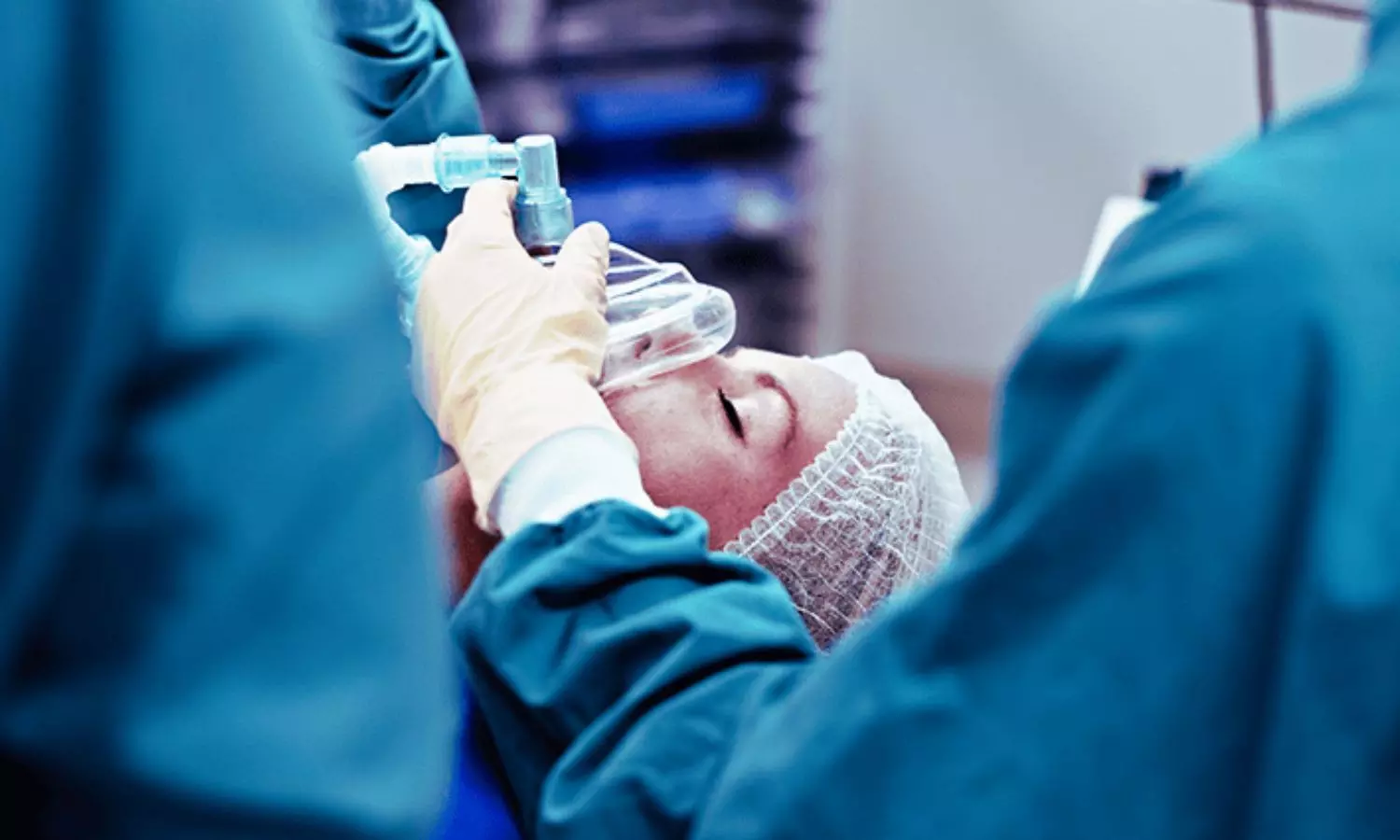
It is crucial to provide excellent hospital care during labor and delivery to decrease avoidable maternal morbidity and mortality. The latest MBRRACE-UK report found that while deaths directly linked to anesthesia are uncommon, enhancing the overall peripartum care provided to 38% of those who passed away between 2018 and 2020 could have impacted the outcome, as only 22% received high-quality care. Recent study examined the variability in obstetric anesthesia practice and care within the UK’s National Health Service. The researchers conducted a survey of 106 out of 107 participating hospitals, representing 69% of all UK obstetric units. The key findings were: Staffing and Training: – 94% of hospitals had a dedicated consultant obstetric anesthetist during working hours, but 25% of out-of-hours duty anesthetists had other clinical commitments outside of obstetrics. – 98% of hospitals offered multidisciplinary team training, mostly using simulation-based methods. Facilities and Resources: – 47% of hospitals had dedicated high-risk antenatal clinics, and 77% provided written patient information on anesthesia options in multiple languages. – 69% used point-of-care testing to estimate hemoglobin concentration. Clinical Practices: – 76% used patient-controlled epidural analgesia during labor, and 26% used programmed intermittent epidural boluses. – 93% used intrathecal diamorphine for elective cesarean deliveries, with a common dose of 300 mcg. – 74.5% routinely used intraoperative patient warming measures. Outcomes and Quality Indicators: – 76% tracked the incidence of post-dural puncture headache, with a median incidence of 0.96%. – 75% and 67% recorded elective and emergency cesarean delivery rates, respectively, but only 13% measured achievement of adequate pain relief 45 minutes after epidural placement

The study demonstrates significant variability in staffing, facilities, clinical practices, and outcome measurements related to obstetric anesthesia across the UK. The authors recommend standardizing anesthetic peripartum care based on national guidelines and systematically measuring quality indicators to ensure safe and equitable care.

Key Points

1. 94% of hospitals had a dedicated consultant obstetric anesthetist during working hours, but 25% of out-of-hours duty anesthetists had other clinical commitments outside of obstetrics.

2. 98% of hospitals offered multidisciplinary team training, mostly using simulation-based methods.

3. 47% of hospitals had dedicated high-risk antenatal clinics, and 77% provided written patient information on anesthesia options in multiple languages. 69% used point-of-care testing to estimate hemoglobin concentration.

4. 76% used patient-controlled epidural analgesia during labor, and 26% used programmed intermittent epidural boluses. 93% used intrathecal diamorphine for elective cesarean deliveries, with a common dose of 300 mcg. 74.5% routinely used intraoperative patient warming measures.

5. 76% tracked the incidence of post-dural puncture headache, with a median incidence of 0.96%. 75% and 67% recorded elective and emergency cesarean delivery rates, respectively, but only 13% measured achievement of adequate pain relief 45 minutes after epidural placement.

6. The study demonstrates significant variability in staffing, facilities, clinical practices, and outcome measurements related to obstetric anesthesia across the UK. The authors recommend standardizing anesthetic peripartum care based on national guidelines and systematically measuring quality indicators to ensure safe and equitable care.

Reference – 

O’Carroll J, Zucco L, Warwick E, et al. (October 04, 2024) A Survey of Obstetric Anaesthesia Services and Practices in the United Kingdom. Cureus 16(10): e70851. DOI 10.7759/cureus.70851

Powered by WPeMatico

Study Reveals Link Between RBC Transfusion and Reduced Mortality in Septic Patients with CKD

China: A recent study published in Scientific Reports has highlighted a significant association between red blood cell (RBC) transfusion and reduced 28-day mortality rates among septic patients suffering from concomitant chronic kidney disease (CKD).

The findings indicate that a patient’s base excess (BE) value, Sequential Organ Failure Assessment (SOFA) score, and estimated glomerular filtration rate (eGFR) play vital roles in determining treatment outcomes and should be factored into decisions regarding RBC transfusion.

Sepsis is a life-threatening response to infection that often complicates the management of patients with chronic kidney disease. CKD patients frequently experience compromised immune function, increasing their susceptibility to infections that can trigger sepsis. The presence of both conditions can lead to lower hemoglobin levels and is linked to a higher mortality rate.

Against the above background, Xingxing Hu, Nanjing University of Chinese Medicine, Nanjing, Jiangsu, China, and colleagues aimed to examine whether RBC transfusions enhance the outcomes of septic patients with concurrent CKD and to assess the criteria for administering RBC transfusions.

For this purpose, the researchers conducted a retrospective cohort study using data from the MIMIC-IV (v2.0) database. The study included 6,604 patients with sepsis and concurrent chronic kidney disease who were admitted to the Intensive Care Unit (ICU). Propensity score matching (PSM) was employed to account for confounding variables.

The study led to the following findings:

  • Multivariate Cox regression analysis revealed an association between RBC transfusion and a decreased risk of 28-day mortality (HR: 0.61).
  • Following a meticulous 1:1 propensity score matching analysis between the two cohorts, the matched population revealed a notable decrease in 28-day mortality within the RBC transfusion group (HR: 0.60).
  • The researchers observed that a SOFA score ≥ 5, a Base Excess value < 3, and an eGFR < 30 may be considered when evaluating the potential need for RBC transfusion.

The findings revealed that RBC transfusion may enhance the 28-day survival rate in septic patients with concurrent CKD. In particular subgroups, factors like base excess value, SOFA score, and estimated glomerular filtration rate significantly influence treatment outcomes. Consequently, these variables should be considered when determining the need for initial RBC transfusion. The researchers suggest the necessity for additional research to validate these findings through randomized clinical trials.

“Our study found a significant link between RBC transfusion and reduced 28-day mortality in septic patients with CKD. However, its generalizability is limited due to reliance on data from a single U.S. academic center, which may not reflect broader practices or patient demographics, along with inherent retrospective limitations,” the researchers concluded.

Reference:

Chen, L., Lu, H., Lv, C., Ni, H., Yu, R., Zhang, B., & Hu, X. (2024). Association between red blood cells transfusion and 28-day mortality rate in septic patients with concomitant chronic kidney disease. Scientific Reports, 14(1), 1-10. https://doi.org/10.1038/s41598-024-75643-3

Powered by WPeMatico

Study Finds Immediate GTR Superior for d-M2 Infrabony Defects Post Third Molar Extraction

China: In a recent retrospective study, the researchers delved into the critical timing of guided tissue regeneration (GTR) following third molar extraction.

The study, published in BMC Oral Health, found that immediate and delayed guided tissue regeneration treatments demonstrated efficacy for managing infrabony defects at the distal aspect of the second molar (d-M2) following third molar extraction.

Immediate GTR performed concurrently with third molar extraction, and delayed GTR, administered three months later, led to a significant reduction in defect depth, the researchers reported. Immediate GTR potentially provides an additional advantage by minimizing the necessity for a second surgery.

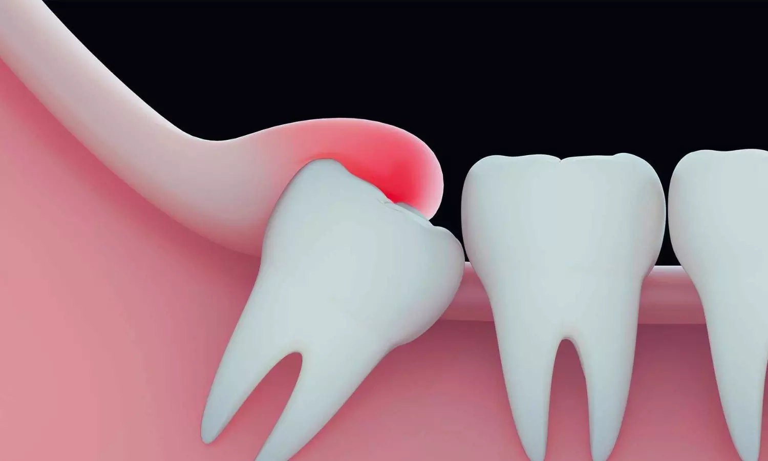
Infrabony defects commonly affect the distal aspect of the second molar (d-M2) following extraction of the adjacent third molar. Guided tissue regeneration (GTR) is a recognized treatment option for addressing these defects post-third molar removal. However, the optimal timing of GTR administration after third molar extraction remains a pivotal consideration in clinical decision-making.

Against the above background, Ying Xuan, Department of Periodontology, Hangzhou Stomatology Hospital, Hangzhou, Zhejiang Province, China, and colleagues aimed to compare delayed and immediate GTR treatments to assist clinical decision-making.

For this purpose, the research team collected D-M2 infrabony defects with a minimum 1-year follow-up. The participants were divided into three groups: the Immediate GTR group, where GTR was performed concurrently with third molar extraction; the Delayed GTR group, where GTR was delayed by at least three months after third molar extraction; and the Control group, which received only scaling and root planing during third molar extraction.

They also evaluated radiographic and clinical parameters related to the infrabony defect before GTR and post-surgery using the Kruskal-Wallis test or one-way ANOVA, followed by post-hoc Dunn’s test or the Bonferroni test for pairwise comparisons.

The following were the key findings of the study:

· A total of 109 d-M2 infrabony defects were assessed.

· No significant differences were found between the two GTR groups, although both of them showed significant reductions in infrabony defect depth: the immediate GTR group (2.77 ± 1.97 mm versus 0.68 ± 1.03 mm) and the delayed GTR group (2.98 ± 1.08 mm versus 0.68 ± 1.03 mm) compared to the control group.

“Guided tissue regeneration has shown effective improvement in infrabony defects at the distal aspect of the second molar (d-M2) following third molar removal, whether performed simultaneously or delayed. Immediate GTR treatment may offer patients reduced discomfort since it requires only one surgical procedure,” the researchers wrote.

“The outcomes of GTR in infrabony defects at the distal aspect of the second molar may be affected by factors such as the morphology of the defects, the specific location of the second molar (M2), and the initial depth of the defects,” they concluded.

Reference:

Tang, SM., Liu, DX., Xiong, ZY. et al. Comparison of immediate vs. delayed guided tissue regeneration in Infrabony defect of second molars after adjacent third molar extraction: a retrospective study. BMC Oral Health 24, 830 (2024). https://doi.org/10.1186/s12903-024-04591-1

Powered by WPeMatico

New study implicates Six protein in early-onset preeclampsia

Preeclampsia is a life-threatening pregnancy complication marked by persistent high blood pressure that is even more serious when it occurs early in the first trimester. The exact cause of early-onset preeclampsia is unknown, and it is difficult to predict, prevent and diagnose. Now, in ACS’ Journal of Proteome Research, researchers report on six proteins that could be used as targets to diagnose and treat the condition.

Preeclampsia’s key symptom is high maternal blood pressure, and serious cases can lead to maternal organ failure, low infant birth weight, or maternal or fetal death. Preeclampsia before 34 weeks of pregnancy has a higher risk of severe outcomes, especially for the fetus. But it’s difficult for health care providers to detect this condition before harmful symptoms appear, because little is known about what causes it. So, Jing Li and colleagues set out to characterize proteins in placenta tissue that may offer clues about the cause of early-onset preeclampsia and serve as targets for early detection or treatment.

The researchers collected placenta tissue from 30 pregnant people, half with early-onset preeclampsia and half with healthy pregnancies. Li and colleagues used mass spectrometry to screen molecular fragments in each sample, followed by a software program to match the fragments to their associated proteins. This process pinpointed 59 proteins that were present in different amounts (either higher or lower) for preeclamptic placenta tissue samples versus healthy placenta tissue samples. The researchers chose 16 of these proteins to target with a different, more sensitive mass spectrometry method, which more precisely measured the amounts of each protein. Of these 16 proteins, six were present in statistically different amounts across tissue sample groups:

  • Preeclamptic placenta tissue had higher levels of monocarboxylate transporter 4, ERO1-like protein alpha and pappalysin-2. These proteins are involved in synthesizing proteins and regulating growth hormones.
  • Preeclamptic placenta tissue had lower levels of desmin, caldesmon and keratine 18. These proteins play key roles in cardiovascular complications, like an enlarged heart; blood flow in placental muscle cells; estrogen signaling and cell health in the uterus lining.

Altogether, the results suggest that cardiovascular complications or the estrogen cycle could be linked to the development of early-onset preeclampsia. The team says that more research is needed, but identifying these six proteins serves as a promising first step toward improved detection and treatment of this life-threatening condition.

Reference:

Zhou, J., et al. (2024). Proteomic Analysis Reveals Differential Protein Expression in Placental Tissues of Early-Onset Preeclampsia Patients. Journal of Proteome Research. doi.org/10.1021/acs.jproteome.4c00404.

Powered by WPeMatico

Ultraprocessed Foods Linked to Increased Risk of Developing Lupus, reveals research

New research suggests that higher daily intakes of ultraprocessed foods (UPFs) are associated with a higher risk of developing systemic lupus erythematosus (SLE). A recently published paper indicates that diets high in UPFs-including foods like chips, sodas, and ready-to-eat processed foods -may also confer an increased risk for SLE, an autoimmune disease. This was published in the journal Arthritis Care & Research by Rossato S. and colleagues.

The researchers included data from the Nurses’ Health Study I (NHSI) and Nurses’ Health Study II (NHSII), two large prospective cohort studies in the U.S. with more than 121,000 female registered nurses enlisted. Participants were between 20 and 50 years of age at enrollment in 1976 (NHSI) and 1989 (NHSII) and had completed comprehensive dietary questionnaires every four years using a validated semiquantitative food frequency questionnaire (SQFFQ). The dietary questionnaire focused on consumption of ultraprocessed foods, which were reported by number of servings/day, grams, or milliliters consumed.

The SLE diagnoses of participants were confirmed by rheumatologists. Tertiles of cumulative average UPF intake will be determined based on the average UPF intake, ranked from highest to lowest. Time-varying Cox regression models will be used to estimate hazard ratios with 95% confidence intervals for the development of SLE adjusting for important covariates that include age, race, BMI, menopausal status, and other lifestyle factors. In addition, risk stratification by BMI and further examination of SLE subtypes-also in the context of the presence and absence of dsDNA antibodies-was performed.

  • This study included 99,000 female participants recruited to NHSI and 106,000 females in NHSII. The mean age of participants in this study using NHSI was 50 years, while, with NHSII, it was 36 years. More than 90% of participants self-reported as White.

  • The highest tertile of UPF intake was significantly associated with an increased risk of SLE, 56% higher among those in the lowest tertile (p= 0.03).

  • The association was stronger with the anti-dsDNA positive SLE cases (p = 0.01). suggesting that higher UPF intake is associated with more severe disease.

  • Among UPF categories, artificially sweetened and sugar-sweetened beverages were most strongly associated with a higher risk of SLE.

  • The associations did not alter after adjustment for many confounders, including BMI, smoking, alcohol consumption, and menopausal status

The results of this study provide novel evidence that increases in UPF intake lead to an increased risk of developing SLE. Since UPFs account for more than 50% of the total calories American adults consume on a daily basis, it is relevant to educate consumers about the health risks associated with such foods. Limiting UPF exposure would reduce the incidence of lupus; however, reduction in overall health risk is also possible.

Reference:

Rossato S, Oakes EG, Barbhaiya M et al. Ultraprocessed Food Intake and Risk of Systemic Lupus Erythematosus Among Women Observed in the Nurses’ Health Study Cohorts. Arthritis care & research, June 2024. 10.1002/acr.25395.

Powered by WPeMatico

Naloxone successfully reverses signs of opioid overdose in over half of users in prehospital setting: Study

The opioid epidemic is a leading cause of morbidity and mortality in the United States, and it is increasingly impacting children and teenagers. Naloxone can reverse the effects of opioid overdose and is being used in hospitals and by emergency responders outside the hospital to save the lives of young people poisoned by opioids.

Research titled, “Naloxone Administration to Pediatric Patients During Emergency Medical Service Events,” finds that among pediatric emergency medical service responses where naloxone was administered, the first dose was successful in improving clinical status in 54.1% of cases. Approximately one-third (32.7%) of pediatric patients received two or more naloxone doses, according to an abstract presented during the American Academy of Pediatrics 2024 National Conference & Exhibition at the Orange County Convention Center from Sept. 27-Oct..

“Emergency medical services clinicians rarely reported that naloxone worsened clinical status, and naloxone improved a patient’s clinical condition in over half of emergency responses in our study,” said lead study author Christopher Gaw, MD, MPH, MBE, FAAP, emergency medicine physician at Nationwide Children’s Hospital. “This finding underscores how naloxone can be a safe and effective antidote when used for suspected opioid poisonings in children and adolescents.”

Researchers examined data from the National Emergency Medicine Service Information System (NEMSIS) on emergency medical service activations for pediatric patients ages 17 and younger in 2022, and found naloxone was administered to teenagers and children at least 6,215 times that year. The study also found that one in five adolescents 13-17 years old were documented as receiving naloxone prior to emergency medical service arrival (20.7%), meaning that somebody on the scene administered naloxone. Naloxone administrations occurred most often in the home or residential setting (61.4%). The research also found that most overdoses occur in adolescents, ages 13-17 (79.4%), followed by children ages 1-5 (10.2%), and boys were also slightly more likely to receive naloxone (55.3%).

The calls for assistance suggest there may be initial confusion in the early emergency medical service response period before a diagnosis is made. In the study, the initial emergency call was for an overdose, poisoning, or ingestion in about a third of the calls for adolescents 13-17 (31.5%) and just 12.8% of calls for infants.

“Our study highlights how EMS clinicians are reporting naloxone use after responding to different types of emergency dispatch calls, such as poisonings, unconsciousness, and problems breathing,” Dr. Gaw said. “These signs and symptoms could represent a possible opioid poisoning, which may explain why naloxone was administered in those situations.”

Reference:

Naloxone successful in over half of users in reversing signs of opioid overdose in the prehospital setting, American Academy of Pediatrics, Meeting: American Academy of Pediatrics 2024 National Conference & Exhibition.

Powered by WPeMatico

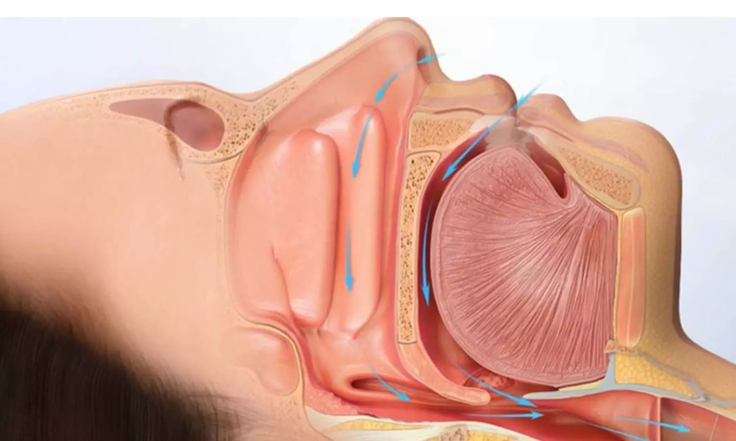
Mouth Closure May Worsen Airflow in Patients with Severe Obstructive Sleep Apnea, unravels study

Researchers found that clenching the mouth at night may help to exacerbate airflow in patients suffering from obstructive sleep apnea (OSA), especially those who are more dependent on mouth breathing. A new study contradicts the recent hype of “mouth taping” as a preventive measure against mouth breathing, concluding that this is the wrong approach to take for certain OSA patients.

Obstructive sleep apnea (OSA) is characterized by repeated episodes of airway obstruction during sleep, which causes disrupted breathing and poor oxygen levels. Most commonly, it is typical in patients with OSA to breathe through the mouth, but more recently, there’s been the hashtag on TikTok of “mouth taping” as a treatment method for OSA-forced nasal breathing. According to a new study by Daniel Vena, PhD from Brigham & Women’s Hospital and Harvard Medical School, this practice raises issues about safety and efficacy.

For the research, the mouths of 54 patients were manually closed during the period of drug-induced sleep to determine their effects on airflow. The outcomes were that mouth closure improved airflow in some patients but worsened it in others, particularly those who relied mainly on mouth breathing.

The study published in JAMA Otolaryngology-Head & Neck Surgery aimed to evaluate the effect of oral closure during sleep on airflow. Altogether 54 adult OSA patients were studied, with a median age of 55 years, BMI of 28.9, and an apnea-hypopnea index of 26.9 events/hour. Researchers induced sleep with propofol and manually closed the patients’ mouths to see whether or not they would alter the airflow.

  • In the 32 patients who experienced mild mouth breathing (oral airflow before maneuver was 0.05 to 2.2 L/min), manually occluding the mouth resulted in a 53.1 percentage-point increase in airflow, equivalent to 2.0 L/min.

  • In the 12 patients who were major mouth breathers (>2.2 L/min oral ventilation during normal sleep), mouth closure reduced ventilation by 40 percentage points or 1.86 L/min (95% CI -3.1 to -0.6).

  • Patients suffering the worst obstruction of the soft palate and destined for mouth breathing demonstrated the biggest drops in flows, because their mouths were closed.

Propofol was administered to sleep-induced patients for routine clinical endoscopies and airflow measurements. Researchers operated to alternately close the mouth and then release it in order to see the effect on breathing. In each patient, at least 15 breath cycles were recorded, alternating between mouth closure and relaxed mouth positioning.

There were a number of limitations to the study. It was an acute outcome trial measuring single breath effects rather than long-term outcomes, so these results may not reflect the longer-term impact of forced mouth closure during sleep. Moreover, the contribution of nasal obstruction to open-mouth breathing was not measured and could be a critical factor in explaining these findings.

Researchers advise doctors to take proper caution in recommending such treatments like using mouth tapes and monitor patients closely so the therapy doesn’t worsen their disease condition. Long-term effects of such therapies on OSA patients should be explored as well.

Reference:

Yang, H., Huyett, P., Wang, T.-Y., Sumner, J., Azarbarzin, A., Labarca, G. P. T., Messineo, L., Gell, L. K., Aishah, A., Hu, W.-H., White, D. P., Sands, S. A., Wellman, A., & Vena, D. (2024). Mouth closure and airflow in patients with obstructive sleep apnea: A nonrandomized clinical trial. JAMA Otolaryngology– Head & Neck Surgery. https://doi.org/10.1001/jamaoto.2024.3319

Powered by WPeMatico

NEET PG 2024 Counselling Schedule Expected in 2 to 3 Days, informs Doctors’ Association

The National Eligibility-and-Entrance Test Postgraduate (NEET-PG) exam aspirants might soon get clarity regarding their admission process as the authorities might release the tentative schedule for the exam in the next two to three days.

Dr. Lakshya Mittal, the president of the United Doctors Front Association (UDFA), mentioned in an X (formerly Twitter) post that the tentative schedule for the NEET-PG 2024 exam is expected to be updated within the next 2-3 days.

Powered by WPeMatico

Manipal Hospitals Buys Hospital Property Worth Rs 415 Crore in Mumbai’s Andheri

Mumbai: Manipal Hospitals, backed by Singapore’s global investment company Temasek Holdings, has purchased a hospital property worth Rs 415 crore in Mumbai’s Andheri.

As per the documents, accessed by Propstack, the Deed of Transfer was registered between the seller Khubchandani Properties and Investment Private Limited and Manipal Health Enterprises Private Limited on October 7, HT has reported. 

The new hospital property, purchased by Manipal Hospitals, is spread across an area of 10,590 sq m. The hospital has been developed on a land parcel spread across an area of almost 2.61 acres and encompasses an area of 20,851.65 sq m (built-up).

Also Read: Manipal Hospitals opens 250-bed Super Specialty Hospital in Pune

As per the latest media report by Hindustan Times, the Block A hospital structure comes with a basement having 80 enclosed parking spaces and 204 mechanical stack parking spaces. It also has a lower ground, a ground floor, and three upper floors, the document showed.

For the property transaction, Manipal Hospitals had to pay around Rs 27.30 Crores as stamp duty. According to the documents, the transaction includes both the hospital building, as well as the land where it stands, Money Control has reported.

Singapore’s Temasek Holdings bought a majority stake in the Manipal Hospitals last year for $2 billion. In September 2023, Manipal Hospitals acquired a 84 per cent stake in Emami Group’s AMRI Hospitals Ltd. According to media reports, currently, the Emami Group owns a 15 per cent stake in AMRI.

Medical Dialogues had previously reported that earlier this year, Manipal Hospitals, the second largest hospital chain in India, announced the successful brand integration of three AMRI Hospitals units in Dhakuria, Mukundapur, and Salt Lake in Kolkata, which were acquired in September 2023.

Manipal Hospitals also invested in the hospital chain Medica for around Rs 1,400 crore. Back in 2021, it acquired hospital chain Columbia Asia having a portfolio of 11 healthcare facilities.

Back in 2022 Manipal Hospitals had announced the commencement of its new 250-bed hospital. Earlier this year in February, the Managing Director and CEO of Manipal Hospitals, Dilip Jose informed The Week that the Company had already two hospitals in Pune, including one in Baner. He had said, “Mumbai is also of keen interest to us, but building a greenfield hospital there is very difficult.”

Also Read: Manipal Hospitals expands footprints in East India, rebrands AMRI units

Powered by WPeMatico