Hyperbaric Oxygen Therapy Enhances Hearing Recovery in Sudden Sensorineural Hearing Loss Patients: Study

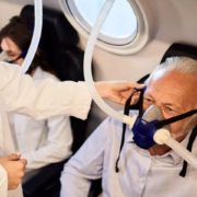
Researchers found that the application of hyperbaric oxygen therapy (HBOT) in addition to current medical treatment significantly improves hearing gain in the case of sudden sensorineural hearing loss (SSNHL). SSNHL, an acute hearing impairment happening in thousands of individuals each year, is a challenging condition with unclear pathogenesis and variable responsiveness to treatment. The study was recently published in the journal Laryngoscope by Issac L. and colleagues.

Systematic review entailed extensive search on major databases, such as PubMed, EMBASE, CENTRAL, MEDLINE, Google Scholar, Web of Science, and ClinicalTrials.gov, for publications up to May 7, 2025. The review was based on the PRISMA (Preferred Reporting Items for Systematic Reviews and Meta-Analyses) guidelines. Two independent reviewers screened and selected eligible studies and extracted data. Meta-analysis was conducted using a random-effects model to combine odds ratios (ORs) for recovery of hearing.

The review consisted of 20 studies, including 16 randomized controlled trials (RCTs) and four prospective non-randomized studies. A total of 1,087 patients who underwent HBOT and 600 medical therapy alone-treated patients were assessed.

Key Findings

• The meta-analysis consisted of ten studies and demonstrated convincing evidence favoring the application of HBOT in conjunction with MT alone, including steroids.

• Patients treated with HBOT and medical therapy had 2.61 times greater chances of hearing improvement compared to those treated with medical therapy only (OR 2.61, 95% CI 1.86–3.68, p < 0.001).

• Subgroup analysis between HBOT + systemic steroids (SS) and SS alone revealed HBOT was linked with better outcomes (OR 2.54, 95% CI 1.63–3.97, p < 0.001).

• When HBOT + SS + intratympanic steroids (ITS) was contrasted with SS + ITS alone, the probability of recovery of hearing was still significantly higher for the HBOT group (OR 2.64, 95% CI 1.39–5.02, p < 0.001).

• It is highly suggestive of the fact that HBOT improves the effectiveness of both systemic and intratympanic steroid therapy for SSNHL.

This meta-analysis and systematic review came to the conclusion that standard medical therapy supplemented with hyperbaric oxygen therapy significantly enhances hearing outcomes in patients with sudden sensorineural hearing loss. These findings support the inclusion of HBOT in the treatment protocol in suitable patients.

Reference:

Alter, I. L., Hamiter, M., Han, J., Leu, C. S., Usseglio, J., & Lalwani, A. K. (2025). Hyperbaric Oxygen and Sudden Sensorineural Hearing Loss: A Systematic Review and Meta-Analysis. The Laryngoscope, 10.1002/lary.32472. Advance online publication. https://doi.org/10.1002/lary.32472

Powered by WPeMatico

Tildrakizumab Shows Significant Efficacy in Psoriatic Arthritis Phase 3 Trials

Phase 3 trials have demonstrated that tildrakizumab 100mg significantly improved ACR20 response rates in patients with active psoriatic arthritis at 24 weeks compared to placebo, marking a promising advancement in PsA treatment.

Treatment with tildrakizumab 100 mg (ILUMYA) resulted in greater improvements in PsA signs and symptoms at
Week 24 compared to treatment with placebo. Both the INSPIRE-1 and INSPIRE-2 studies achieved the primary
endpoint, with a higher proportion of patients in the INSPIRE-1 and INSPIRE-2 studies treated with tildrakizumab
achieving ACR20 responses at week 24, compared to those receiving placebo (p < 0.05).


“We are excited to share that both the INSPIRE-1 and INSPIRE-2 clinical trials have successfully met their primary
endpoints. These top-line results reinforce the therapeutic potential of ILUMYA as a treatment option for patients
with active psoriatic arthritis. We extend our sincere gratitude to the patients, healthcare professionals and
administrators whose contributions made the studies possible. We look forward to sharing the complete clinical data
in the near future,” said Marek Honczarenko, MD, PhD, Senior Vice President and Head of Global Specialty
Development at Sun Pharma.

Safety data in the studies was consistent with the well-documented safety profile of ILUMYA, which is approved for
the treatment of adults with moderate-to-severe plaque psoriasis who are candidates for systemic therapy or
phototherapy. No new safety signals were identified in the INSPIRE-1 and INSPIRE-2 studies.

Findings from the INSPIRE studies will be presented at upcoming medical conferences and published in a peerreviewed medical journal. Use of tildrakizumab 100 mg (ILUMYA) in psoriatic arthritis is not approved, and its
safety and efficacy have not been evaluated by regulatory authorities.

About the INSPIRE-1 and INSPIRE-2 Phase 3 studies

The INSPIRE-1 and INSPIRE-2 are both 52-week global, multicenter, randomized, double-blind, single-dose,
placebo-controlled Phase 3 studies aimed at assessing the efficacy and safety of tildrakizumab 100 mg (ILUMYA®)
in adult patients with active psoriatic arthritis. The INSPIRE-1 enrolled patients having prior exposure to an antiTNF agent while the INSPIRE-2 enrolled anti-TNF naïve patients. A total of over 800 adult patients were enrolled
for the two studies from clinical sites in the US, Europe and Asia. Patients were randomized to receive either
tildrakizumab 100 mg (ILUMYA®) or placebo.
An induction dose was not administered in either study. Study
participants were permitted to use concomitant methotrexate or leflunomide, provided the dose remained stable
throughout the trial. In the INSPIRE-1 and INSPIRE-2 studies, tildrakizumab 100 mg (ILUMYA®) was administered
at Week 0 and then every 12 weeks. In contrast, in Phase 3 studies for chronic plaque psoriasis, the treatment schedule
included doses in Week 0, Week 4, and then every 12 weeks thereafter. ILUMYA® is approved by the regulatory
Agencies for the indication of chronic plaques psoriasis.
The primary endpoint for both studies is the proportion of participants achieving an ACR20 response at Week 24.
The key secondary efficacy endpoints at 24 weeks include ACR50, ACR70, and PASI75 and improvement. Learn
more about the studies at clinicaltrials.gov (INSPIRE-1 NCT04314544 and INSPIRE-2 NCT04314531).
The ACR20 is a composite measure defined as a 20 percent improvement in both the number of tender and number
of swollen joints and a 20 percent improvement in three of the following five criteria: patient global assessment,
physician global assessment, functional ability measure (most often HAQ-DI), visual analog pain scale, and
erythrocyte sedimentation rate or C-reactive protein (CRP). ACR50 and ACR70 are the same measurement with
improvement levels of 50 percent and 70 percent, respectively.

About Psoriatic Arthritis (PsA)

Psoriatic arthritis (PsA) is a chronic (long-lasting or recurring) disease related to the immune system. It causes
swelling, pain, and stiffness in joints and entheses (places where tendons and ligaments connect to bones). Psoriatic
arthritis can develop at any age. Psoriatic arthritis can occur regardless of the severity of psoriasis (mild, moderate,
or severe). Roughly 1 in 3 people living with psoriasis also have psoriatic arthritis [3]; typically, with psoriasis
developing before psoriatic arthritis. While as many as 2.4 million Americans live with psoriatic arthritis, more
than 15% of people living with psoriasis may also have undiagnosed psoriatic arthritis.

About ILUMYA® (tildrakizumab-asmn)

ILUMYA (tildrakizumab-asmn) is a humanized lgG1/k monoclonal antibody designed to selectively bind to the p19
subunit of interleukin-23 (IL-23) and inhibit its interaction with the IL-23 receptor, leading to inhibition of the release
of pro-inflammatory cytokines and chemokines. ILUMYA is indicated for the treatment of adults with moderate-to-severe plaque psoriasis who are candidates for systemic therapy or phototherapy, in the United States and other countries.

Powered by WPeMatico

Mfn2 and Preeclampsia: Study Exploring a Potential Game Changer in Maternal Health: Study

Recent study investigates the expression of mitofusin 2 (Mfn2) in placentas and peripheral blood from patients with early-onset preeclampsia (eoPE) and late-onset preeclampsia (loPE), exploring its potential as a diagnostic and therapeutic target for preeclampsia. Preeclampsia, a serious condition occurring after 20 weeks of gestation, presents significant morbidity and mortality risks, particularly differentiating between eoPE (before 34 weeks) and loPE (after 34 weeks) due to varying placental and maternal pathophysiologies.

Role of Mfn2 in Preeclampsia

Mfn2, a mitochondrial outer membrane protein critical for mitochondrial dynamics, was previously noted to be downregulated in preeclampsia, associated with increased apoptosis and mitochondrial dysfunction. The study emphasizes the differences in placental pathology between eoPE and loPE, highlighting impaired remodeling of uterine spiral arteries in eoPE, leading to ischemia and hypoxia. In contrast, loPE presents with maintained or higher placental perfusion levels.

Mfn2 Expression Analysis

Quantitative assessments revealed a significant decrease in Mfn2 mRNA levels in placentas from eoPE patients compared to both loPE and normal control groups, while loPE did not show significant downregulation compared to controls. Peripheral blood analyses indicated higher levels of Mfn2 protein in both preeclampsia groups compared to controls, with eoPE showing the highest levels. Notably, Mfn2 levels were positively correlated with the severity of preeclampsia, such as systolic and diastolic blood pressure and proteinuria, with stronger correlations in eoPE. The findings suggest a stratification in the role of Mfn2 between the two conditions, establishing that the reduction of Mfn2 in eoPE is indicative of significant mitochondrial dysfunction, while the elevated serum levels in both preeclampsia types may reflect a compensatory response to placental mitochondrial damage. Moreover, these elevated Mfn2 levels were negatively correlated with birth weight and Apgar scores, indicating poor pregnancy outcomes associated with elevated Mfn2 levels.

Conclusion and Future Directions

In summary, this investigation highlights Mfn2’s potential as a biomarker for preeclampsia severity and progression, distinguishing the distinct roles of mitochondrial dysfunction in eoPE and loPE. The findings support the hypothesis that early identification of impaired Mfn2 expression could facilitate enhanced diagnostic and therapeutic strategies for preeclampsia management, advocating for further research to validate these findings in larger cohorts and longitudinal studies.

Key Points

– -Expression Analysis of Mfn2-: Significant downregulation of Mfn2 mRNA levels was identified in placentas from patients with early-onset preeclampsia (eoPE) when compared to both late-onset preeclampsia (loPE) and normal control samples, while loPE did not exhibit significant differences in Mfn2 levels compared to controls.

– -Peripheral Blood Findings-: Elevated Mfn2 protein levels were observed in peripheral blood from both eoPE and loPE patients compared to controls, with eoPE showing the highest serum Mfn2 levels. This suggests a potential compensatory mechanism in response to placental mitochondrial damage.

– -Correlation with Disease Severity-: Mfn2 levels exhibited positive correlations with the severity of preeclampsia indicators such as systolic and diastolic blood pressure, as well as proteinuria. These correlations were found to be more robust in the eoPE cohort, indicating that altered Mfn2 expression may reflect the severity of the condition.

– -Implications for Pregnancy Outcomes-: Elevated Mfn2 levels in both types of preeclampsia were negatively correlated with birth weight and Apgar scores, suggesting that higher Mfn2 may be associated with adverse pregnancy outcomes and poorer fetal health.

– -Differentiation of Pathophysiology-: The study emphasizes the divergent mechanisms of placental pathology between eoPE and loPE, with eoPE characterized by impaired remodeling of uterine spiral arteries, leading to ischemic conditions, whereas loPE maintains or improves placental perfusion levels.

– -Future Research Directions-: The findings propose Mfn2 as a potential biomarker for assessing preeclampsia severity and progression, underscoring the need for further investigations in larger cohorts and longitudinal studies to validate Mfn2’s utility in diagnostic and therapeutic approaches for preeclampsia.

Reference –

Dandan Sun et al. (2025). A Mitochondrial Regulator Protein, Mitofusin 2, Is Elevated In The Maternal Blood Of Women With Preeclampsia. *BMC Pregnancy And Childbirth*, 25. https://doi.org/10.1186/s12884-025-07663-4.

Powered by WPeMatico

75 percent MBBS fee hike at Odisha private medical colleges

Bhubaneswar: Medical education in Odisha is going to get costlier as an announcement has been made regarding a steep fee hike for the MBBS and PG medical courses in the private medical colleges across the State.

MBBS fees at three private medical colleges, including Hi-Tech Medical College Bhubaneswar, DRIEMS Medical College Cuttack, and Hi-Tech Medical College Rourkela, have increased the MBBS fees by approximately 75 percent.

The Odisha Joint Entrance Examination (OJEE) Authority released the revised fee structure after a notification was issued in this regard by the Department of Skill Development and Vocational Education.

The Government has implemented the fee hike based on the Odisha Professional Educational Institutions (Regulation of Admission & Fixation of Fee) Act, 2007, and based on the recommendation of the Fee Structure Committee in its 7th, 8th, 9th, and 12th meeting held on 10.11.2023, 30.09.2024, 26.11.2024, and 14.07.2025, respectively. It will be implemented w.e.f. academic session 2025-2026.

Also Read: No MBBS, PG medical fee hike this year in Maharashtra Private Medical Colleges

MBBS Fee Hike: 

As per the notifications issued on the 11th and 16th of August, 2025, the MBBS fees at DRIEMS Institute of Health Sciences and Hospital, Cuttack have been increased from Rs 6,50,000 (as per the previous fee structure) to Rs 9,08,000 for the upcoming three years (from the academic year 2025-2026 to 2027-2028).

At Hi-Tech Medical College & Hospital, Bhubaneswar, MBBS fees have been increased from Rs 6,50,000 to Rs 11,56,000. For the Hi-Tech Medical College & Hospital, Rourkela, the MBBS fee has been hiked from Rs 5,50,000 to Rs 8,54,000 from the academic year 2025-2026.

SI. No.

Name of the Institute

Name of the Course

Previous fee structure

Fee Structure prescribed by the Government w.e.f. academic session 2025-26 to 2027 28

1.

DRIEMS Institute of Health Sciences and Hospital, Cuttack

MBBS

Rs.6,50,000/-

Rs.9,08,000/-

2.

Hi-Tech Medical College Hospital, Bhubaneswar

MBBS

Rs.6,50,000/-

Rs. 11,56,000/

3.

Hi-Tech Medical College & Hospital, Rourkela

MBBS

Rs. 5,50,000/-

Rs. 8,54,000/-

Fee Hike in PG Clinical & Para-Clinical Courses: 

As per the notification issued on August 16, the fees for PG Anatomy/Physiology/Biochemistry at Hi-Tech Medical College and Hospital, Bhubaneswar has been increased from Rs 4,50,000 to Rs 6,93,000. Similarly, fees for PG Para-clinical and Clinical courses at the institute has been increased from Rs 7,15,000 to Rs 8,67,000 and Rs 17,33,500 respectively. 

In the case of Hi-Tech Medical College & Hospital, Rourkela, the fees for PG Para-Clinical and PG Clinical courses have been fixed at Rs 6,41,000 and Rs 12,81,000 w.e.f. from the academic session 2025-2026.

As per the latest media report by The Daily Jagran, the Odisha Parents’ Federation has strongly opposed the fee hike. Criticising the move, the President of the Federation, Vasudev Bhatt, compared the fee hike to the imposition of an “American-style 75 percent tax” on education. He called the fee increase a form of educational commercialization and warned to launch a strong protest by the Federation if the decision is not reversed.

Also Read: Allahabad HC stays Mid-Session MBBS Fee Hike

Powered by WPeMatico

9-year-old dies of Rare Brain Infection in Kerala

Following the death of a 9-year-old girl from amoebic encephalitis— a rare and often fatal brain infection caused by free-living amoeba commonly found in contaminated water, health authorities in Kozhikode have issued a high alert.

The child was first admitted to a local hospital last week with a high fever. As her condition worsened rapidly, she was shifted to Kozhikode Medical College. Despite treatment, she succumbed on the same day.

According to health officials, this is the fourth case reported from Kozhikode
this year — raising concerns about possible contaminated water sources in the
district. Efforts are now underway to trace the exact pond or water body where
the infection may have been contracted. Doctors explain that the infection is
usually caused by Naegleria fowleri, often referred to as the “brain-eating
amoeba.”

It thrives in warm freshwater lakes, rivers, and ponds. Infection
occurs when contaminated water enters the nose, usually during swimming or
diving, before reaching the brain. What makes it dangerous?

Symptoms such as severe
headache, fever, vomiting, stiff neck, and confusion appear suddenly,
progressing rapidly to coma. The fatality rate stands at over 97%, even with
treatment. Is it treatable?

Doctors use antifungal drugs like amphotericin B,
often in combination with miltefosine and other medicines, but survival remains
rare. Early detection is critical. How can you stay safe?

Avoid swimming or
watersports in warm freshwater bodies. Never use tap water in nasal rinsing
devices (like neti pots) — only distilled or sterilized water. Ensure proper
chlorination of swimming pools. Health experts emphasize: If you develop sudden
fever or headache after swimming in freshwater, seek medical help immediately.

Powered by WPeMatico

Health Bulletin 18/August/2025

Here are the top health stories of the day:

Tirunelveli Medical College doctor removed over alleged fabrication of Road Accident Case

A doctor at Tirunelveli Medical College Hospital (TvMCH), accused of fabricating medical records by falsely stating that the road accident victim was intoxicated at the time of the incident, has been removed from his position.

The action followed a petition filed by the patient, Bala Sundararajan, to the District Collector, Dr. K.P. Karthikeyan, and the Chief Minister’s Special Cell, seeking action against the doctor.

For more details, check out the full story on the link below: 

Tirunelveli MCH doctor removed for falsely implicating patient


9-year-old dies of Rare Brain Infection in Kerala

Following the death of a 9-year-old girl from amoebic encephalitis— a rare and often fatal brain infection caused by free-living amoeba commonly found in contaminated water, health authorities in Kozhikode have issued a high alert.

The child was first admitted to a local hospital last week with a high fever. As her condition worsened rapidly, she was shifted to Kozhikode Medical College. Despite treatment, she succumbed on the same day.

According to health officials, this is the fourth case reported from Kozhikode this year, raising concerns about possible contaminated water sources in the district. Efforts are now underway to trace the exact pond or water body where the infection may have been contracted. Doctors explain that the infection is usually caused by Naegleria fowleri, often referred to as the “brain-eating amoeba.”

It thrives in warm freshwater lakes, rivers, and ponds. Infection occurs when contaminated water enters the nose, usually during swimming or diving, before reaching the brain. What makes it dangerous?

Symptoms such as severe headache, fever, vomiting, stiff neck, and confusion appear suddenly, progressing rapidly to coma. The fatality rate stands at over 97%, even with treatment. Is it treatable?

Doctors use antifungal drugs like amphotericin B, often in combination with miltefosine and other medicines, but survival remains rare. Early detection is critical. How can you stay safe?

Avoid swimming or watersports in warm freshwater bodies. Never use tap water in nasal rinsing devices (like neti pots) — only distilled or sterilized water. Ensure proper chlorination of swimming pools. Health experts emphasize: If you develop sudden fever or headache after swimming in freshwater, seek medical help immediately.

Tracheostomy without consent, consultation- Consumer Court dismisses Rs 25 lakh claim against ENT surgeon, Karnataka Hospital

Reiterating the observations of the National Consumer Court that no medical negligence can be attributed against a competent surgeon if an emergency operation was conducted to save the patient, the State Consumer Disputes Redressal Commission, Karnataka, recently exonerated an ENT surgeon and a private hospital from charges of medical negligence while treating a patient diagnosed with Ludwig Angina.

The complainant alleged that his wife died after the ENT surgeon performed a Tracheostomy without consent and without consulting other doctors. However, the consumer court noted that the patient’s oxygen level had dipped, and the primary duty of the ENT surgeon was to make an airway, which could be done only by operating on the patient by Tracheostomy.

For more details, check out the full story on the link below:

Tracheostomy without consent, consultation- Consumer Court junks Rs 25 lakh claim against ENT surgeon, Karnataka Hospital


Obesity Becoming Major Health Threat, PM Modi Calls for Action

Prime Minister Narendra Modi on Friday said obesity is emerging as a major challenge for the country, and urged everyone to contribute to the fight against it.

Addressing the nation from the ramparts of the Red Fort on the 79th Independence Day, he warned that according to experts, one in every three people will suffer from obesity in the coming years and reiterated his previous suggestion to buy 10 per cent less oil for cooking.

“When I speak of fitness, when I speak of sports, I also wish to place before you a matter of concern. Obesity is becoming a very grave crisis for our nation, and every family in our country should take it seriously. We must protect ourselves from obesity, the news agency PTI reported.

For more details, check out the full story on the link below:

Also Read: Obesity a Grave Crisis, says PM Modi in Independence Address


Powered by WPeMatico

Woman loses Rs 2.10 lakh to Fake UK doctor on social media

Lucknow: A healthcare
worker from Lucknow has been duped of Rs. 2.10 lakh by a man posing as an
England-based doctor on social media on the pretext of sending her gifts. The cybercrime wing has registered a case and launched an investigation into the incident.

According to India Today,
the victim has been identified as a resident of Bigahu village in Kakori, and
she came across a man identifying himself as “Dr. Sameer” on Facebook on July
20. Introducing himself as a doctor from England, he initiated conversations
around health-related issues to gain her trust. Days later, he added her to a WhatsApp
group and claimed he was sending her a gift. Though Sandhya initially refused,
she eventually relented after repeated messages from him.

Soon afterwards, the
victim received a phone call from someone claiming to be a customs officer at
Mumbai Airport. The caller told her that to release the parcel supposedly being
sent by Dr. Sameer, she would have to pay taxes and processing charges.
Believing the claim, she transferred Rs. 2.10 lakh in three separate payments.
However, when the promised package never arrived, she realised that she had
been conned and approached the police.

Kakori Inspector Satish
Rathore confirmed that a formal complaint has been lodged and efforts are
underway to identify and apprehend the culprits. Police also issued a public
advisory urging people to remain vigilant on social media, avoid sharing personal
information with unknown individuals, and be wary of fraudulent schemes
involving parcels or gifts.

Medical
Dialogues had earlier reported that in a similar cyber fraud case, a doctor at Ameen
Hospital in Rayachoti allegedly fell victim to a drug parcel scam, losing a
staggering Rs 2 crore to scammers posing as police officers. The scam began when the doctor received a phone call from a number showing the
photo of a police officer. The caller claimed that a drug package had been sent
in his name from London and that a case had been filed against him.

Powered by WPeMatico

Saliva testing may reveal early signs of diabetes and obesity, reveals research

Measuring elevated levels of insulin in blood, called hyperinsulinemia, is a proven way to measure metabolic health and can show risk of developing future health concerns, including Type 2 diabetes, obesity and heart disease.

Now, a team of UBC Okanagan researchers has found that measuring insulin levels in saliva offers a non-invasive way to do the same test-without the need for needles or lab-based blood work.

Dr. Jonathan Little, Professor with UBC Okanagan’s School of Health and Exercise Sciences, says that a simple spit test goes a bit further. It can also be used to detect early metabolic changes linked to obesity and other health risks.

The study, recently published in Applied Physiology, Nutrition, and Metabolism, included 94 healthy participants with a range of body sizes. After a period of fasting, each participant drank a standardized meal-replacement shake, then provided saliva samples and underwent a finger-prick blood glucose test.

“People living with obesity had much higher insulin levels in their saliva than those who were slightly overweight or had lower body weight-even though their blood sugar levels were the same,” he says. “This suggests that saliva testing could be a simple, non-invasive way to identify people at risk of Type 2 diabetes before symptoms appear.”

Type 2 diabetes affects about 400 million people worldwide and is diagnosed by high blood glucose levels. But Dr. Little notes that prediabetes conditions—such as insulin resistance and hyperinsulinemia-may develop 10 to 20 years before a person is diagnosed.

“If hyperinsulinemia can be detected before blood glucose levels start to rise, people at risk for Type 2 diabetes could be identified early, allowing for lifestyle changes and other treatments to be introduced long before glucose levels rise.”

Taking preventive steps at an early stage is important because hyperinsulinemia is a known predictor of several chronic conditions, including Type 2 diabetes, hypertension, cardiovascular disease, stroke, cancer, and most recently, it has been linked to obesity.

Co-author Dr. Hossein Rafiei explains that the study aimed to help develop a practical non-invasive test for hyperinsulinemia, but they also found an interesting result following the consumption of the meal-replacement drink.

Dr. Rafiei’s previous research at UBC Okanagan showed that saliva insulin levels closely follow plasma insulin levels across the day following high and low-carbohydrate mixed meals.

“This suggests that saliva insulin may help distinguish between high and low plasma insulin responses, and could play a role in predicting the severity of hyperinsulinemia and possibly insulin resistance.”

During the study, participants provided saliva tests 30, 60 and 90-minutes after drinking the beverage.

Dr. Rafiei notes that, interestingly, some participants with lower body weight also experienced large saliva insulin spikes after the meal. This suggests they may be at heightened risk for Type 2 diabetes, even without excess weight and having normal blood glucose levels.

“The finding that some people who are lean have high insulin is intriguing,” says Dr. Rafiei. “This indicates that saliva insulin may be more useful than measuring someone’s weight or waist size.”

The study also looked at the relationship between waist circumference, BMI, age and sex, and found that waist size had the strongest link to saliva insulin levels.

“These findings suggest that waist circumference could be a more reliable indicator of hyperinsulinemia than age or overall body weight when using saliva insulin,” he says. “Our results also suggest that saliva insulin may be better than blood glucose at distinguishing between those who are more metabolically healthy and those who are more likely to live with hyperinsulinemia.”

Reference:

Hossein Rafiei and Jonathan Peter Little. 2025. Saliva insulin concentration following ingestion of a standardized mixed meal tolerance test: influence of obesity status. Applied Physiology, Nutrition, and Metabolism. 50: 1-8. https://doi.org/10.1139/apnm-2024-0532.

Powered by WPeMatico

Amiodarone Linked to Higher Risk of Thyrotoxicosis in AF Patients: Study

A new study published in the journal of Heart Rhythm showed that in individuals with atrial fibrillation, amiodarone therapy was independently linked to a higher incidence of thyrotoxicosis.

A common treatment for potentially fatal arrhythmias is amiodarone. Despite the fact that amiodarone has few adverse effects, 15-20% of patients may experience thyroid dysfunction. Amiodarone-induced thyrotoxicosis (AIT) is a serious side effect that can exacerbate heart failure and lead to arrhythmia recurrence.

While type II AIT is a destructive thyroiditis that reacts to glucocorticoids, type I AIT is often treated with thionamides and occurs in people who already have thyroid disease. There is a mixed kind that is linked to increased mortality, particularly in older persons with heart disease. For individuals who are intolerant or resistant to medical therapy, thyroidectomy is regarded as a last resort.

Research on the possibility of amiodarone-treated individuals developing thyrotoxicosis instead of hypothyroidism is yet lacking. Thus, this study evaluated the relationship between amiodarone and thyrotoxicosis in individuals who were diagnosed with hypothyroidism concurrently with atrial fibrillation (AF).

This research used the Clalit Health Services database to do a population-based retrospective cohort analysis. 2 distinct cohorts, 1 for hypothyroidism and the other for normal thyroid function, were identified from patients who received a new diagnosis of AF between 2010 and 2023. Amiodarone exposure was investigated as a time-dependent variable using Cox proportional hazard regression, which allowed participants to switch between exposure groups throughout follow-up.

The hypothyroidism cohort comprised 23,854 AF patients, of whom 107 (66 of 8,212 amiodarone users and 41 of 15,622 non-users) had thyrotoxicosis during follow-up.

This represents a crude incidence rate of 3.43 and 0.63 per 1000 person-years for amiodarone users and non-users, respectively. Amiodarone had an adjusted-HR of 5.18 (95% CI, 3.48-7.69) and was independently linked to an elevated incidence of thyrotoxicosis in this sample.

With an adjusted-HR of 15.02 (95% CI, 13.56-16.64) and an incidence rate of 19.78 and 1.14 per 1000 person-years for amiodarone users and non-users, respectively, the amiodarone impact was more pronounced in individuals with normal thyroid function. Overall, although amiodarone has a less impact on thyrotoxicosis risk in hypothyroidism patients, the risk is still high and should be used with caution.

Source:

Ryan, D., Gershinsky, R., Gronich, N., Yahav, A., Barnett-Griness, O., Schliamser, J. E., Saliba, W., & Danon, A. (2025). Association between amiodarone and thyrotoxicosis in patients with atrial fibrillation and hypothyroidism. Heart Rhythm: The Official Journal of the Heart Rhythm Society. https://doi.org/10.1016/j.hrthm.2025.08.003

Powered by WPeMatico

Thigh Muscle Fat May Signal Greater Metabolic and Heart Disease Risk: Study Shows

Spain: A study in The Journal of Clinical Endocrinology & Metabolism suggests that intermuscular fat — though not visible like abdominal visceral fat — is metabolically active and may contribute to chronic inflammation, insulin resistance, and muscle dysfunction, increasing the risk of metabolic and cardiovascular diseases.

The research, led by Alba Camacho-Cardenosa from the Sport and Health University Research Institute (iMUDS), University of Granada, examined how fat stored between muscle fibres in different parts of the body relates to blood sugar control and cardiometabolic health in adults with excess weight.
The multicentre cross-sectional study involved 189 adults (50% women, average age 46.8 years) with overweight or obesity, all with a mean BMI of 32.9 kg/m². Magnetic resonance imaging (MRI) was used to measure intermuscular adipose tissue (IMAT) in the abdominal and mid-thigh regions. Participants wore continuous glucose monitoring (CGM) devices for 14 days to track blood sugar levels throughout the day and night. Researchers also calculated a cardiometabolic risk score based on fasting HDL cholesterol, triglycerides, glucose, waist circumference, and blood pressure.
The following were the key findings of the study:
  • Fat stored in the abdominal muscles showed no significant link to glucose control or cardiometabolic risk.
  • Higher levels of mid-thigh IMAT were consistently associated with elevated 24-hour, daytime, and nighttime glucose levels.
  • Increased mid-thigh IMAT was also linked with higher cardiometabolic risk scores.
  • Participants with greater thigh IMAT than abdominal IMAT had significantly higher average blood glucose levels.
  • Greater thigh IMAT accumulation was linked to worse overall cardiometabolic profiles.
“These results indicate that the location of intermuscular fat matters — and that hidden fat in the lower body may be a stronger signal of cardiometabolic risk than abdominal IMAT in people with obesity,” the authors noted.
Vicente Javier Clemente-Suárez, PhD, professor of sports sciences at the European University of Madrid, commented on the broader implications, stating that intermuscular fat plays a significant role in cardiovascular disease development. He emphasised the need for both clinicians and the public to look beyond conventional measures such as BMI and waist circumference. According to him, the findings also highlight the potential dangers in “patients with apparent normal weight but hidden metabolic risk.”
The researchers acknowledged some limitations, including the cross-sectional design, which prevents conclusions about causality. All participants were of Caucasian descent, which may limit applicability to other ethnic groups with different fat distribution patterns. Additionally, while two types of CGM devices were used, the team noted that accuracy in this relatively stable, non-diabetic group was consistent with other real-time systems.
“The study highlights the importance of assessing fat deposits in specific body regions when evaluating cardiometabolic risk. Mid-thigh intermuscular fat, in particular, may offer a more sensitive marker of early risk than traditional measures, potentially enabling earlier interventions to prevent metabolic and cardiovascular complications,” the authors concluded.
Reference:
Gatti, A., Concepción, M., Manuel, V., J, J., Cabeza, R., Idoate, F., L, J., García Pérez, P. V., Ruiz, J. R., & Labayen, I. Impact of Abdominal and Thigh Intermuscular Adipose Tissue on Glucose and Cardiometabolic Risk in Adults With Obesity. The Journal of Clinical Endocrinology & Metabolism. https://doi.org/10.1210/clinem/dgaf362

Powered by WPeMatico