Scientists develop mRNA vaccine that protects mice against intestinal C. difficile bacteria
Powered by WPeMatico
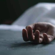
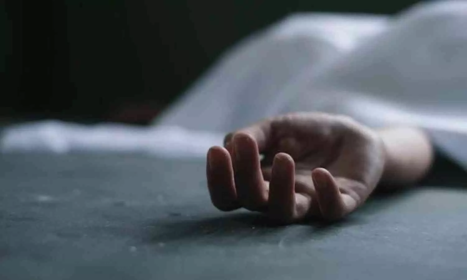
Kolkata: Junior doctors in West Bengal have initiated a hunger strike, protesting the brutal rape and murder of the postgraduate trainee doctor at RG Kar Medical College Hospital. The fast began on Saturday evening, following the state government’s failure to address their demands within a stipulated deadline.
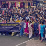
With three days left before the Durga Puja festivities start, the doctors had on Friday begun a sit-in demonstration at the Dorina Crossing in Dharmatala in the heart of Kolkata, setting a 24-hour deadline for the state government to fulfil their demands.
According to the PTI report, a junior doctor said, “The state government has failed the deadline and hence we are starting the fast unto death, which will continue till our demands are fulfilled. To maintain transparency, we have installed CCTV cameras at the dais where our colleagues are holding the fast.”
Also Read:West Bengal: Doctors give 24 hour ultimatum to govt, threaten hunger strike
“We joined duty last night but will not eat anything,” he said, adding that at present six junior doctors were sitting on fast.
The six doctors who were sitting on the fast were identified as Snigdha Hazra, Tanaya Panja and Anustup Mukhopadhyay of Kolkata Medical College and Hospital, SSKM’s Arnab Mukhopadhyay, Pulastha Acharya of NRS Medical College and Hospital, and Sayantani Ghosh Hazra of KPC Medical College.
The state would be held responsible if any doctor fell ill during the fast, the junior doctor said.
“We have the support of the people, and that is the reason we are not scared of any sort of hindrances by the administration. We will continue our hunger strike until our demands are met,” he added, news agency PTI reported.
A large number of common people and a few celebrities were present at the protest site in the evening.
The junior doctors had on Friday night called off their ‘total cease work’, which had crippled healthcare services at state-run medical colleges and hospitals.
Earlier in the day, the medics alleged the police were not allowing them to set up the dais.
Kolkata Police had denied the junior doctors’ request for permission for the sit-in, stating that the road witnesses heavy traffic flow.
The protesting doctors had also alleged that they were baton-charged by the police on Friday night.
Promising “necessary action”, Kolkata Police in an e-mail asked them to identify the police personnel involved and lodge a complaint.
“With reference to the allegation of physical assault, the matter is being enquired. However, you are requested to direct the doctor/person who has allegedly been assaulted to lodge a formal complaint at the concerned Police Station, necessary action will be taken in accordance with the law,” the mail said.
The protesters emphasised that securing justice for the deceased woman medic remains their foremost priority.
Among the other nine demands, they called for the immediate removal of Health Secretary NS Nigam, as well as accountability for the alleged administrative incompetence and corruption within the Health Department, adds PTI.
Other demands include the establishment of a centralised referral system for all hospitals and medical colleges in the state, the implementation of a bed vacancy monitoring system, and the formation of task forces to ensure essential provisions for CCTV, on-call rooms, and washrooms at their workplaces.
Furthermore, they are advocating increased police protection in hospitals, the recruitment of permanent women police personnel, and the swift filling of vacant positions for doctors, nurses, and other healthcare workers.
The junior doctors began their strike in response to the tragic incident that occurred on August 9, when a fellow medic was raped and murdered. They temporarily halted their protests on September 21 after receiving assurances from the state government regarding their concerns.
Powered by WPeMatico
Shahjahanpur: In a shocking incident, a 24-year-old second year medical student of Varun Arjun Medical College was found dead lying in a pool of blood behind the hostel on the campus under suspicious circumstances on Sunday morning.
The police are investigating the possibility of suicide or criminal activity, given that the student was found in suspicious circumstances. Superintendent of Police Rajesh S said that Prima facie it appears that either he fell on his own or someone pushed him.
Also read- Motilal Nehru Medical College MS orthopaedics medico allegedly commits suicide, 3 doctors booked
The student was a native of Gorakhpur who was living on the ground floor of the hostel which is a three-storey building. It is unclear how the incident had taken place and the police are investigating the matter.
“He was a second-year MBBS student at the college. He is a resident of Gorakhpur. Today his body was found lying behind the hostel, after which the police were informed,” the college principal, Colonel (retired) Dr Ravindra Nath Shukla, told PTI.
When the police arrived at the campus, they conducted preliminary investigation and sent his body for post-mortem. As part of the investigation, the police have sealed the student’s room and are reviewing CCTV footage from the area.
To gather more details about the matter, the police are questioning fellow students and local residents. “We are doing everything we can to get to the bottom of this,” SP Yash told India today.
Shocked over the sudden mysterious death of the student, one of the student told the daily, “This is terrifying. We don’t know what happened, but it’s very unsettling to think that something like this could happen right here.”
Also read- MGM Indore MBBS student commits suicide, parents allege harassment by roommates
Powered by WPeMatico
Researchers reported that high serum 25-hydroxyvitamin D [25 (OH)D] levels significantly reduced the risk of anemia, thereby suggesting that maintaining the level of this type of vitamin D high may have a protective role against the blood disorder. This conclusion comes from a new analysis that confirms the possibility of regulating 25(OH)D levels through supplementation or lifestyle changes as a therapeutic strategy to reduce anemia, especially among high-risk populations. A new study was recently published in the International Journal of General Medicine by Bi S. and colleagues.
Anemia is a leading public health issue across the entire globe, with a universal prevalence in all regions, by age group, and among all demographic groups. According to the WHO, there are about 1.6 billion anemic individuals worldwide-thus it poses as a critical health issue. This is a recent research that was concerned with placing 25(OH)D as an inhibitor in the course of developing anemia among patients. Various researchers have discovered a significant role for reducing inflammation and hepcidin inhibition, which orchestrates the iron metabolism; therefore, the mechanisms by which 25(OH)D may affect anemia are still not well understood.
This study, carried out by Chunyan Wang’s group at The Second Hospital, Cheelo College of Medicine, Shandong University, aimed to determine the causal relationship between 25(OH)D levels and anemia risk through cross-sectional and two-sample Mendelian randomization. Such studies could contribute to furthering our understanding of the protective properties of 25(OH)D and the possible impact of 25(OH)D on preventing anemia.
Cross-sectional recruitment took place from the local database of 7,160 inpatients in China for the period January 2021 to June 2023. The study population is predominantly female with a mean age of 56.5 years. The percentage of anemia within this cohort of patients was at 24.3%. Patients have been stratified into groups based on serum 25(OH)D concentration for better understanding the correlation between vitamin D levels and anemia within the patient population:
• Adequate ≥75 nmol/L
• Insufficient 50–75 nmol/L
• 25–50 nmol/L: deficient
• ≤25 nmol/L: severe deficiency
The researchers performed a multivariable logistic regression to adjust for demographic factors, comorbid conditions, and results from laboratory investigations with an aim to probe the association of 25(OH)D levels with risk of anemia. Additionally, to establish a possible causal relationship of the 25(OH)D level and incidence of anemia, two-sample bidirectional Mendelian randomization analysis was conducted.
The main outcome of the study was a strong inverse association between serum 25(OH)D concentrations and anemia risk:
For every one-unit increase in serum concentrations of 25(OH)D, the anemia risk decreased by 5% (odds ratio [OR], 0.95; 95% confidence interval [CI], 0.94–0.97; P <.001).
The prevalence of anemia in the study population was 24.3%.
There was a nonlinear dose-response relationship identified with the 25(OH)D concentration threshold below which the risk for anemia increased significantly at 48.716 nmol/L .
Mendelian randomization supported the protective causal relationship between higher levels of 25(OH)D and a 5% decrease in the risk of anemia for a standard deviation increase in the concentration of 25(OH)D.
Serum 25-hydroxyvitamin D [25(OH)D] levels had been lowered as an independent factor in the risk of developing anemia. This may open novel lines of thought into the pathogenesis of the condition. Wang et al’s research study adds one among many others while documenting the numerous studies supporting the protective role of vitamin D against the development of anemia. In this high-risk population, regulating levels of 25(OH)D through supplementation or lifestyle might be a useful approach to reducing anemia.
Reference:
Bi S, Zhang J, Wei N, Zhou Q, Wang C. Association Between Serum 25-Hydroxyvitamin D Level and Risk of Anemia: An Observational and Mendelian Randomization Study. Int J Gen Med. 2024;17:3893-3905. Published 2024 Sep 6. doi:10.2147/IJGM.S479039
Powered by WPeMatico
USA: The ADMIRE pivotal trial has provided significant insights into using pulsed-field ablation (PFA) for treating paroxysmal atrial fibrillation, demonstrating safety and effectiveness. This innovative approach utilizes a variable-loop PFA catheter, which facilitates shorter procedure times and reduces exposure to fluoroscopy.
“The ADMIRE trial confirmed that the variable-loop PFA catheter is both safe and effective, demonstrating short procedure and PFA application times, along with low levels of fluoroscopy exposure,” the researchers wrote in Circulation.
Atrial fibrillation (AF) is a common cardiac arrhythmia that can lead to severe complications, including stroke and heart failure. Paroxysmal atrial fibrillation, characterized by intermittent episodes, is particularly challenging to manage. Traditional ablation methods have their drawbacks, including longer recovery times and higher radiation exposure for patients and medical staff.
Clinical trials of early PFA systems for treating atrial fibrillation have shown promising potential to minimize complications associated with traditional thermal methods while preserving efficacy. However, the absence of a fully integrated mapping system—commonly used in contemporary electrophysiology procedures—limits lesion creation and workflow efficiency. To address this, a novel variable-loop PFA catheter has been developed, which integrates with an electroanatomic mapping system. This advancement enables real-time nonfluoroscopic procedural guidance, lesion indexing, and feedback on the proximity between tissue and catheter.
In light of this context, Vivek Y. Reddy and colleagues from the Helmsley Electrophysiology Center at Mount Sinai Fuster Heart Hospital in New York, NY, conducted the ADMIRE trial. This multicenter, single-arm study, which received an investigational device exemption from the Food and Drug Administration, evaluated the long-term safety and effectiveness of the integrated pulsed field ablation (PFA) system in a large population of drug-refractory patients with symptomatic paroxysmal atrial fibrillation in the United States.
Using the PFA catheter in conjunction with a compatible electroanatomic mapping system, patients with drug-refractory symptomatic paroxysmal atrial fibrillation underwent pulmonary vein isolation.
The primary safety endpoint was the occurrence of major adverse events within seven days post-ablation. The primary effectiveness endpoint was a composite measure that included 12 months of freedom from documented episodes of atrial tachyarrhythmia (such as atrial fibrillation, atrial tachycardia, and atrial flutter), failure to achieve pulmonary vein isolation, use of a non-study catheter for isolation, repeat procedures (excluding one redo during the blanking period), initiation of a new or previously ineffective class I or III antiarrhythmic drug at a higher dose after the blanking period, or the need for direct current cardioversion after the blanking phase.
Based on the study, the researchers reported the following findings:
In conclusion, the ADMIRE pivotal trial confirms that pulsed field ablation with the variable-loop catheter is a promising treatment option for patients with paroxysmal atrial fibrillation.
“With shorter procedure times and reduced fluoroscopy exposure, this innovative technique could significantly improve patient outcomes and quality of life for those affected by this common cardiac condition,” the researchers wrote.
Reference:
Reddy VY, Calkins H, Mansour M, et al. Pulsed-field ablation to treat paroxysmal atrial fibrillation: safety and effectiveness in the ADMIRE pivotal trial. Circulation. 2024; Epub ahead of print.
Powered by WPeMatico
A new study published in the European Heart Journal showed that semaglutide is a helpful additional medication for the individuals who are obese and have high blood pressure.
Even with the availability of efficient medicines, meeting blood pressure objectives is difficult and poses a serious threat to world health. There are several ways in which obesity and hypertension interact. The processes most likely include changes in how the body handles salt, an inflammatory environment, and activation of the sympathetic nervous system. Treatments for hypertension now available focus on pathways that interact with the ones impacted by being overweight. But addressing the obesity illness could be a useful tactic to manage blood pressure, especially for the ones with resistant hypertension (RH). Thereby, determining the impact of semaglutide therapy on systolic BP (SBP) among individuals with hypertension, SBP in individuals with RH, SBP while baseline body mass index (BMI) is taken into account, and modifications to anti-hypertensive drugs were among the objectives of this study.
Individual patient data (IPD) from 3 RCTs that looked at how semaglutide 2.4 mg affected body weight over a 68-week period was included. The trial participants were divided into 3 groups based on the hypertension diagnosis, where the one group had apparent resistant hypertension (RH), other group with baseline SBP > 130 mmHg (HTN130) or > 140 mmHg (HTN140) and the last group was treatment or baseline measurement (HTN). The main comparison was between the semaglutide and placebo groups’ in-trial changes in SBP. Treatment intensity score was used to quantify and compare changes in antihypertensive medication across groups. Analysis of covariance was used in these analyses.
The study found that there was a difference in SBP change of −4.95 mmHg between the treatment (n = 2109) and placebo (n = 1027) groups. The HTN, HTN130, HTN140, and RH had the largest differences, with −4.78 mmHg (95% CI −5.97 to −3.59), −4.93 mmHg, and −4.09 mmHg for HTN140, respectively. Weight loss was a major mediating factor in the reduction in SBP. The anti-hypertensive therapy intensity score dropped for semaglutide users when compared to placebo. Overall, the outcome of this analysis showed a drop in SBP that was equivalent in persons with normotension and hypertension. The weight loss impact of semaglutide acted as a mediator for the decrease in SBP.
Reference:
Kennedy, C., Hayes, P., Cicero, A. F. G., Dobner, S., Le Roux, C. W., McEvoy, J. W., Zgaga, L., & Hennessy, M. (2024). Semaglutide and blood pressure: an individual patient data meta-analysis. In European Heart Journal. Oxford University Press (OUP). https://doi.org/10.1093/eurheartj/ehae564
Powered by WPeMatico
China: A recent nationwide study has revealed a compelling link between iron status and insulin resistance (IR) among adults in the United States. The research, which examined a large and diverse sample of the adult population, highlights a strong correlation suggesting that variations in iron levels may significantly influence insulin resistance—a key factor in the development of type 2 diabetes.
The study, published in The Journal of Clinical Endocrinology & Metabolism, revealed that higher serum iron (SI) levels might actually protect against insulin resistance.
Among a cohort of 2,900 adults, researchers discovered that elevated serum iron levels were associated with reduced insulin resistance. In contrast, higher iron intake and increased serum transferrin receptor (sTfR) levels were correlated with greater IR. Transferrin saturation was identified as the most reliable predictor of IR, surpassing other iron-related indicators in its predictive accuracy.
Evidence connecting markers of iron status to insulin resistance remains sparse. To fill this knowledge gap, Haiqing Zhang, Shandong University, Jinan, Shandong, China, and colleagues aimed to investigate the relationship between iron status and IR among U.S. adults.
For this purpose, the researchers analyzed data from 2,993 participants of the National Health and Nutrition Examination Survey (NHANES) spanning the periods 2003-2006 and 2017-2020. A HOMA-IR value of ≥2.5 defines insulin resistance.
The team employed weighted linear and multivariable logistic regression analyses to explore the linear relationships between iron status and IR. They utilized restricted cubic splines (RCS) to uncover potential non-linear dose-response associations. Additionally, stratified analyses were conducted based on age, sex, body mass index (BMI), and physical activity (PA). Finally, receiver operating characteristic (ROC) curve analysis assessed the predictive value of iron status for IR.
The following were the key findings of the study:
“All biomarkers showed a significantly reduced risk of insulin resistance as iron levels increased. These findings enhance our understanding of the relationship between iron status and IR, offering a robust foundation for further investigation into the mechanisms driving this connection,” the researchers concluded.
Reference:
Liu, X., Zhang, Y., Chai, Y., Li, Y., Yuan, J., Zhang, L., & Zhang, H. Iron status correlates strongly to Insulin Resistance among U.S. Adults: A nationwide population-base study. The Journal of Clinical Endocrinology & Metabolism. https://doi.org/10.1210/clinem/dgae558
Powered by WPeMatico
Norway: Recent case reports in the Journal of Endodontics have highlighted the rare but serious risk of anaphylaxis following treatment with endodontic materials containing polyethylene glycol (PEG). Two patients experienced severe allergic reactions after dental procedures involving these materials, raising important concerns within the dental community.
“While rare, clinicians should be aware that PEG in endodontic filling materials can trigger anaphylaxis. It is important to conduct a PEG allergy assessment when anaphylaxis is suspected,” the researchers wrote.
Anaphylaxis is a life-threatening allergic reaction that can occur rapidly, making early recognition and treatment critical. While anaphylactic reactions to PEG are uncommon, they are likely underreported and can lead to severe consequences for patients, especially those who remain undiagnosed. These cases emphasize the need for heightened awareness among dental professionals regarding the potential for PEG to trigger such reactions.
Marie Alnæs, Department of Clinical Medicine, University of Bergen, Bergen, Norway, and colleagues report two cases of anaphylaxis following the use of an endodontic temporary filling material that contains polyethylene glycol.
The first case involved a healthy man in his 30s who developed generalized itching, nausea, and cold sweats after undergoing endodontic treatment. He subsequently lost consciousness, with a blood pressure reading of 70/40 mm Hg when assessed in the ambulance. Fortunately, he recovered after receiving emergency medical care. The second case involved a patient around 50 years old, who also experienced skin itching following treatment. Tragically, this patient lost consciousness and suffered cardiac arrest upon arrival at the hospital, leading to his death.
The authors emphasize that these case reports highlight the need for increased awareness regarding the potential for endodontic materials containing polyethylene glycol (PEG) to cause anaphylaxis. They recommend that PEG allergy assessments be included in evaluations of patients experiencing anaphylaxis related to endodontic treatment. Confirmed PEG allergies can have significant implications for patients, particularly those who remain undiagnosed, as they face the risk of further anaphylactic reactions upon re-exposure.
Given the associated risks, the authors advise against using endodontic filling materials containing PEG, suggesting that alternative materials without PEG should be preferred. In cases where the benefits of using PEG-containing materials outweigh the risks, they recommend a 30-minute observation period post-treatment. Furthermore, they advocate for clear labeling in the instructions, explicitly stating that PEG in endodontic materials can trigger anaphylactic reactions.
“Thorough investigations were required to identify the source of the allergic reactions. Reports of allergies to polyethylene glycol are on the rise and can have serious consequences for patients,” the researchers concluded.
Reference:
Alnæs, M., Storaas, T., Vindenes, H. K., Guttormsen, A. B., Brudevoll, S., & Björkman, L. (2024). Anaphylaxis after treatment with an endodontic material containing polyethylene glycol. Journal of Endodontics. https://doi.org/10.1016/j.joen.2024.09.002
Powered by WPeMatico
USA: A recent multicenter, retrospective, single-arm observational study has unveiled promising results for the use of droxidopa in critically ill patients experiencing persistent hypotension.
The study, published in the Journal of Intensive Care Medicine, suggested that droxidopa may be a valuable adjunct therapy for facilitating vasopressor weaning, with comparable effects observed in patients receiving the medication through feeding tubes.
Persistent hypotension is a significant challenge in the management of critically ill patients, often leading to prolonged reliance on vasopressor agents. These medications can have adverse effects and complications, underscoring the need for alternative strategies to facilitate safe withdrawal.
According to the study authors, persistent reliance on vasopressors is a frequent factor contributing to delays in patient discharge from the intensive care unit (ICU). They note that adjunct oral agents can accelerate the timeline for discontinuing vasopressor treatment. Considering this, Andrew J Webb, Department of Pharmacy, Massachusetts General Hospital, Boston, USA, and colleagues sought to examine the application of droxidopa for facilitating vasopressor weaning in critically ill patients experiencing prolonged hypotension.
For this purpose, this retrospective, single-arm observational study focused on adult patients admitted to ICUs at two academic centers between June 2016 and July 2023 who received droxidopa for vasopressor weaning. Patients who had received droxidopa before admission or for other indications were excluded from the analysis.
The primary outcome measured was the time to vasopressor discontinuation, defined as the duration for which vasopressors were stopped and remained off for at least 24 hours. Secondary outcomes included the rates of tachycardia and hypotension following the initiation of droxidopa, norepinephrine equivalents before and after treatment, concomitant use of oral agents, and dosing details. Additionally, a subgroup analysis was performed on patients receiving droxidopa via feeding tubes.
The following were the key findings of the study:
“The findings indicate that droxidopa could serve as an effective adjunct therapy for vasopressor weaning in critically ill patients. Further prospective studies are needed to assess this intervention comprehensively,” the researchers concluded.
Reference:
Webb, A. J., Casal, G. L., Newman, K. A., Culshaw, J. R., Northam, K. A., Solomon, E. J., Beargie, S. M., Johnson, R. B., Lopez, N. D., Hayes, B. D., & Roberts, R. J. (2024). Droxidopa for Vasopressor Weaning in Critically Ill Patients with Persistent Hypotension: A Multicenter, Retrospective, Single-Arm Observational Study. Journal of Intensive Care Medicine. https://doi.org/10.1177/08850666241270089
Powered by WPeMatico
Researchers in a study found that moisture on surgical wounds in the chosen 24-h postoperative period was not associated with an increase in the incidence of infection or other wound complications. It thereby rejects the conventional doctrine, which is the dry and undisturbed principle of the dressings in the time frame 24-72 hours post-surgery, unless there is a heightened possibility of moisture associated with most daily activities. Christen S. and colleagues reported it in the Journal of the American Academy of Dermatology.
Many standard patient instructions after surgical wound care include keeping the initial dressings clean and dry and free of contamination, and maintained for 24 to 72 hours to avoid the development of infection and to promote healing of the wound, which is in sharp contrast to the energetic levels required to meet daily activities like bathing, leisure, and optimum levels of functioning. The randomized controlled trial comparatively assessed standard keeping of wounds dry and covered for the period of 48 hours to early post-operative (6 hours) water exposure.
This was a randomized controlled, investigator-blinded, comparative study to assess infection rates and other outcomes. In total, 437 patients were randomized to early water exposure (6 hours post-operatively, n = 218) and standard care (48 hours post-operatively, n = 219). Primary outcome: Culture-proven infection. Other outcomes that were measured: bleeding rate, bruising rate, and scar assessment according to the Patient and Observer Scar Assessment Scale.
The key findings of the study were:
• The incidence of culture-proven infection was comparable between the early water exposure group, (1.8%), and the standard care group, (1.4%); this difference was not significant with (p > 0.99).
• There were no differences between groups in rates of bleeding or bruising.
• The scar results, recorded with the POSAS, did not reveal any significant differences between both groups, thus showing similar cosmetic results.
The study has revealed that allowing surgical wounds to get wet as early as six hours after surgery does not increase the rate of complications or infections. Hence, this increases flexibility in the post-operative care routine for patients, hence helping them to return back to normal activities earlier than thought.
This study was conducted in a single academic center. Generalizability of the results is therefore limited, and further studies are required in different clinical settings for the confirmation of these findings.
Surgical wounds can be safely exposed to water immediately after the operation without increasing the risk of infections or compromising the final cosmetic outcome. Evidence suggesting that post-operative care recommendations should be revised to allow greater patient convenience and comfort.
Reference:
Samaan, C., Kim, Y., Zhou, S., Kirby, J. S., & Cartee, T. V. (2024). Early post-operative water exposure does not increase complications in cutaneous surgeries: A randomized, investigator-blinded, controlled trial. Journal of the American Academy of Dermatology. https://doi.org/10.1016/j.jaad.2024.05.098
Powered by WPeMatico