Poor sleep quality raises risk of delirium after surgery, study finds

People who experience poor sleep in the month before surgery may be more likely to develop postoperative delirium, according to new research being presented at the ANESTHESIOLOGY® 2024 annual meeting.

Postoperative delirium is a change in mental function that can cause confusion and occurs in up to 15% of surgical patients. In certain high-risk patients, such as those with hip fractures, the incidence can be even higher. It is a significant complication in older adults. Pain, age, stress, anxiety and insomnia are known to contribute to the risk for postoperative delirium. The researchers believe this study is the first to assess sleep quality before surgery as it relates to postoperative delirium.

“Good sleep quality before surgery is crucial for the recovery period,” said Faegheh Miryousefiata, M.D., MPH, lead author of the study and clinical researcher at the Cleveland Clinic Foundation Outcomes Research Consortium. “People may not get good sleep because they are nervous right before surgery, but it is really important to get the best sleep possible, especially for those over 65, who are at greater risk for postoperative delirium.”

The study included 150 patients having general anesthesia for non-cardiac surgeries, mostly abdominal and gynecologic procedures, of more than two hours. The day before the surgery, the researchers had the patients assess their sleep quality using the Pittsburgh Sleep Quality Index (PSQI), which evaluates sleep duration, disturbance, efficiency and overall quality, length of time to fall asleep, daytime dysfunction due to sleepiness and use of sleep medication. Scores for each category range from 0 (no difficulty) to 3 (severe difficulty), with total scores ranging from 0 to 21. A score of 5 or above indicated poor sleep. After surgery, they assessed postoperative delirium using the Intensive Care Unit Confusion Assessment Method (ICU-CAM) and the Three-minute Diagnostic Confusion Assessment Method (3D-CAM). They checked patients’ hospital records for three days after surgery for postoperative delirium.

Researchers determined:

  • 44 patients had poor sleep and 11 of those (25%) experienced delirium
  • 106 patients had good sleep and 19 of those (17.9%) experienced delirium
  • When comparing all patients who experienced delirium, there was a 39.66% higher rate of delirium among those who had poor sleep, compared to those who had good sleep

“Doctors should make it their mission to let patients know it’s important to get good sleep before surgery to protect their brains, as well as provide some tips,” said Yasin Tire, M.D., senior author of the study and an anesthesiologist at Konya City Hospital, University of Health Science, Konya, Turkey.

To improve sleep quality in general, as well as before surgery, the researchers recommend:

  • Going to bed and waking up at the same time every day
  • Creating a bedtime ritual, such as reading a book, taking a warm bath or practicing relaxation techniques
  • Keeping the bedroom cool, dark and quiet
  • Avoiding phones, tablets and computers at least an hour before bed
  • Staying active during the day, but not exercising too close to bedtime
  • Using stress-management techniques such as mindfulness or deep breathing

Reference:

Poor sleep quality raises the risk of delirium after surgery, study finds, American Society of Anesthesiologists, Meeting: Anesthesiology 2024.

Powered by WPeMatico

Clonidine effective in treating newborn opioid withdrawal syndrome: AAP

A new study published in the journal of Pediatrics showed that there was no significant difference in the duration of pharmacologic therapy or the final neurobehavioral performance between the groups treated with morphine and clonidine. Overdose deaths, maternal deaths, and newborn abstinence syndrome (NAS), which has both immediate and long-term effects, have all increased as a result of the opioid pandemic in the US. Misuse of opioids or their prescription usage for chronic pain management or treatment for opioid use disorder (MOUD) can expose a fetus.

The observed deleterious consequences of in utero opioid exposure on the developing brain include neonatal stroke, hydrocephaly, decreased brain cell number, small head size, postnatal head development retardation, lower brain sizes, and related long-term cognitive deficiencies and behavioral disorders. To ascertain if clonidine which is a non-opioid α−2-adrenergic agonist would be an effective treatment for newborn opioid withdrawal syndrome (NOWS), Henrietta Bada and her colleagues carried out this investigation.

Prenatal opioid exposure, gestational age ≥35 weeks, absence of any other medical conditions, and need for medication were among the requirements for enrolment in this randomized clinical study. Neurobehavioral performance and treatment duration were the main outcomes measured.

In all, 1107 patients underwent enrolment screening (645 were deemed ineligible, 91 parents or staff were unavailable, 216 rejected, and 155 gave their approval). Out of the 155 neonates, 120 needed therapy and were randomly assigned to receive either 0.06 mg/kg/dose of morphine or 1 µg/kg/dose of oral clonidine every three hours.

Every 12 to 24 hours, the dosages for infants who showed no improvement were raised by 25%. Adjunctive treatment was administered to the ones who did not improve by the fourth dosage increment. With median treatment durations of 15 and 17, respectively, morphine and clonidine did not differ in length.

More babies treated with clonidine required adjunct treatment (adjusted odds ratio = 8.85) when compared to 10% in the morphine group. Following therapy, there was no difference in the NICU Network Neurobehavioral Scales summary scores between babies treated with clonidine and the babies treated with morphine.

Overall, the duration of pharmacologic therapy and the ultimate evaluation of neurobehavioral function did not differ substantially between the groups treated with clonidine and morphine in a randomized experiment. To increase effectiveness and reduce the need for supplementary treatment in NOWS, more research and dedicated studies are required to determine the ideal dosage and frequency of clonidine administration.

Reference:

Bada, H. S., Westgate, P. M., Sithisarn, T., Yolton, K., Charnigo, R., Pourcyrous, M., Tang, F., Gibson, J., Shearer-Miller, J., Giannone, P., & Leggas, M. (2024). Clonidine as Monotherapy for Neonatal Opioid Withdrawal Syndrome: A Randomized Trial. In Pediatrics. American Academy of Pediatrics (AAP). https://doi.org/10.1542/peds.2023-065610

Powered by WPeMatico

Patients taking muscle relaxants at elevated risk of COPD exacerbation, unravels research

A new study published in the Chest Journal found that individuals using methocarbamol or cyclobenzaprine were more likely to experience exacerbations of their chronic obstructive pulmonary disease (COPD). In accordance with recent data, using gabapentinoids (pregabalin and gabapentin) may increase the frequency and severity of exacerbations of chronic obstructive pulmonary disease. Therefore, Irakli Lemonjava and her colleagues set out to look into any possible links between exacerbations of COPD and regularly prescribed muscle relaxants like methocarbamol and cyclobenzaprine.

The patients over the age of 18 who were diagnosed with COPD after January 1, 2000 were included in the study. They were divided into two groups where Group A consisted of patients taking either methocarbamol or cyclobenzaprine, and Group B consisted of patients not taking either of these drugs. This study focused on COPD exacerbations that occurred 1, 3, and 5 years after the index event and were classified by the international classification of diseases code J44.1.

The first day that a patient met all the chosen criteria, such as receiving a diagnosis of COPD and starting a medicine of interest, was considered the index event for that patient. Analysis did not include patients whose results occurred prior to the study timeframe. In order to account for demographic factors and ten different organ system illnesses, including the ones affecting the cardiovascular and pulmonary systems, the study used propensity score matching. Following propensity score matching, there were 399,886 people in Group A and 429,743 people in Group B.

COPD exacerbation occurred in 21,922 (5.482%) of Group A patients at 1-year follow-up, while it occurred in 21,513 (5.006%) of Group B patients, representing a 9.5% relative risk increase. At 3- and 5-year follow-ups, the risk gradually rose to 17.6% and 20.2%, respectively. At the 1-, 3-, and 5-year follow-up periods, the relative risk increase for COPD exacerbations was 9.5%, 17.6%, and 20.2% for patients on cyclobenzaprine or methocarbamol, respectively.

The warning range for the use of central nervous system depressing medicines among patients with COPD is expanded by the possibility of an increased risk of exacerbation linked to the use of routinely prescribed muscle relaxant pharmaceuticals. Thereby, physicians should weigh the possible danger of exacerbating COPD against the purported therapeutic advantages of these muscle relaxants. Alternative forms of therapy have to be taken into account.

Reference:

LEMONJAVA, I., GUDUSHAURI, N., MARTINEZ MANZANO, J. M., & AZMAIPARASHVILI, Z. (2024). EXPLORING THE ASSOCIATION BETWEEN ELEVATED COPD EXACERBATION RISK AND COMMONLY PRESCRIBED MUSCLE RELAXANTS. In CHEST (Vol. 166, Issue 4, p. A4691). Elsevier BV. https://doi.org/10.1016/j.chest.2024.06.2827

Powered by WPeMatico

Higher doses of buprenorphine may improve treatment outcomes for people with opioid use disorder: Study

Adults with opioid use disorder who receive a higher daily dose of the opioid addiction treatment medication buprenorphine may have a lower risk of subsequent emergency department visits or use of inpatient services related to behavioral health (such as for mental health and substance use disorders) than adults receiving the recommended dose, according to an analysis funded by the National Institutes of Health (NIH). These findings suggest that higher buprenorphine doses could be more effective in managing opioid use disorder, which may be particularly relevant for improving treatment for those who use fentanyl, a major driver of the overdose crisis.

Researchers reviewed insurance claims data from over 35,000 people who were diagnosed with opioid use disorder and began buprenorphine treatment between 2016 and 2021. They found that among all people who started treatment with buprenorphine, 12.5% experienced an emergency department or inpatient visit related to behavioral health within the study period. After adjusting for patient demographics and medical history available in the data, researchers then analyzed how long it took for people receiving different doses of buprenorphine to use emergency care or have an inpatient stay after starting treatment.

The recommended target dose for buprenorphine in the U.S. Food and Drug Administration (FDA)’s approved labeling is 16 mg per day. Researchers found that those taking higher daily doses of buprenorphine (>16 to 24 mg) took 20% longer to have a subsequent emergency department or inpatient health care visit related to behavioral health within the first year after receiving treatment, compared to those receiving >8 to 16 mg a day. Those taking daily doses of more than 24 mg of buprenorphine went 50% longer before having a subsequent emergency or inpatient health care visit related to behavioral health within the first year after receiving treatment, compared to those receiving >8 to 16 mg a day.

“As the overdose crisis evolves, particularly with the rise of fentanyl, it is crucial to investigate how to best adapt and deliver the life-saving and evidence-based treatments for opioid use disorder that we have available,” said Nora D. Volkow, M.D., director of NIH’s National Institute on Drug Abuse (NIDA). “The findings add to the growing evidence that higher doses of buprenorphine may have meaningful health impacts for people with opioid use disorder.”

In 2022, of the nearly 108,000 overdose deaths reported in the U.S., almost 70% were primarily due to fentanyl, a synthetic opioid that is approximately 50 times stronger than heroin. The ubiquity of fentanyl in the drug supply and associated overdose deaths have raised questions about whether existing dosing guidelines for buprenorphine should be modified to better address the unique challenges posed by such a potent opioid. Higher doses of buprenorphine may be necessary to effectively manage the more severe withdrawal symptoms, cravings, and tolerance associated with fentanyl use.

“Preventing or delaying the need for high-intensity, urgent health care among people with opioid use disorder has tremendous benefits on health and recovery,” said Bradley D. Stein, M.D., Ph.D., a study co-author and director of the RAND-USC Schaeffer Opioid Policy Tools and Information Center. “As we continue to gather data across studies, findings suggest that higher doses of buprenorphine may have the ability to significantly improve treatment in the era of fentanyl, as both fatal and nonfatal overdoses remain unacceptably high.”

The authors also note that addressing barriers to accessing higher doses, such as state laws and insurance policies, will be important in ensuring that all patients receive effective care. In addition, revisiting guidelines that serve as barriers to higher doses could be beneficial, as these may limit access to potentially life-saving treatment for patients at high risk of relapse or overdose.

Notably, the data used in the study were pulled from a single commercial insurance company, which does not include uninsured people and those with Medicaid or fee-for-service Medicare coverage, and the sample of people included in the analysis was 75% non-Hispanic white. Further research is needed to explore the effects of higher buprenorphine doses in more diverse populations, including those with different insurance statuses or in different clinical settings. In addition, the authors note that future research should also investigate additional outcomes such as the long-term effects of high-dose buprenorphine on opioid use disorder treatment retention and overall health outcomes using similar data.

These findings build upon accumulating evidence of the safety and efficacy of higher doses of buprenorphine. Studies have shown that more than 16 mg of buprenorphine is safe and well tolerated in people with opioid use disorder in emergency department and outpatient treatment settings, and that higher buprenorphine doses are associated with improved retention in treatment for opioid use disorder.

Reference:

Axeen S, Pacula RL, Merlin JS, Gordon AJ, Stein BD. Association of Daily Doses of Buprenorphine With Urgent Health Care Utilization. JAMA Netw Open. 2024;7(9):e2435478. doi:10.1001/jamanetworkopen.2024.35478

Powered by WPeMatico

Repeated low-level red light exposure could arrest progression of myopia, reports study

A new study published in the American Journal of Ophthalmology showed that repeated low-level red light treatment (RLRL) successfully delayed the progression of myopia in children suffering from severe myopia (–6.00 D or worse). The frequency of myopia has sharply grown in recent decades, particularly in East Asian nations. A significantly elongated eye and alterations in other ocular components that may raise the risk of complications like retinal detachment, scleral thinning, glaucoma, and localized posterior ectasia of the sclera are characteristics of high myopia, which is defined as myopic refractive error greater than -6.00 D. An other method for controlling and preventing myopia is RLRL therapy, which emits visible red light at 650 nm.

From March 2022 to December 2022, 292 high myopia children (6 to 12 years old) with cycloplegia spherical equivalent refraction (SE) ≤ -6.00 D, astigmatism < 2.50 D, and anisometropia < 1.50 D were recruited. December 2023 saw the completion of the follow-up. The control group (SVS) and the intervention were assigned at random to the eligible individuals. The RLRL therapy was given twice daily for 3 minutes each, separated by at least 4 hours. The change in axial length from baseline to 12 months was the main result. Changes in SE, choroidal thickness (ChT), and retinal thickness (RT) in various circular sectors were examples of secondary outcomes.

Following a 12-month course of therapy, the RLRL group saw AL and SE changes of -0.11 ± 0.25 mm and 0.18 ± 0.63 D, while the control group experienced changes of 0.32 ± 0.09 mm and -0.80 ± 0.42 D. At 12 months, axial shortening > 0.05 mm was found to be 59% in the RLRL and 0% in the control group. The ChT and RT from a single center were examined, and at 12 months, ChT thickened in every area in the RLRL group. In the parafoveal and perifoveal circles, RT was elevated. At 12 months, all sectors of ChT and only perifoveal RT were noticeably thinner in the control group.

Significant relationships between changes in the ChT central foveal circle and RT perifoveal circle at one month and AL changes at 12 months were found using the multivariate linear regression model. Overall, RLRL might successfully shorten the AL and slow the evolution of myopia in patients with extreme myopia.

Source:

Liu, G., Liu, L., Rong, H., Li, L., Liu, X., Jia, Z., Zhang, H., Wang, B., Song, D., Hu, J., Shi, X., Du, B., & Wei, R. (2024). Axial Shortening Effects of Repeated Low-level Red-light Therapy in Children with High Myopia: A Multicenter Randomized Controlled Trial. In American Journal of Ophthalmology. Elsevier BV. https://doi.org/10.1016/j.ajo.2024.10.011

Powered by WPeMatico

Riboflavin intake potential beneficial role in CKD patients, indicates study

Riboflavin intake potentially benefits CKD patients, indicates a study published in Medicine.

Current studies have not clarified the relationship between riboflavin intake and all-cause mortality in patients with chronic kidney disease (CKD). This study aims to investigate whether there is an association between riboflavin intake and the risk of all-cause mortality in patients with CKD. This was a retrospective cohort study with data extracted from the National Health and Nutrition Examination Survey (NHANES). The study used Cox regression analysis to calculate hazard ratio (HR) and 95% confidence interval (CI) to assess the association between riboflavin intake and risk of all-cause mortality. Subgroup analyses were performed regarding gender, CKD stage, hypertension, hyperlipidemia and cardiovascular disease (CVD). A total of 3750 patients were ultimately included in the analyses. After excluding potential confounders, lower riboflavin intake was associated with a higher risk of all-cause mortality (Q1: HR = 1.33, 95% CI: 1.05–1.69). A similar association was also found in patients at mild/moderate stage (HR = 1.32, 95% CI: 1.05–1.66), in females (HR = 1.35, 95% CI: 1.01–1.81), with hypertension (HR = 1.37, 95% CI: 1.07–1.75), CVD (HR = 1.48, 95% CI: 1.08–2.03), and dyslipidemia (HR = 1.29, 95% CI: 1.01–1.66). This study found an association between low riboflavin intake and a high risk of all-cause mortality, indicating a potential beneficial role of riboflavin in CKD patients.

Reference:

Ren, Xiaoxu MDa,b; Wang, Ronga; Liu, Fen MDb; Wang, Quanzhen MDb; Chen, Hairong MDb; Hou, Yunfeng MDb; Yu, Lifeng MDb; Liu, Xiangchun MDc; Jiang, Zhimingb,*. Association between riboflavin intake and the risk of all-cause mortality of patients with chronic kidney disease: A retrospective cohort study. Medicine 103(39):p e39417, September 27, 2024. | DOI: 10.1097/MD.0000000000039417

Keywords:

Riboflavin, intake, potential, beneficial, role, CKD, patients, indicates, study, medicine, Ren, Xiaoxu MDa,b; Wang, Ronga; Liu, Fen MDb; Wang, Quanzhen MDb; Chen, Hairong MDb; Hou, Yunfeng MDb; Yu, Lifeng MDb; Liu, Xiangchun MDc; Jiang, Zhimingb

Powered by WPeMatico

Global Trends suggest significant changes in maternal and fetal health in recent decades increasing birthweights over time

Recent presented research paper, authored by Giulia Bonanni, Chiara Airoldi, and Vincenzo Berghella, aims to assess global trends in mean birthweights at term as reported in peer-reviewed literature. The study conducted a systematic review of literature on this subject and included data from over 183 million births worldwide. The authors mention that there have been significant changes in maternal and fetal health in recent decades, influencing birthweight dynamics. While genetic factors and in utero influences are primary contributors to birthweight, additional predictors like age, parity, and maternal anthropometry have been identified. The study used electronic searches in PubMed, Embase, and Web of Science without any language or geographic restrictions, including ecological and observational studies reporting mean birthweight at term as a continuous numerical variable over time. The study retrieved 6447 articles and ultimately assessed 29 studies meeting the inclusion criteria. The majority of the selected studies were hospital-based, while 44.8% utilized national data, and a small proportion used municipality, community, or regional data. Geographically, North America had the highest representation, followed by Asia, Europe, South America, and Oceania. The univariate and multivariate linear models revealed a significant increase in mean birthweight at term over time. The regression model focusing on records from 1950 onward reported a robust annual increase in mean birthweight. Subgroup analyses were also performed to focus on national data sources and data collected from 1950 onward, both of which yielded significant relationships. The authors acknowledge limitations in the study, such as variations in study quality, the diversity of data sources, and sample size discrepancies. The study also mentions the need for future research to use precise gestational age distinctions and predetermined time frames to gain a deeper understanding of the trend in birthweight and its implications for maternal and child health. The authors also provide a detailed discussion of the implications of their findings, including the impact of changing birthweights on childbirth practices and the rising rate of cesarean delivery. They highlight the need for ongoing research to further understand this trend, address the study limitations, and provide a more nuanced perspective on the implications for maternal and child health.

Key Points

1. The research paper aims to assess global trends in mean birthweights at term by conducting a systematic review of literature on the subject, including data from over 183 million births worldwide. The authors note the significant changes in maternal and fetal health in recent decades and the influence on birthweight dynamics by genetic factors, in utero influences, and additional predictors like age, parity, and maternal anthropometry.

2. The study used electronic searches in PubMed, Embase, and Web of Science to retrieve 6447 articles and ultimately assessed 29 studies meeting inclusion criteria. These studies were primarily hospital-based, with 44.8% utilizing national data and a small proportion using municipality, community, or regional data. Geographically, North America had the highest representation, followed by Asia, Europe, South America, and Oceania.

3. The univariate and multivariate linear models revealed a significant increase in mean birthweight at term over time, with a robust annual increase reported from 1950 onward. Subgroup analyses focusing on national data sources and data collected from 1950 onward also yielded significant relationships.

4. The study acknowledges limitations such as variations in study quality, diversity of data sources, and sample size discrepancies, highlighting the need for future research to use precise gestational age distinctions and predetermined time frames to gain a better understanding of the trend in birthweight and its implications for maternal and child health.

5. The authors discuss the implications of their findings, including the impact of changing birthweights on childbirth practices and the rising rate of cesarean delivery, and stress the need for ongoing research to further understand this trend and its implications for maternal and child health. 6. In conclusion, the paper provides a comprehensive analysis of global trends in birthweights at term, emphasizing the need for further research to address study limitations and provide a more nuanced perspective on the implications for maternal and child health.

Conclusion

Overall, the study offers a comprehensive analysis of global trends in birthweights at term, exhibiting a robust approach to data analysis and a systematic review of the relevant literature. The study concludes by stressing the need for further research addressing the limitations highlighted in the study and providing a more nuanced perspective on the implications for maternal and child health.

Reference –

Bonanni G, Airoldi C, Berghella V. Birthweights at term have increased globally: insights from a systematic review of 183 million births. Am J Obstet Gynecol. 2024 Oct;231(4):395-407.e4. doi: 10.1016/j.ajog.2024.03.002. Epub 2024 Mar 7. PMID: 38460833.

Powered by WPeMatico

Preterm births on the rise with ongoing racial and economic disparities: JAMA

Preterm births have increased by more than 10 percent over the past decade, with racial and socioeconomic disparities persisting over time, according to a new study analyzing more than five million births.

The study, published in the journal JAMA Network Open also found that some factors that increase the risk for preterm birth-such as diabetes, sexually transmitted infections, and mental health conditions-became much more common over the past decade, while other factors that protect against preterm birth declined.

“Our findings not only show that preterm births are on the rise, but provide clues as to why this may be the case,” said Laura Jelliffe-Pawlowski, the study’s lead author, an epidemiologist and professor at the NYU Rory Meyers College of Nursing and professor emeritus of epidemiology, biostatistics, and of global health sciences in the University of California San Francisco (UCSF) School of Medicine. “Understanding patterns of and factors related to preterm birth is important for informing clinical care and the development of public health programs to address this critical need.”

Babies born preterm or prematurely-before the 37th week of pregnancy-are more likely to experience a range of short and longer-term problems, including a higher risk for illness, intellectual and emotional difficulty, and death. Certain factors are known to increase the risk of preterm birth, including mothers having high blood pressure, diabetes, an infection, or smoking. In addition, Black, Native American, and Hawaiian and Pacific Islander expectant mothers are at higher risk for preterm birth, which is thought to be driven by a long history of structural racism experienced by these groups.

To understand recent trends around preterm births, Jelliffe-Pawlowski and her colleagues looked at more than 5.4 million singleton births (not twins or other multiples) from 2011 to 2022 in the state of California. They examined how preterm birth rates changed over time and explored patterns in risk and protective factors within racial/ethnic and socioeconomic groups. Health insurance type was used as a proxy for socioeconomic status, comparing public insurance (MediCal, California’s Medicaid program) with nonpublic insurance (including private insurance and coverage through the military and the Indian Health Service).

A growing risk and “alarm bells”

The researchers found that preterm births increased by 10.6 percent over the decade studied, from 6.8 percent in 2011 to 7.5 percent in 2022-echoing a report from the Centers for Disease Control and Prevention (CDC) released earlier this year that also found an increase in preterm birth across the nation from 2014 to 2022.

Rates of preterm birth grew across nearly all groups, but varied by racial/ethnic and socioeconomic group. The highest rates of preterm birth were among Black mothers with public insurance (11.3 percent), while the lowest rates were among white mothers who had nonpublic insurance (5.8 percent). Preterm birth rates decreased slightly over time among Black mothers with nonpublic insurance, from 9.1 percent in 2011 to 8.8 percent in 2022, but were still significantly higher than rates among white mothers. In contrast, preterm birth rates jumped from 6.4 percent to 9.5 percent among Native American mothers with nonpublic insurance.

“We found stark differences in terms of what it looks like to be a Black or Native American pregnant person compared with a white individual who is of middle or higher income,” said Jelliffe-Pawlowski.

Expanding on the CDC report’s findings by looking at risk and protective factors over time, the researchers determined that several factors were linked to an increased risk for preterm birth, including diabetes, high blood pressure, previously having a preterm birth, having fewer than three prenatal care visits, and housing insecurity. Notably, the rates of preexisting diabetes, sexually transmitted infections, and mental health conditions more than doubled during the decade studied.

“These patterns and changes in risk factors should be setting off alarm bells,” said Jelliffe-Pawlowski.

Several factors were found to protect against preterm birth among low-income expectant mothers, including receiving prenatal care and participation in WIC, the supplemental nutrition program supporting women and children. Unfortunately, the researchers observed a decline in WIC participation across most low-income racial/ethnic groups over the period studied.

What can be done to improve birth outcomes

The researchers note that their findings underscore the need to improve pregnancy care and promote treatments that address risk factors associated with preterm birth-which are often underutilized during pregnancy, especially among mothers of color.

“We need to do a better job of sharing information with pregnant people about risk factors for preterm birth and interventions that may be able to help them address this risk. Some providers report not wanting to scare or overwhelm pregnant people, but pregnant people tend to report wanting to have this information,” said Jelliffe-Pawlowski. “For those who are at increased risk due to factors like hypertension or previous preterm birth, for example, providers should be having conversations about how taking low-dose aspirin might be helpful to them and their growing baby. This also extends to things like screening for sexually transmitted infections and offering mental health care in a non-judgmental, supportive way.”

“There is also important work to be done to improve structural issues and respectful care in WIC to increase participation,” added Jelliffe-Pawlowski.

Jelliffe-Pawlowski and her colleagues are also working to develop a digital platform called Hello Egg to help expectant mothers better understand their risk for preterm birth, identify interventions that may be helpful to them, and create a healthy pregnancy plan co-developed with prenatal providers. Jelliffe-Pawlowski and the team at the start-up, EGG Healthy Pregnancy, aim to conduct a large study to see if using the platform boosts a pregnant person’s knowledge; a key outcome will be seeing if this information sharing leads to increases in the use of interventions and, ultimately, to a reduced risk of preterm birth and other adverse outcomes. 

Reference:

Jelliffe-Pawlowski LL, Baer RJ, Oltman S, et al. Risk and Protective Factors for Preterm Birth Among Racial, Ethnic, and Socioeconomic Groups in California. JAMA Netw Open. 2024;7(9):e2435887. doi:10.1001/jamanetworkopen.2024.35887

Powered by WPeMatico

Glass-ionomer sealant and fluoride varnish application equally effective for preventing occlusal caries: Study

Glass-ionomer sealant and fluoride varnish application equally effective for preventing occlusal caries suggests a study published in the Journal of Dentistry.

A study was done to compare the clinical efficacies of 5% sodium fluoride varnish (NaFV) and glass-ionomer sealant (GIS) in preventing occlusal caries in primary second molars (PSMs). A total of 736 children were recruited from 18 kindergartens and randomised into either Group 1- receiving NaFV quarterly, or Group 2 – single placement of GIS at baseline. Interventions were performed on 1431 and 1264 PSMs in Group 1 and Group 2, respectively. The primary outcome was the development of ICDAS≥4 dentine caries at the occlusal surfaces of PSMs. Results: A total of 736 children (383 in Group 1; 353 in Group 2) received the intervention. After 18-24 months, 479 children with 1764 PSMs were reviewed. Despite a low GIS retention rate of 2.4% by 18-24 months, only 17.1% of PSM in the GIS group developed caries into dentine. No significant difference was identified when compared to the proportion of PSMs with caries into dentine in the NaFV group (17.0%). Regression analysis with GEE revealed that only baseline dft score and plaque level had a significant influence on development of ICDAS≥4 dentine caries in PSMs. Quarterly-applied NaFV and a single placement of medium viscosity GIS have similar clinical efficacies in preventing occlusal caries among preschool children. There is no significant difference between quarterly-applied NaFV and GIS in preventing occlusal caries at PSMs. Dental clinicians can choose to use either of these preventive methods after considering their own and parents’ preferences, children’s cooperation and other practical factors.

Reference:

Lam PPY, Lo ECM, Yiu CKY. Effectiveness of glass-ionomer sealant versus fluoride varnish application to prevent occlusal caries among preschool children over 18-24 months – A randomised controlled trial. J Dent. 2024 Sep 30:105356. doi: 10.1016/j.jdent.2024.105356. Epub ahead of print. PMID: 39357618.

Keywords:

Glass-ionomer, sealant, fluoride, varnish, application, equally ,effective, preventing, occlusal, caries, study, Journal of Dentistry, Lam PPY, Lo ECM, Yiu CKY, Sealants; fluoride varnish; glass-ionomer sealants; preschool children; randomised controlled trial.

Powered by WPeMatico

What is role of dinoprostone vaginal insert in induction of labour?9

Induction of Labour

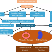
Induction of labour is the deliberate initiation of the labour
process before it occurs naturally or spontaneously. An estimated 5% to 10% of
women reach gestational periods beyond 294 days or 42 completed weeks,
classifying them as post-term pregnancies. This demographic notably contributes
to the increased frequency of induced labour. Although induction of labour
stands as a prevalent intervention in obstetrics, it carries inherent risks and
necessitates careful consideration before implementation. Labour progresses
through three stages, with the initial phase marked by the cervix gradually
dilating, causing characteristic pain. As the cervix dilates, mucus that
protected against bacteria is often expelled. This dilation also weakens
support for the fetal membranes, potentially leading to their rupture and initiating
active labour. Ideally, regular uterine contractions begin when cells form low
resistance connections, allowing electrical signals to pass smoothly across the
uterus. If contractions start prematurely or if the cervix is not adequately
prepared, prostaglandins released from the membranes and uterine decidua
stimulate labour, leading to a slower dilation phase, which can be challenging
for the mother and increase infection risk. Induced labour is indicated in
women who have prolonged pregnancy, premature rupture of membrane (PROM)
(Preterm at ≥34 weeks in absence of other obstetric indications and term at ≥37
weeks.), intrauterine fetal death and maternal request.

Caesarean Section

The prevalence of caesarean sections has risen notably in both
developed and developing nations. The WHO systematic review suggests that
caesarean section rates of 10-15% are associated with decrease in maternal,
neonatal and infant mortality. When life is expected to be normal, why
shouldn’t childbirth be normal too?

Unnecessary caesarean sections are recognized to elevate
health risks for both the mother and the new-born, while also imposing
financial strains on healthcare budgets. The rising trend in caesarean
deliveries is influenced by healthcare providers’ safety perceptions,
obstetricians’ convenience preferences and healthcare system structures.
Mothers who deliver vaginally tend to recover faster postpartum and are better
equipped to care for their new-borns. Prostaglandins play pivotal roles in
parturition, specifically focusing on myometrial contraction. Elevated levels
of uterine prostaglandins or increased myometrial responsiveness to
prostaglandins induce contraction and initiate labour by promoting cervical
ripening. Hence, prostaglandins have been widely used for the induction of
labour. Induction of labour can be done by mechanical methods and
pharmacological methods.

Mechanical Modalities

Mechanical methods include hygroscopic dilators, which
functions by absorbing fluids from endocervical and local tissues. Balloon
devices exert mechanical pressure directly onto the cervix during inflation.
Membrane stripping increases enzyme activity, dilates the cervix and detaches
membranes from the uterus. Amniotomy can increase the release of prostaglandins
locally. Possible risks include cord problems, infections, fetal heart rate
changes, bleeding from placenta issues and fetal injury.

Pharmacological
Methods

1. Prostaglandins

Prostaglandins are naturally produced hormones in the body
and are important during the labour. Prostaglandins, produced both locally in
the cervix and uterus as well as from the fetal membranes, play a critical role
in cervical ripening and other processes of parturition, including uterine
contractility and the induction of labour. They are frequently used when the
ripening of cervix has not occurred with a Bishop score<6. It supports
cervical ripening and promotes the cervix to soften and stretch in preparation
for childbirth. Numerous prostaglandin formulations have been utilized for
labour induction, encompassing prostaglandin F2 alpha (PGF2α, dinoprost),
prostaglandin E2 (PGE2, Dinoprostone), prostaglandin E1 (PGE1) and misoprostol,
a synthetic analogue of PGE1.

2. Oxytocin

Oxytocin, a natural hormone, aids in uterine contractions
during labour. Its synthetic forms are used for induction globally. IV oxytocin
is administered as the cervix dilates. Dosage typically starts low (0.5-2.0
mU/minute) and increases every 15-60 minutes, with higher doses (up to 6.0
mU/minute) increasing every 15-40 minutes.

3. Mifepristone (Progesterone receptor antagonists)

Progesterone plays a crucial role in all stages of
pregnancy. It prevents uterine muscle contraction and helps maintain cervical
structure. When labour begins, progesterone withdrawal is necessary. Progesterone
receptor antagonists can induce labour by mimicking this withdrawal.

4. Nitric Oxide (NO) donors

Nitric oxide (NO) donors have been used to ripen the cervix
for first-trimester pregnancy terminations. Studies indicate that nitric oxide
metabolites rise in cervical fluid after ripening or manipulation, indicating
NO donors could be beneficial for labour induction. 9

5. Dinoprostone

PGE2, alternatively referred to as Dinoprostone, is an
endogenous compound that plays a pivotal role in labour induction. PGE2 prompts
myometrial contractions through direct stimulation, binding to EP1-4 G
protein-coupled receptors (GPCRs), initiating diverse downstream events
contingent on EP subtype and cell-specific expression patterns.

Dinoprostone is available in 2 formulations: a vaginal
insert and a cervical gel. Dinoprostone exhibits a sustained and controlled
onset of action and duration of effect, with a half-life ranging from 2.5-5
minutes. Both formulations necessitate cold storage to maintain chemical stability.
While the cervical gel enables faster release of Dinoprostone compared to the
vaginal insert, the latter provides a more gradual elevation in plasma PGE2
levels and a prolonged duration of action.

The vaginal insert offers easy retrieval compared to gel,
administered at a rate of 0.3 mg/h for 24 hours, it proves superior compared to
cervical gel owing to its ease of removal, diminished invasiveness and reduced
necessity for vaginal examinations. Dinoprostone gel often requires repeated
doses, leading to potential discomfort for the patient. Moreover, in cases of
hyperstimulation, where excessive uterine contractions occur, the
administration of the gel lacks an effective reversal mechanism, thereby posing
challenges in managing this complication.

Dinoprostone Vaginal Insert

PGE2 is pivotal in facilitating cervical ripening and the
onset of parturition. The localized actions of PGE2 encompass alterations in
cervical consistency, dilation and effacement. The Dinoprostone vaginal insert
comprises 10 mg of Dinoprostone uniformly distributed within the matrix of a
thin, flat polymeric hydrogel drug delivery device. The delivery mechanism is
engineered to sustain a controlled and consistent release of Dinoprostone from
the reservoir. In women with intact membranes, the release rate averages approximately
0.3 mg per hour. In women experiencing premature rupture of membranes, the
release of Dinoprostone may occur at an accelerated pace and exhibit greater
variability. The utilization of a Dinoprostone insert is associated with a
significantly higher likelihood of achieving vaginal delivery within a 24-hour
timeframe when compared to the application of Dinoprostone gel. Furthermore,
the Dinoprostone insert demonstrates superiority in facilitating vaginal
delivery within this time frame, accompanied by shorter hospital stays and
reduced incidence of postpartum haemorrhage compared to the gel formulation.

Need for Consensus

Given the extensive pre-existing data on Dinoprostone
vaginal insert and the ongoing emergence of clinical evidence, there is a
critical necessity for a clinical consensus regarding its role in initiating
and intensifying labour induction. These imperatives underscore the need for a
practical document tailored to provide guidance for healthcare professionals
(HCPs) regarding the diverse indications of Dinoprostone vaginal insert. Such a
consensus serves as an indispensable resource, synthesizing current knowledge
and offering actionable recommendations to empower HCPs in optimizing obstetric
care and treatment approaches.

A group of gynaecologists from India have discussed the
various methodology for induction of labour and the role of Dinoprostone
vaginal insert for the use in induction of labour. Experts framed statements
based on available scientific evidence, experience and collective judgement
from practical experience with Dinoprostone vaginal insert. Objective related
to Dinoprostone vaginal insert were discussed and each expert shared their
view, which led to group discussions. Consensus was reached when agreement with
the statement exceeded 80% within the group.

Expert Opinion on Dinoprostone Vaginal Insert

1. Predictors of success for IOL

For a successful induced labour, it’s crucial to have a
Bishop score lower than 6. Other important factors include a lower BMI, having
had fewer than 5 previous deliveries, gestational age of >39 weeks and
ensuring the baby’s weight is up to 3.2 kg.

2. Indication for dinoprostone vaginal insert

It was unanimously recommended that promotional material
refrain from outlining specific indications for the use of Dinoprostone Vaginal
Insert. The decision to employ Dinoprostone in a particular patient should be
left to the discretion of individual healthcare providers, as they possess the
requisite clinical judgment to assess its appropriateness on a case-by-case
basis.

3. Benefits of dinoprostone vaginal insert over other IOL
agents

Dinoprostone Vaginal Insert is distinguished by its capacity
to initiate labour through a gentle process facilitated by the controlled release
of Dinoprostone. A notable advantage lies in its “easy reversibility due
to retrievability,” a feature unparalleled by other methods such as
misoprostol or Dinoprostone Gel. This attribute holds significant clinical
importance as it markedly reduces the risk of uterine hyperstimulation. The
rapid clearance of Dinoprostone upon removal of the insert, owing to its short
half-life of 2.5- 5 minutes, further contributes to the safety profile of this
approach.

4. Cost is not a major concern

If patient has successful induction of delivery with the
Dinoprostone vaginal insert, the cost of hospitalisation is reduced to a
fraction of that of caesarean section (C/S). On the other hand, Dinoprostone
failed patients will have to bear greater cost of C/S with the uncertainty of
complications, such as maternal-fetal morbidity risk, possibility of NICU cost
and trauma of the mandatory C/S delivery in future, etc.

5. Dinoprostone vaginal insert over misoprostol

Misoprostol exhibits dual pharmacological effects: cervical
ripening and oxytocic action, inducing contractions. However, during IOL, the
desired effect is solely cervical ripening, without the oxytocic effect. Herein
lies the advantage of Dinoprostone vaginal insert over Misoprostol.
Additionally, Misoprostol lacks the capability for reversing hyperstimulation,
unlike Dinoprostone vaginal insert, which can be easily retrieved. This ease of
reversal is facilitated by the short half-life of Dinoprostone (2.5-5 minutes) compared
to Misoprostol’s half-life of approximately 30-40 minutes.

6. Dinoprostone vaginal insert over conventional gel

Both Dinoprostone vaginal insert and gel can be used in
cases of premature rupture of membranes (PROM) cases. Furthermore, the
potential for reversing uterine hyperstimulation is feasible with Dinoprostone
vaginal insert, a capability not afforded by gel.

Key Recommendations for Deploying Dinoprostone for Cervical
Ripening

1. Utilization Guidelines for Dinoprostone

(a) The application of Dinoprostone is recommended when the
Modified Bishop Score is less than 6.

(b) In addition to the specified maternal medical conditions
for Induction of Labour (IOL), the pregnancy should have progressed to at least
37 weeks.

(c) Advanced Maternal Age (above 35 years) and/or High Body
Mass Index (BMI) may diminish the effectiveness of agents which is been used
for IOL.

2. Implementation Protocol for Dinoprostone

(a) The Dinoprostone vaginal insert should be stored in a
freezer from procurement until just prior to insertion.

(b) Prior to inserting the Dinoprostone Vaginal insert, an
intravaginal saline wash of 20 mL 0.7% should be administered.

(c) Dinoprostone should only be removed from cold storage
once the patient is positioned and the vaginal wash is completed.

(d) The duration between retrieval from cold storage and
insertion must not exceed 30 seconds.

(e) Following the insertion of Dinoprostone, cervical
ripening may take up to 24 hours.

(f) If intravenous Oxytocin supplementation is required, it
should be administered no sooner than 30 minutes after the removal of
Dinoprostone.

Summary

1. When natural processes fail to initiate labor in women at
term, Dinoprostone facilitates natural delivery by promoting cervical ripening
and uterine contractions.

2. Dinoprostone Vaginal Insert should be administered in an
in-patient setting with meticulous supervision, mandating non-discharge
post-insertion.

3. Dinoprostone Vaginal Insert necessitates storage within a
freezer, maintaining temperatures between – 10◦C and -25◦C, emphasizing the
criticality of freezer storage over refrigeration.

4. Concurrent administration of Dinoprostone Vaginal Insert
with oxytocin is contraindicated. Oxytocin initiation should be deferred until
30 minutes postremoval of Dinoprostone, permitting simultaneous use with
mechanical methods like Foley’s or Balloon Catheter.

5. Timely removal of Dinoprostone Vaginal Insert upon the
establishment of painful uterine contractions marks best practice.

6. Augmenting the efficacy of Dinoprostone Vaginal Insert
necessitates pre-insertion cleansing with 20 mL 0.9% saline wash, elevating
vaginal pH to enhance Dinoprostone release.

7. Swift insertion of Dinoprostone Vaginal Insert within 30
seconds post-freezer removal underscores the importance of patient positioning
prior to removal. Optimal placement in the posterior vaginal fornix
transversely aligns with recommended technique.

8. When opening the package via perforation, it is necessary
to push the tape from the bottom promptly during pack tearing to expedite the
process and save time.

9. An educational initiative targeting post-graduate
students is recommended to enhance awareness, particularly focusing on cold
chain maintenance and administration protocols, aiming for a lifelong
improvement in expertise.

10. The typical duration for labour induction with
Dinoprostone Vaginal Insert spans 14-16 hours, albeit varying between 10 to
20-22 hours for select individuals.

11. Post-insertion, patients receiving Dinoprostone Vaginal
Insert can anticipate a waiting period of up to 24 hours for the initiation of
labour.

12. Data was presented on the out-patient use of DVI for
IOL. It was concluded that, unlike the western countries, the Indian obstetric
scenario is not yet ready for out-patient deployment of DVI for IOL due to the
limited awareness.

Source: Pandya, Kakkar and Gupta / Indian Journal of
Obstetrics and Gynecology Research 2024;11(3):325–329

https://doi.org/10.18231/j.ijogr.2024.062

Powered by WPeMatico