Coaxial SIL Drains Less Painful Yet Equally Effective as PVC Drains after video-assisted thoracoscopic surgery lung resections: Study

A new study has confirmed that coaxial silicone (SIL) drains, due to their softer material, result in less patient discomfort while providing drainage efficacy comparable to that of traditional PVC drains after video-assisted thoracoscopic surgery (VATS) lung resections.

Chest drains are routinely used after video-assisted thoracoscopic surgery (VATS) lung resections to evacuate fluid and air from the pleural space. We compared the impact of coaxial silicone (SIL) drains vs. standard polyvinyl chloride (PVC) drains on postoperative pain, drainage efficacy, and short-term treatment outcome following VATS lobectomy.

The prospective randomized study included 80 patients who underwent VATS lobectomy for lung cancer between September 2020 and June 2023. Patients were randomized into two groups based on the type of chest drain used postoperatively: 40 in the experimental group (coaxial SIL drain Fr 24) and 40 in the control group (standard PVC drain Fr 24). The researchers collecting the data and the caregivers were not blinded to the group allocation. The primary objective was to evaluate pain over the initial 2 postoperative days by assessing analgesic consumption, respiratory muscle strength [measured as maximal inspiratory pressure (MIP) and maximal expiratory pressure (MEP)], and pain intensity using the visual analog scale (VAS). MIP, MEP, and VAS were measured both at rest and during physical activity.

 Sixty-nine patients were included in the final analysis: 35 in the experimental group and 34 in the control group. The groups were comparable in terms of drainage efficacy and short-term treatment outcome, but pain was significantly lower in the experimental group (coaxial SIL drain). Diclofenac consumption was significantly lower in the experimental group (P=0.004), with a trend toward lower consumption of other analgesics. All respiratory muscle strength measurements were higher in the experimental group, with significant differences in static MIP on the second postoperative day (P=0.046), both static (P=0.02) and dynamic (P=0.050) MEP on the first postoperative day, and static MEP on the second postoperative day (P=0.02). Static VAS (S-VAS) on the first postoperative day was statistically significantly lower in the experimental group (P=0.003). Dynamic VAS (D-VAS) was comparable between the groups.

This study confirmed the hypothesis that coaxial SIL drains, owing to their softer material, cause less pain while maintaining efficacy comparable to standard PVC drains.

Reference:

Boris Greif, Janez Žgajnar, Tomaž Štupnik,  Impact of chest tube type on pain, drainage efficacy, and short-term treatment outcome following video-assisted thoracoscopic surgery lobectomy: a randomized controlled trial comparing coaxial silicone drains and standard polyvinyl chloride drains, Journal of Thoracic Disease, DOI:10.21037/jtd-24-1489 

Powered by WPeMatico

Cognitive tests in infancy can offer insight into intelligence at age 30 and beyond: Study

Watching a baby babble, play and interact with others can provide useful insight into what their cognitive ability might be like decades later, according to new University of Colorado Boulder research published in the journal PNAS.

The study of more than 1,000 twins found that tests as early as 7 months of age can help predict performance on cognitive measures at age 30. It also found that an infant’s environment plays a more significant role in shaping life-long cognition than scientists realized. It could even influence risk of dementia later in life, the authors said.

“Our findings highlight the enduring consequences of the very early childhood environment on cognitive ability and suggest that early life is a critical developmental period that we should be paying attention to,” said lead author Daniel Gustavson, assistant research professor at the Institute for Behavioral Genetics (IBG).

What twins can teach us

Similar to “IQ,” general cognitive ability (GCA) is a single, composite measure of a person’s capacity to learn, reason, understand and problem-solve.

Prior research has shown that much of our GCA is established by childhood. Give an 8-year-old a battery of tests to determine their GCA and their score will look remarkably similar at age 30. Measures of intelligence at age 20 are highly correlated with those at age 62, and IQ doesn’t change much between age 11 and 90.

But few scientists have looked back further to see what-if anything-signals in infancy can tell us about cognition in adulthood and old age.

Gustavson and senior author Chandra Reynolds, a professor of psychology and neuroscience, looked at data from 1,098 participants in the Colorado Longitudinal Twin Study. IBG launched the study in 1985, enrolling baby twins from Colorado’s Front Range to assess the role that genes vs. environment play in various aspects of development.

Researchers have since collected reams of data, via periodic laboratory samples, home visits, surveys, interviews and behavioral tests.

“We have co-authors on this paper who have been involved since the start and watched these twins grow up,” said Gustavson.

As early as 7 months old, researchers assessed seven measures of cognition, including vocalization, ability to stay on task, and “novelty preference”-whether the infants preferred to play with new toys over ones they were familiar with.

Age-appropriate cognitive assessments have been done at five points, so far.

The team found that looking at cognitive tests in infancy could predict about 13% of the variance in scores at age 30. Two measures-novelty preference and task orientation-were the strongest predictors. This early life “signal” is not huge, the authors note.

“We certainly do not want to imply that cognition is somehow fixed by seven months old,” Gustavson said. “But the idea that a very simple test in infancy can help predict the results of a very complicated cognitive test taken 30 years later is exciting.”

Nature, nurture or both?

To explore what role genetics vs. environment plays, the study compared GCA score differences between identical twins, who share 100% of their genes, and fraternal twins, who only share half of their genes. In general, if there is greater similarity among identical twins than fraternal twins, this suggests that genes play a strong role in that trait.

They also analyzed the twins’ DNA collected via blood or saliva.

As expected, genes played a big role in influencing general cognitive ability, with genetic influences measured by age 7 accounting for about half of the variation in scores at age 30.

But environment also had a significant and lasting impact.

“One of the most exciting findings was that 10% of the variability in adult cognitive ability was explained by environmental influences before year one or two,” said Gustavson.

The older the children got, the more influence genes had and the less environment had.

“This suggests that even the pre-preschool environment matters,” Gustavson said.

Reynolds, who studies age-related diseases including Alzheimer’s and dementia, says the findings could have implications not only for how youth do in school or how adults perform at work but also how prone they may be to age-related cognitive decline.

“Cognitive aging is a life-long process, not just something that begins in mid-life,” she said. “It could be that certain interventions, like strong educational foundations in early life could help maximize what people are capable of and help them keep that cognitive gas in the tank for as long as possible.”

A polygenic score for intelligence

The study also confirms that “polygenic scores” can be a useful tool.

Polygenic scores are single numbers that aggregate a person’s genetic variants to estimate predisposition to a trait, like intelligence.

“There are thousands of genes that influence intelligence, so you are never going to find an ‘intelligence gene’, but we have found many with tiny effects that when put together can have an impact,” Gustavson said.

For the study, the researchers used genetic data from nearly 1 million individuals gathered via large datasets like 23 and Me to give each of the adult twins a polygenic score based on their own DNA, for cognitive ability.

Remarkably, the twins’ scores closely matched what would be expected based on their tests when they were babies.

“Studies like ours show us that both family-based and genomic-based datasets are valuable in answering questions about how genetic and environmental influences change across the lifespan,” said Gustavson. 

Reference:

D.E. Gustavson,G.A. Borriello,M.A. Karhadkar,S.H. Rhee,R.P. Corley,S. Rhea,L.F. DiLalla,S.J. Wadsworth,N.P. Friedman,& C.A. Reynolds, Stability of general cognitive ability from infancy to adulthood: A combined twin and genomic investigation, Proc. Natl. Acad. Sci. U.S.A. 122 (21) e2426531122, https://doi.org/10.1073/pnas.2426531122 (2025).

Powered by WPeMatico

High Uric Acid Levels Tied to Elevated Risk of Skin Cancer, Study Finds

China: A new study published in Clinical, Cosmetic and Investigational Dermatology has uncovered a significant association between elevated serum uric acid (SUA) levels and an increased risk of both malignant melanoma (MM) and non-melanoma skin cancer (NMSC). Conducted by Dr. Yihao Niu and colleagues from the Faculty of Chinese Medicine, Macau University of Science and Technology, the research utilized large-scale data from the National Health and Nutrition Examination Survey (NHANES). It employed Mendelian randomization techniques to evaluate causality.

Serum uric acid, a metabolic byproduct of purine breakdown, has previously been linked to various health conditions, including cardiovascular disease and metabolic syndrome. However, its role in skin cancer development has not been established. This study aimed to fill that gap by analyzing SUA levels with skin cancer incidence among 1,219 individuals—336 diagnosed with melanoma (MM) and 883 with non-melanoma skin cancer (NMSC)—from the NHANES dataset spanning 1999 to 2020.

Multivariable regression models were used to adjust for potential confounding factors, including age, sex, race, lifestyle habits, and existing comorbidities.

The following were the key findings of the study:

  • Each 1 mg/dL increase in serum uric acid (SUA) was linked to a 19% higher risk of malignant melanoma.
  • A similar rise in SUA was associated with a 13% higher risk of non-melanoma skin cancer.
  • These associations were more evident among women and individuals of Mexican American ethnicity.
  • Despite the observed associations, Mendelian randomization analysis did not confirm a causal relationship between SUA levels and skin cancer.
  • Causality was assessed using methods like inverse variance weighting (IVW), MR-Egger, and the weighted median approach based on genome-wide association study (GWAS) data.
  • The findings suggest that SUA may act as a risk marker rather than a direct cause of skin cancer.
  • Restricted cubic spline (RCS) models revealed a non-linear relationship, with skin cancer risk increasing more sharply beyond certain SUA thresholds.
  • Elevated SUA levels were more strongly linked to malignant melanoma risk in older adults (aged 60 and above).
  • In contrast, non-melanoma skin cancer was found to be more prevalent in younger individuals with higher SUA levels.

The study’s authors emphasized the importance of interpreting SUA levels as part of a broader clinical picture, noting that systemic inflammation, oxidative stress, and immune dysregulation—all of which can be influenced by uric acid—may contribute to carcinogenesis.

The researchers note that while elevated SUA does not appear to be a direct cause of skin cancer, its strong association with MM and NMSC highlights its potential utility as a biomarker for identifying individuals at increased risk. They call for further investigations that integrate basic science, longitudinal data, and refined genetic tools to clarify the mechanisms at play and to explore whether lowering SUA might offer protective benefits.

Reference:

Niu Y, Shi S, Chen Y, Yang S, Yan P, Kou J. Association Between Serum Uric Acid Levels and Skin Cancer: Results from NHANES Data and Mendelian Randomization Analysis. Clin Cosmet Investig Dermatol. 2025;18:1437-1452

https://doi.org/10.2147/CCID.S517389

Powered by WPeMatico

IL-17 Inhibitors Increase Risk of Candida and Fungal Infections in Psoriasis Patients: Study

Researchers have found in a recent analysis reveals IL-17 inhibitors significantly increase the risk of Candida infections and other superficial fungal infections in patients with moderate-to-severe psoriasis, compared to placebo and other biologics.

Biologic agents have become a key treatment option for moderate-to-severe plaque psoriasis; however, the associated risk of superficial fungal infections, such as Candida and dermatophytes infections, remains unclear. This study aims to systematically assess the impact of different biologic agents on these infection risks and to compare the differences between them. Research questions and keywords were developed based on the Population, Intervention, Control and Outcome (PICO) framework. A systematic search of PubMed, EMBASE, the Cochrane Library and Web of Science was conducted for randomised controlled trials (RCTs) published up to December 2024, using the keywords ‘psoriasis’, ‘biologics’, ‘anti-IL-17’, ‘anti-IL-12/23’, ‘anti-TNF’, ‘superficial fungal infections’, ‘dermatophyte infections’, ‘Candida’ and ‘onychomycosis’. Meta-analyses were performed using RevMan 5.4 and STATA 16.0 software. Results: A total of 644 records were identified, with 29 articles included in the final analysis. Meta-analysis indicated that compared with placebo, interleukin-17 (IL-17) inhibitors notably raised the risk of Candida infections (OR = 2.39, 95% CI = 1.84–3.11, p < 0.00001), whereas tumour necrosis factor-alpha (TNF-α) inhibitors (OR = 1.75, 95% CI = 0.53–5.82, p = 0.36) and interleukin-12/23 (IL-12/23) inhibitors (OR = 1.11, 95% CI = 0.27–4.63, p = 0.88) showed no significant differences. Cross-comparison demonstrated that IL-17 inhibitors had a higher risk of Candida infection compared to TNF-α inhibitors (OR = 2.23, 95% CI = 1.08–4.57, p = 0.03) and IL-12/23 inhibitors (OR = 4.21, 95% CI = 2.71–6.55, p < 0.00001). For dermatophyte infections, the overall risk associated with biologic agents was increased (OR = 1.89, 95% CI = 1.19–3.01, p = 0.007), IL-17 inhibitors showed a higher risk compared to IL-12/23 inhibitors (OR = 2.70 95% CI = 1.29–5.63, p = 0.008). Overall, biologic agents significantly increased the risk of superficial fungal infections compared to placebo (OR = 2.10, 95% CI = 1.73–2.55, p < 0.00001). Biologic agents, particularly IL-17 inhibitors, notably increase the risk of superficial fungal infections in psoriasis patients. In clinical practice, targeted monitoring protocols should be established, including regular follow-up to promptly detect superficial fungal infections and initiate antifungal treatment as necessary.

Reference:

H. Liu, L. Zhou, Z. Song, R. Zhang, and Y. Kang, “ Biologic Therapy and Superficial Fungal Infection Risk in Moderate-to-Severe Psoriasis: A Meta-Analysis,” Mycoses 68, no. 6 (2025): e70081, https://doi.org/10.1111/myc.70081.

Keywords:

IL-17, Inhibitors, Increase, Risk, Candida, Fungal Infections, Psoriasis Patients, Study, H. Liu, L. Zhou, Z. Song, R. Zhang, and Y. Kang, Mycoses

Powered by WPeMatico

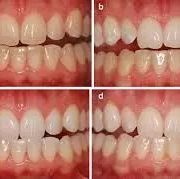
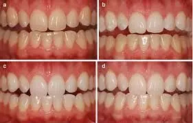
Antioxidants Preserve Bleached Enamel Color but Acidic Gels May Pose Enamel Risk: Study

Researchers have found in a new study that antioxidant agents maintained color stability after tooth bleaching and showed high antioxidant activity. However, QUI and QC gels had acidic pH, which may risk enamel damage. Therefore clinically, these antioxidants can aid bond strength recovery without compromising esthetic outcomes, but acidic formulations should be used cautiously.

This study evaluated the color stability of enamel submitted to 10% hydrogen peroxide (HP) followed by antioxidants agents, and the pH and antioxidant activity (AA%) of these agents. Bovine enamel-dentin blocks were randomly distributed into groups (n=10/group): GNC (negative control: no treatment); GPC (positive control: bleaching only); TOC_10% (HP+10% α-tocopherol); GT_10% (HP+10% green tea extract); GS_5% (HP+5% grape seed extract); SA_10% (HP+10% sodium ascorbate); QUI_10% (HP+10% quinoa extract); and QC_1% (HP+1% quercetin). Color (ΔE00) and whiteness index (ΔWID) changes were analyzed using a digital spectrophotometer. The pH and AA% were determined using a pH meter and the DPPH method, respectively. Data were analyzed by ANOVA/Tukey’s and Dunnett’s tests (α=0.05). Results At 14 days post-bleaching, GNC promoted the lowest ΔWID and ΔE00 (p<0.05), and no differences were found between GPC and the remaining groups submitted to the antioxidant agents (p>0.05). QC_1% and QUI_10% exhibited acidic pH levels (3.64 and 4.75, respectively), whereas TOC_10% and GS_5% exhibited alkaline pH (7.07 and 7.64, respectively). No differences in AA% were found between the agents (p>0.05), ranging from 92.6 to 97.6%. The antioxidant agents did not interfere in bleached enamel color stability, showing satisfactory antioxidant activity. However, QUI and QC gels displayed acidic pH. Clinical significance: The antioxidants evaluated showed high AA% and no impact on post-bleaching color stability, suggesting that their capacity to recover bond strength demonstrated elsewhere would not compromise the esthetic efficacy of tooth bleaching. However, those with acidic pH should be used with caution due to potential enamel damage.

Reference:

Alves RO, Nunes GP, Martins TP, Alves de Toledo PT, Ragghianti MHF, Delbem ACB. Effect of Quercetin-Doped Hydrogen Peroxide Gels on Enamel Properties: An In Vitro Study. Gels. 2025 Apr 27;11(5):325. doi: 10.3390/gels11050325. PMID: 40422345; PMCID: PMC12111415.

Keywords:

Antioxidants, Preserve, Bleached, Enamel, Color, Acidic, Gels, May, Pose, Enamel Risk, Study , Journal of Applied Oral Science, Antioxidants; Tooth bleaching; Color; Hydrogen peroxide, Alves RO, Nunes GP, Martins TP, Alves de Toledo PT, Ragghianti MHF, Delbem ACB

Powered by WPeMatico

Low-Energy Total Diet Replacement Does Not Worsen Eating Disorder Symptoms in High-Risk Patients: Lancet

Researchers have found in a new study that a 12-week low-energy total diet replacement program did not exacerbate eating disorder symptoms in individuals with overweight or obesity and type 2 diabetes who were at high risk of developing such disorders. The study was conducted by Elena T. and fellow researchers published in The Lancet Psychiatry journal.

Low-calorie TDR diets, usually formula products of nutritionally complete fare like soups, shakes, and bars, are most frequently employed for inducing fast weight loss in those with obesity. Their strict composition and emphasis on weight loss have, however, fueled worry over their capacity to induce disordered eating, particularly in susceptible individuals. That fear was addressed by this trial in a rigorous assessment of the safety and psychological effects of TDR in a high-risk group.

The trial was a randomized, controlled, non-inferiority trial in England involving participants with type 2 diabetes, overweight, and exhibiting signs of eating disorder symptoms. Participants were allocated (1:1) to a low-energy TDR program or standard care for diabetes.

Intervention comprised:

  • 12 weeks of low-energy diet with formula products

  • 8 weeks of stepped reintroduction of foods

  • 4 weeks of weight maintenance advice tailored to participants’ preferences

Behavioral treatment was provided remotely. The main outcome was modification in the Eating Disorder Examination Questionnaire (EDE-Q) global score at 6 months. Safety was measured by tracking the development of any new cases of eating disorder. The non-inferiority bound was +1 SD (0.72) for the EDE-Q score.

Participant Profile

  • From March 8 to September 12, 2023, 56 participants were enrolled altogether

  • 28 in intervention group and 28 in control group

  • Mean age: 49.9 years (SD 8.1)

  • Gender: 35 (63%) women, 20 (36%) men, 1 (2%) non-binary

  • Ethnicity: 54 (96%) White, 2 (4%) Asian

  • Mean BMI: 39.6 kg/m² (SD 7.8)

  • Mean baseline EDE-Q score: 3.3 (SD 0.4)

Key Findings

At 6 months, when TDR program concluded:

  • Mean weight loss in the intervention group was –13.9 kg (SD 11.2)

  • Control group lost –3.7 kg (SD 7.9)

  • Between-group difference in weight: –10.2 kg (95% CI: –14.2 to –6.2)

Symptoms of eating disorders improved:

  • Between-group difference in EDE-Q global score: –0.8 points (95% CI: –1.4 to –0.3), establishing non-inferiority

  • No participant was suspected of having a new eating disorder

  • At 12 months, while between-group differences in weight change were no longer statistically significant, EDE-Q score improvement was maintained, suggesting psychological benefit over the long term.

Engagement in a remotely delivered, structured total diet replacement (TDR) program did not exacerbate eating disorder symptoms in individuals with type 2 diabetes and obesity who were at elevated risk for disordered eating. The program resulted in significant weight reduction and even demonstrated evidence of decreasing eating disorder scores. This study justifies the safe implementation of TDR with adjunctive behavioral treatment in clinical practice.

Reference:

Tsompanaki, E., Aveyard, P., Park, R. J., Jebb, S. A., & Koutoukidis, D. A. (2025). An intensive weight loss programme with behavioural support for people with type 2 diabetes at risk of eating disorders in England (ARIADNE): a randomised, controlled, non-inferiority trial. The Lancet. Psychiatry, 12(7), 483–492. https://doi.org/10.1016/S2215-0366(25)00126-9

Powered by WPeMatico

New PET tracer delivers high-quality images of brain inflammation activity: Study

A newly developed PET radiotracer has shown the ability to produce high-quality images of real-time brain inflammation, according to research presented at the Society of Nuclear Medicine and Molecular Imaging 2025 Annual Meeting. By identifying activated immune cells in the brain, the tracer may pave the way for earlier diagnosis and more personalized treatment of a range of neurological diseases including Alzheimer’s, Parkinson’s, ALS, and multiple sclerosis.

Neuroinflammation-an immune response triggered by infection, toxin buildup, or injury in the central nervous system-is a key driver in the progression of many neurodegenerative and psychiatric disorders. Imaging neuroinflammation plays a critical role in diagnosis, monitoring, and treatment.

“Current clinical PET imaging for neuroinflammation primarily uses tracers that target TSPO, a downstream marker that is broadly expressed across multiple cell types,” said Jiahui Chen, PhD, associate scientist in the Department of Radiology and Imaging Sciences at Emory University School of Medicine in Atlanta, Georgia. “Our study presents ¹⁸F-PDE-1905, a novel PET tracer specifically developed to target phosphodiesterase 4B (PDE4B)-a crucial intracellular enzyme that regulates inflammatory signaling within microglia, the immune cells of the central nervous system.”

Researchers began by analyzing a genomics database to assess PDE4B expression in neuroinflammatory diseases using bioinformatics tools. They then developed a mouse model of neuroinflammation and performed dynamic PET imaging using the ¹⁸F-PDE-1905 alongside the TSPO-specific tracer ¹⁸F-D2-LW223. Follow-up analyses were conducted to evaluate protein expression and confirm its correlation with the PET imaging findings.

Bioinformatics analysis revealed elevated PDE4B levels in both Parkinson’s disease and multiple sclerosis patients, as well as in corresponding mouse models. PET imaging showed significantly higher uptake of ¹⁸F-PDE-1905 in the brains of diseased mice compared to controls, indicating increased tracer activity in neuroinflammatory conditions. When compared to the TSPO-specific tracer ¹⁸F-D2-LW223, ¹⁸F-PDE-1905 demonstrated superior image quality and greater brain distribution, underscoring its promise for imaging neuroinflammation.

“By directly targeting PDE4B, ¹⁸F-PDE-1905 provides a more specific and upstream view of microglial activation—an early and critical factor in the progression of many neurological diseases,” said Chen. “For patients, this could mean earlier and more accurate diagnoses, better tracking of treatment effectiveness, and more personalized therapies based on direct measures of neuroinflammation. Ultimately, ¹⁸F-PDE-1905 has the potential to drive a major shift toward precision-guided care in neurodegenerative disorders.”

Reference:

Jiahui Chen, Yabiao Gao, Xin Zhou,Visualization of phosphodiesterase 4B in neuroinflammation mouse models with positron emission tomography, Journal of Nuclear Medicine.

Powered by WPeMatico

Higher Visceral Fat-to-Muscle Ratio Linked to Severe COPD Exacerbations and Heart Risks: Study Shows

China: A recent prospective cohort study published in Respirology has revealed a significant association between the visceral fat-to-muscle ratio (VMR) and the risk of severe exacerbations in individuals with chronic obstructive pulmonary disease (COPD). Led by Yuanyuan Li from the School of Public Health at Xinjiang Medical University in Urumqi, China, the study emphasizes the potential role of VMR as a predictive marker for COPD-related hospitalizations.

The research was conducted as part of the Xinjiang Multi-Ethnic Cohort study, spanning from May 2018 to December 2023. It included 631 patients diagnosed with COPD. At baseline, participants underwent bioelectrical impedance analysis to assess visceral fat and muscle mass, from which VMR was calculated. Over the study period, researchers tracked severe COPD exacerbations that required hospitalization and evaluated the relationship between VMR and these exacerbations using logistic regression and zero-inflated Poisson regression models.

The study revealed the following findings:

  • Patients with a higher visceral fat-to-muscle ratio (VMR) had a significantly increased risk of experiencing severe COPD exacerbations during both one-year and five-year follow-up periods.
  • Each unit increase in VMR was linked to a 34% higher risk of hospitalization due to COPD exacerbation within one year and a 44% increase over five years.
  • VMR proved to be a more reliable predictor of severe exacerbations compared to conventional metrics like body mass index (BMI) and other obesity-related measures.
  • Subgroup analyses showed that the association between elevated VMR and exacerbation risk was especially marked in women and individuals classified as overweight.
  • In women, the odds of having a severe exacerbation rose by 89% within one year and by 99% over five years for every one-unit rise in VMR.
  • Among overweight participants, each unit increase in VMR corresponded to an 80% increased risk of exacerbation at one year and 88% at five years.
  • Additional analysis indicated that for every one-point increase in VMR, the number of COPD exacerbations rose by 46%.
  • These associations remained consistent even after controlling for potential confounding factors like smoking history and removing underweight individuals from the analysis.
  • The results were further validated through sensitivity analyses and remained unchanged even when considering competing risks, such as deaths from causes unrelated to COPD.
  • Although the study’s main focus was on severe COPD exacerbations, it also referenced related findings where elevated VMR levels were associated with greater cardiovascular risks in COPD patients.
  • In those cases, each standard deviation increase in VMR was tied to a 50% higher likelihood of experiencing a major adverse cardiovascular event (MACE), and patients in the highest VMR quartile had a five-fold greater risk of MACE compared to those in the lowest quartile.

The authors concluded that VMR is a valuable risk indicator for COPD exacerbations. Proactively measuring and monitoring VMR could help clinicians better identify high-risk patients and tailor interventions to reduce hospitalizations and improve outcomes in COPD management.

Reference:

Li, Y., Wang, L., Li, Z., Luo, T., Sun, Q., Lynn, H. S., & Dai, J. (2025). Association Between the Visceral Fat-to-Muscle Ratio and Severe Exacerbation of COPD: A Prospective Cohort Study. Respirology, 30(5), 398-407. https://doi.org/10.1111/resp.14883

Powered by WPeMatico

New: Recommendations for antirheumatic treatment in men and women planning a family

The European Alliance of Associations for Rheumatology-published points-to-consider on the use of antirheumatic drugs in reproduction, pregnancy, and lactation. Since then, new evidence has become available, so the points have been reviewed and updated–including an upgrade to full recommendations.

Rheumatic and musculoskeletal diseases (RMD) often affect people during the years when they might be planning a family. The underlying nature of these diseases can affect people’s fertility, and may make women more at risk of some poor pregnancy outcomes, such as having small or premature babies. Some of the antirheumatic treatments used can also affect fertility, and may not be safe to use during pregnancy. What drugs are used for a person’s RMD when they are planning a family is therefore a consideration for both men and women, and impact show treatment might need to be tailored around conception, as well as during pregnancy and breastfeeding.

A series of points-to-consider were released in 2016, but since then treatment approaches have evolved –moving towards a treat-to-target concept that can help to avoid some of the negative impacts of active disease on fertility and pregnancy outcomes. Alongside this, there has been new relevant data published about antirheumatic drugs in the context of pregnancy and breastfeeding –as well as in male reproductive health. To address this, EULAR has reviewed the evidence and upgraded the advice to full recommendations.

The new work, published in the April 2025 issue of the Annals of the Rheumatic Diseases, includes 12 individual recommendations and five overarching principles. These principles emphasise that everyone with an RMD–both men and women –should be offered early and regular counselling about their reproductive health and the need to adjust therapy in relation to pregnancy. But treatment of the RMD before conception and during and after pregnancy should still aim at remission or low disease activity. Importantly, the potential risk to the child should be weighed against the risk to the mother of having a period of untreated disease. Given the known benefits, women should not be discouraged from breastfeeding and there are compatible medications they can take during this period. Finally, the choice of treatment before, during, and after pregnancy should be a shared decision-making process between the treating healthcare providers and the patient.

The 12 new recommendations focus on three areas. First, the specific antirheumatic drugs that can be used for women before and during pregnancy, including which are compatible and which should be discontinued before attempting to conceive a child. They also look at which vaccines can be used in infants who have been exposed to antirheumatic drugs in the womb. Second, the drugs that are compatible with breastfeeding, and thirdly drug choices for men wishing to father a child. 

EULAR hopes that these recommendations will help to improve treatment and outcomes for people with an RMD during their reproductive years. It will be important to share the updated knowledge widely, as it will be helpful to many healthcare professionals outside rheumatology, including those working in internal medicine, gynaecology and obstetrics, family medicine, paediatrics, and pharmacology.

Reference:

Rüegg L, et al. EULAR recommendations for use of antirheumatic drugs in reproduction, pregnancy, and lactation: 2024 update. Ann Rheum Dis 2025;doi: 10.1016/j.ard.2025.02.023.

Powered by WPeMatico

Metabolic Syndrome May Drive Treatment Resistance in Psoriatic Arthritis, Study Finds

Italy: A recent study published in RMD Open: Rheumatic & Musculoskeletal Diseases highlights a critical link between metabolic syndrome (MetS) and treatment resistance in patients with psoriatic arthritis (PsA). Conducted by Dr. Damiano Currado and colleagues from the University of Rome Campus Biomedico, the research reveals that the presence of MetS significantly increases the risk of developing a difficult-to-treat (D2T) PsA phenotype.

Psoriatic arthritis is a chronic inflammatory condition often marked by joint pain, skin involvement, and a range of musculoskeletal symptoms such as enthesitis and dactylitis. While many patients respond well to current therapies, a subset faces persistent symptoms and poor therapeutic outcomes, termed the “difficult-to-treat” phenotype. This study aimed to examine whether metabolic dysfunction contributes to this challenging clinical profile.

The cross-sectional study was carried out at the Rheumatology Clinic of Fondazione Policlinico Campus Bio-Medico of Rome and involved 182 patients diagnosed with PsA. Disease activity and the presence of metabolic syndrome were assessed using established criteria. The D2T classification was based on modified definitions drawn from rheumatoid arthritis guidelines and adapted for PsA.

The key findings were as follows:

  • 42.9% of the total psoriatic arthritis (PsA) cohort met the criteria for metabolic syndrome (MetS).
  • Among patients with the difficult-to-treat (D2T) phenotype, 81.8% had comorbid MetS.
  • In contrast, only 29.4% of patients in the non-D2T group had MetS.
  • Statistical analysis showed a strong association between MetS and the D2T phenotype, with an odds ratio of 7.56.
  • Patients with MetS were over seven times more likely to show treatment resistance.
  • Path analysis confirmed MetS as an independent factor contributing to the development of the D2T phenotype.

These findings emphasize the role of metabolic health in influencing the course and severity of PsA. The authors emphasize that managing metabolic dysfunctions—such as obesity, hypertension, and insulin resistance—may be crucial in improving therapeutic response and reducing disease burden.

While the study provides valuable insight, the authors acknowledge certain limitations. These include reliance on D2T criteria adapted from rheumatoid arthritis, a single-center design, and the inability to capture certain variables like treatment adherence or patient-reported outcomes. Moreover, as a cross-sectional study, the data cannot establish causality between MetS and the D2T phenotype.

The research represents the first to establish a clear association between metabolic syndrome and treatment resistance in PsA. It suggests that integrated care approaches targeting both PsA and its metabolic comorbidities could enhance treatment outcomes. Strategies such as lifestyle modifications, interdisciplinary collaboration with metabolic specialists, and the use of therapies with minimal metabolic impact may offer a more effective path forward for patients facing this difficult-to-manage form of PsA.

The authors concluded, “Further studies are needed across diverse patient populations to confirm these findings and help refine clinical definitions and treatment pathways for D2T PsA.”

Reference:

Damiano Currado, Francesca Trunfio, Francesca Saracino, Lyubomyra Kun, Annalisa Marino, Erika Corberi, Antonio Orlando, Ludovica Lamberti, Leonardo Frascà, Marta Gatti, Onorina Berardicurti, Marta Vomero, Vasiliki Liakouli, Roberto Giacomelli, Luca Navarini – Patients with psoriatic arthritis and comorbid metabolic syndrome show a difficult-to-treat phenotype: another mosaic tile in the definition of a still undefined subset of patients: RMD Open 2025;11:e005717.

Powered by WPeMatico