Neutrophil-to-HDL Cholesterol Ratio and associated with Gallstone Disease Risk: Insights from NHANES 2017-2020 Data

A recent study revealed that the neutrophil-to-high-density
lipoprotein cholesterol ratio (NHR) could be a useful tool to identify individuals
at risk of developing gallstone disease (GSD). NHR can be used as an easy and
cost-effective tool for the early detection of individuals at GSD as per a
study published in the journal Lipids in Health and Disease.

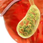
Gallstone disease (GSD) is one of the most frequent and expensive
gastrointestinal disorders caused by cholesterol, bile pigment, and mixed stones,
with cholesterol stones outranking the three. They are caused by the oversaturation
of calcium salts and cholesterol in bile, leading to cholesterol deposits. Gallstones
have an increased risk of cancer. The neutrophil to high-density lipoprotein
cholesterol ratio (NHR) is a novel comprehensive marker of inflammatory
responses and lipid metabolism that can detect cardiovascular disease,
depression, acute biliary pancreatitis, and hepatocellular carcinoma. Previous research
has shown that inflammatory reactions can promote gallstone formation by
altering protein and lipid metabolic pathways and increasing bile acid salt
concentrations by involving neutrophils and lipids. As there is ambiguity on
the association between NHR and gallstones, researchers from Zhejiang Chinese
Medical University, China conducted a study on the relationship between
gallstone disease (GSD) and the neutrophil-to-high-density lipoprotein
cholesterol ratio (NHR) in American patients with gallstones.

The study was conducted by collecting data from the National
Health and Nutrition Examination Survey (NHANES) between 2017–2020. Data was collected
through interviews, health exams, and lab tests. Out of 15,560 participants,
7,894 were included in the study. NHR calculation was done by dividing the neutrophil
count by HDL-C levels and the participants were divided into 3 NHR categories
based on the count. Gallstone data was reported by the participants based on
the physician’s diagnosis. Unadjusted and adjusted logistic regression models
were used to evaluate the association between NHR and GSD.

Key findings:

  • Among the 7894 participants analyzed in this
    study, the prevalence of GSD was 10.98%, and the average NHR value was 3.41 ±
    0.06.
  • An obvious positive association between NHR and
    the likelihood of GSD was found as per the fully adjusted multivariable
    logistic regression analysis (OR = 1.09, 95% CI: 1.01, 1.16; P = 0.0197).
  • Subset analyses confirmed the consistency of
    this association and interaction tests across various subgroups, including
    those categorized by smoking status and asthma.
  • Furthermore, smoothed curve fitting and
    threshold effect analyses revealed a nonlinear relationship with a threshold of
    2.86.

Thus, the study concluded that a higher NHR is associated
with a greater likelihood of having GSD and NHR can be used as a low-cost
method to screen patients at risk of GSD. NHR can also be used in early
diagnosis so that preventive measures can be taken to reduce the risk of gallstones.

Further reading: Ma
D, Ma H, Li Y, Yang L. Association between Neutrophil-to-high-density
lipoprotein-cholesterol ratio and gallstones: insights from the national health
and nutrition examination survey (2017-2020). Lipids Health Dis.
2024;23(1):355. Published 2024 Oct 31. doi:10.1186/s12944-024-02349-w.

Powered by WPeMatico

Which is better TTE or TEE for Grading RV Diastolic Dysfunction?

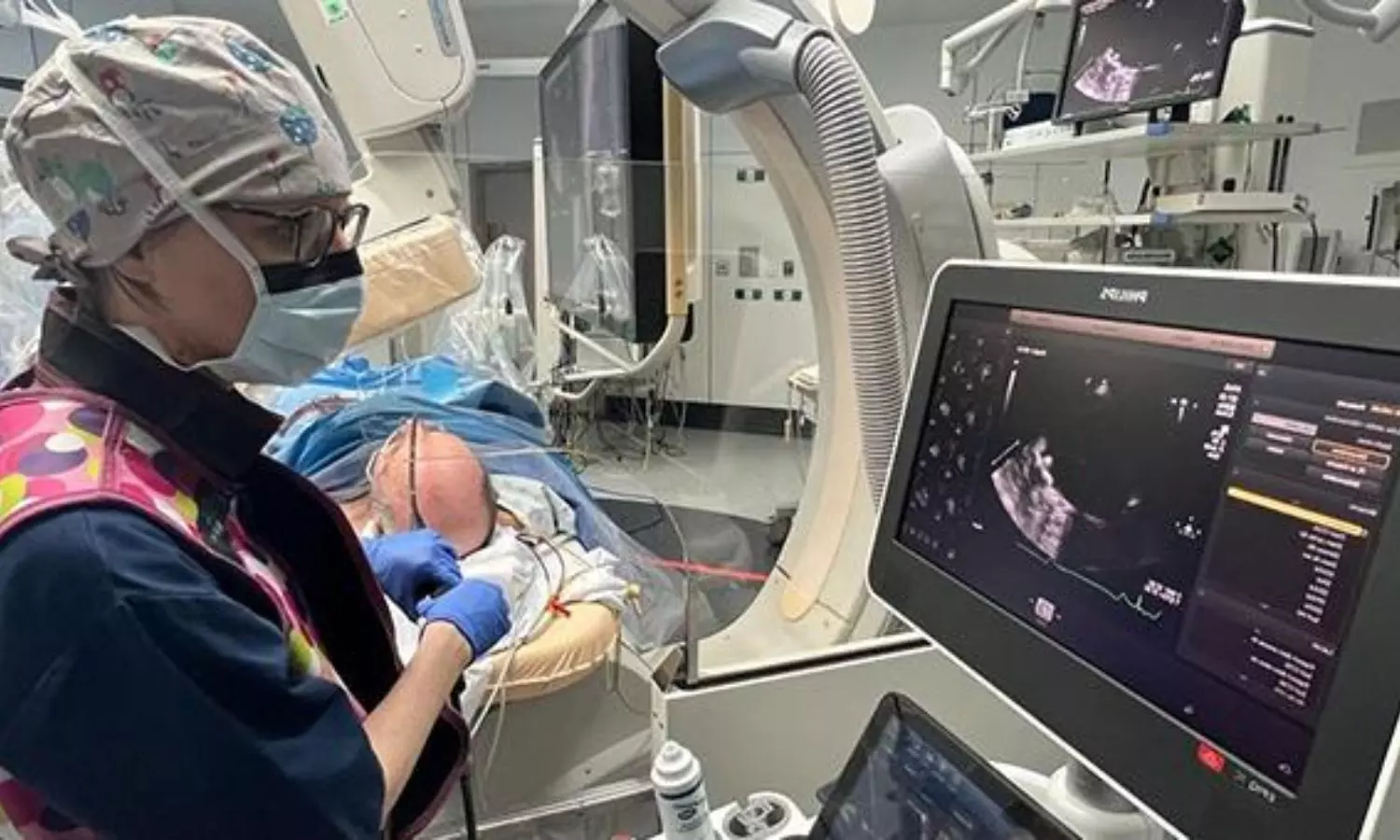
Assessing right ventricular diastolic function is crucial in cardiac surgery, as it significantly impacts long-term prognosis. Typically, RV diastolic dysfunction is evaluated using criteria from the American Society of Echocardiography (ASE) or the British Society of Echocardiography (BSE) via transthoracic echocardiography (TTE). Nevertheless, transesophageal echocardiography (TEE) is the primary method for perioperative evaluation during cardiac surgery. Recent study aimed to assess the agreement between transthoracic echocardiography (TTE) and transesophageal echocardiography (TEE) in grading right ventricular diastolic dysfunction (RVDD) using criteria from the American Society of Echocardiography (ASE) and the British Society of Echocardiography (BSE).

The study included 81 patients undergoing cardiac surgery. TTE and TEE measurements were taken within 10 minutes of each other, under similar hemodynamic, anesthetic, and ventilatory conditions. RVDD grading was performed separately using TTE, TEE mid-esophageal right ventricular inflow-outflow (MERVIO) view, and TEE deep transgastric right ventricular inflow-outflow (DTGRVIO) view, based on both ASE and BSE criteria. According to the ASE criteria, disagreement of ≥1 RVDD grade was seen in 53.1% of patients when comparing TTE and TEE-MERVIO, and in 39.5% when comparing TTE and TEE-DTGRVIO. The weighted kappa was 0.14 and 0.3, respectively, indicating poor to fair agreement. Using the BSE criteria, disagreement occurred in 11.1% and 14.8% of patients when comparing TTE with TEE-MERVIO and TEE-DTGRVIO, respectively, with unweighted kappa of 0.25 and 0.26, indicating fair agreement.

Limits of Agreement and Rater Consistency

The study found wide limits of agreement between individual 2D and Doppler parameters measured by TTE and TEE. There was almost perfect agreement between independent raters for RVDD grading using both TTE and TEE. In conclusion, the study revealed at best only fair agreement between TTE and TEE in grading RVDD. The authors recommend further research to develop a TEE-based algorithm for grading RVDD and to evaluate the prognostic effectiveness of perioperative TEE for predicting adverse clinical outcomes associated with RVDD.

Key Points

1. The study aimed to assess the agreement between transthoracic echocardiography (TTE) and transesophageal echocardiography (TEE) in grading right ventricular diastolic dysfunction (RVDD) using criteria from the American Society of Echocardiography (ASE) and the British Society of Echocardiography (BSE).

2. The study included 81 patients undergoing cardiac surgery, with TTE and TEE measurements taken within 10 minutes of each other under similar hemodynamic, anesthetic, and ventilatory conditions. RVDD grading was performed separately using TTE, TEE mid-esophageal right ventricular inflow-outflow (MERVIO) view, and TEE deep transgastric right ventricular inflow-outflow (DTGRVIO) view, based on both ASE and BSE criteria.

3. Using the ASE criteria, there was a disagreement of ≥1 RVDD grade in 53.1% of patients when comparing TTE and TEE-MERVIO, and in 39.5% when comparing TTE and TEE-DTGRVIO, with poor to fair agreement as indicated by the weighted kappa values.

4. Using the BSE criteria, disagreement occurred in 11.1% and 14.8% of patients when comparing TTE with TEE-MERVIO and TEE-DTGRVIO, respectively, with fair agreement as indicated by the unweighted kappa values.

5. The study found wide limits of agreement between individual 2D and Doppler parameters measured by TTE and TEE, but almost perfect agreement between independent raters for RVDD grading using both TTE and TEE.

6. The authors conclude that the study revealed at best only fair agreement between TTE and TEE in grading RVDD, and they recommend further research to develop a TEE-based algorithm for grading RVDD and to evaluate the prognostic effectiveness of perioperative TEE for predicting adverse clinical outcomes associated with RVDD.

Reference –

Singh N C, Biswas I, Kumar B, et al. (October 07, 2024) The Agreement Between Transthoracic and Transesophageal Echocardiography in the Assessment of Right Ventricular Diastolic Dysfunction Grades in Adult Patients Undergoing Cardiac Surgery: A Prospective Observational Study.

Cureus 16(10): e70976. DOI 10.7759/cureus.70976

Powered by WPeMatico

Add on Estradiol to 2% minoxidil offers no additional advantage in Female pattern hair loss, suggests study

A new study published in the Clinical and Experimental Dermatology journal showed that estradiol with minoxidil (EMX) may raise the risk of irregular menstruation in individuals with female pattern hair loss (FPHL) which is also known as female androgenetic alopecia (AGA). The variables that influence miniature follicle development and the pathophysiology of FPHL are yet unknown. There is proof that genetic, hormonal, and environmental factors also play a part in male AGA, in addition to the most obvious impact of androgens. Women who have thick, healthy hair are more likely to feel secure, confident, and capable of change. They are also more likely to engage with others.

The prevalence of female pattern hair loss is high and has a detrimental effect on one’s quality of life. The most effective treatment for FPHL is minoxidil, however it is more difficult to treat than male pattern hair loss. Several trials treated FPHL with 17α-estradiol solution, either by alone or in combination with minoxidil, with varying degrees of success. Thus, this study evaluated the safety and effectiveness of topical 17α-estradiol 0.01% with minoxidil 2% against minoxidil 2% in the treatment of FPHL, by Nermeen Ibrahim Bedair and colleagues.

For this study, 43 women with FPHL were instructed to apply a spray-on solution comprising either 17α-estradiol 0.01% plus minoxidil 2% (EMX group) or minoxidil 2% alone (MX group) blindfolded twice a day. The course of treatment lasted six months. After baseline and after the conclusion of the therapy, trichoscopic and clinical evaluations were conducted.

Both groups displayed indicators of improvement at the conclusion of the therapy session. The EMX group outperformed the MX group in terms of improvement metrics, however, this difference was not statistically significant. Menstrual abnormalities were more common among the patients of EMX group. Overall, the findings of this study suggest that a 0.01% 17α-estradiol and 2% minoxidil solution does not appear to provide a statistically significant benefit over minoxidil alone, and there may be a greater chance of related menstrual abnormalities in  the treatment of FPHL.

Reference:

Bedair, N. I., EL-Komy, M. H. M., Mohamed, R. E., Shamma, R. N., & Amer, M. A. (2024). Efficacy and safety of combined topical ethinylestradiol with minoxidil versus topical minoxidil in female pattern hair loss. A trichoscopic randomized controlled clinical study. In Clinical and Experimental Dermatology. Oxford University Press (OUP). https://doi.org/10.1093/ced/llae436

Powered by WPeMatico

Elective Single-Embryo and Transfer Mechanisms leading to embryonic splitting and its risk factors, explains study

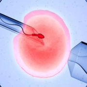
Multiple gestations are the most frequent complications
associated with assisted reproductive techniques (ART). To decrease the rate of
twin and higher order multiple gestations, the elective single-embryo transfer
(eSET) is currently encouraged. However, blastocyst stage SET was still found
to be associated with multiple gestations. The present data revealed that the
frequency of embryo splitting post-eSET is 1.36% with the rate of monozygotic
twin and triplet pregnancies being 0.9-3.1% and 0.048%, respectively. However,
dizygotic twin and triplet pregnancies can also be encountered post-eSET. Both
monozygotic and dizygotic pregnancies can occur in the setting of fresh embryo
transfers, in natural or modified natural frozen embryo transfers (FET), and
rarely in medicated FET cycles. Such events challenge the twinning dogma
proposed by Corner.

Incidence of Zygotic Splitting after SET

Single-embryo transfer is becoming the most favored method
of ART conclusion in recent years as it decreased the complications associated
with multiple gestations. However, multiple gestations in the form of mono- or
dizygotic twinning are still encountered. The classic definition of when one
embryo undergoes fission into 2 or more genetically identical embryos is called
monozygotic splitting, while when 2 different embryos implant, a dizygotic
pregnancy ensures.

The findings of monochorionic and multiple chorionic
pregnancies in blastocyst eSET confirmed the fact that embryo splitting took
place after the transfer. However, not all pregnancies are monozygotic. As per
Osianlis et al., the calculated dizygotic rate in their paper was 0.5% with an
overall Di-Di birth rate of 1%. Based on these numbers, they concluded that 50%
of the multizygotic pregnancies are due to actual embryo splitting while the
other 50% could be explained by concomitant natural conception at the same time
of the ART conception. On another level, the Japanese ART national registry
database along with a survey done by Yamashita et al. documented 122 triplet
pregnancies, of which 46 were single gestational sac pregnancies, 18 were
double gestational sac pregnancies, and 59 were with 3 gestational sacs. It is
worth mentioning that the trichorionic pregnancies had zero fetuses in 9 cases,
1 fetus in 12 cases, 2 fetuses in 9 cases, and three fetuses in 29 cases. One
quadruplet case was also documented.

Chorionicity

Chorionicity refers to the placenta the origin of which can
be determined accurately. Zygosity on the other hand, which is the origin of
the fetus, can be predicted in half of the cases as multiple gestation
pregnancies can originate from one or multiple embryos especially when the sex
of the babies is discordant. Given this fact, same-sex twins or triplets could
be true monozygotic or dizygotic in origin. The only way to accurately diagnose
the zygosity is to do DNA fingerprinting which is expensive and thus not
performed in daily practice.

In contrast, monochorionic multiple gestations are always
monozygotic. Originally, it was thought that the earlier the embryonic
division, the more separate and independent the fetuses were. In other words,
cleavage stage divisions were believed to result in dichorionic diamniotic
pregnancies while blastocyst stage divisions resulted in monochorionic
monoamniotic pregnancies. According to Konno et al., dichorionic pregnancies
were found to be more common with ART. As such, authors can conclude that SET
can result in monozygotic (monochorionic and multichorionic) as well as
dizygotic pregnancies (multichorionic pregnancies).

Risk Factors

Naturally occurring twinning, especially the dizygotic form,
is believed to be linked to a genetic predisposition most commonly located on
chromosome 3. Some ethnicities were found to be more predisposed to dizygotic
twinning where the rate reached 50/1000 in Nigeria. This contrasts with the
naturally occurring monozygotic twinning which was found to be nonaffected by
the ethnicity or the genetic makeup of the couple.

ART on the other hand has increased the incidence of
monozygotic twining. It has been shown that the patient’s young age might
predispose to zygotic twinning while unexplained infertility was found to be
protective. It was proposed that ART associated embryo manipulations such as
FET per se, blastocyst culture, and assisted hatching could be risk factors for
zygotic splitting while the zona manipulation of the oocyte in the form of
intracytoplasmic sperm insemination (ICSI) was not. Interestingly, there was no
difference in the splitting rate neither between the cleavage stage and the
blastocyst stage transfers nor between fresh and frozen embryo transfer cycles.

Another risk factor for splitting is a lower inner cell mass
(ICM) grading of B or C. It is thought that loose intercellular connections may
induce the ICM fission. This has been documented through the time-lapse imaging.
The quality of the culture media is also thought to stimulate zygotic
splitting. An increase in the free radicals’ concentration due to increased
glucose concentration in the culture media used for prolonged culture could
lead to ICM splitting at the site of glucose-induced apoptosis of certain
regions of the ICM.

The new sequential culture systems with antioxidant activity
might explain the lack of increase of the rate of embryo splitting despite the
major increase in the number of IVF cycles and embryo transfers worldwide. When
coupled with the improvement in the embryologists training and experience, the
rate of splitting associated with a blastocyst transfer has been found to
decrease. The OR for embryo splitting decreased from 2.2 to 1.7 when comparing
the periods of 2007 to 2010 and 2010 to 2014. Embryo biopsy on the other hand
was not found to increase the risk of embryo splitting contrary to what was
believed before.

Suggested Mechanisms of Division

It has been shown that blastomeres from a 4-cell stage
embryo can develop into an ICM and trophectoderm; hence, any division after
this stage could give rise to 2 or more embryos with an implantation potential.
Of the suggested mechanisms, abnormal cellular axis formation and cytoplasm
folding in the secondary oocyte prior to fertilization or during the actual
fertilization lead to duplication. It is speculated that gonadotropin
stimulation might disrupt the fine balance and gradients of signalling
molecules affecting the polarity of the oocyte. This is thought to lead to the
formation of 2 cells referred to as daughter cells or tertiary oocytes that
could be fertilized. This is speculated to be caused by the displacement of the
meiotic spindle due to oocyte aging postovulation. This disruption might lead
to the duplication of the axes and formation of 2 embryos upon fertilization or
the fission of the ICM into 2 at the blastocyst stage. This theory would be
replaced later on by the formation of 2 zygotes postfertilization of the
secondary oocyte and not 2 blastomeres.

Other studies advocated the fission to happen closer to the
cleavage stage, and thus, the sequence of events happening during hatching
would then explain the type of the twin gestation. If both blastocysts were
released at the same time, then the resultant pregnancy would be a dichorionic
diamniotic twin gestation.

If on the other hand the blastocysts fused with the
conservation of 2 separate ICMs prior to hatching, then monochorionic
diamniotic twins would appear. If complete fusion of the trophectoderm and the
ICM happened, then monochorionic monoamniotic twins would be created.

Another suggestion was that the ICM would split due to
mechanical compression during hatching through the manipulated zona pellucida
of the embryo which is also referred to as atypical hatching. This atypical
hatching is referred to as 8-shaped hatching, which usually takes place when
the embryo is squeezing out through the hatch of the hardened zona pellucida
due to prolonged culture to blastocyst stage and in cryopreserved-thawed
blastocysts especially with the application of the day 3 prehatching protocol.
It is speculated that this phenomenon might also be the culprit for the
monozygotic triplet gestations that have been documented post-SET.

Another possible explanation to dual or even more ICM is the
nature of human blastomere plasticity. Studies have shown that isolated
trophectoderm cells when cultivated could give rise to a whole new embryo with
an implantation potential. As such, if a blastomere gets separated from the
trophectoderm into the blastocele due to low-grade compaction of the
trophectoderm, this blastomere could give rise to an ICM. Theoretically, each
ICM should give rise to a separate fetus with the surrounding amnion while the
chorion develops during the implantation. The mechanism of chorionic
differentiation between mono and higher order chorionicity in monozygotic
pregnancies is still unknown.

Triplets: Possible Explanation

The explanation of embryo splitting into three is
challenging since triplets after SET is a very rare event. What is known so far
is that to have implantation, an embryo with an intact ICM should be present.
The number of the ICM that the embryo has will define the number of fetuses
that will be seen on the pregnancy ultrasound.

The chorionicity of the pregnancy will depend on the number
of zygotes present at the time of implantation. In theory, the chorion should
rise from trophectoderm cells; thus, it would be logical to consider that the
higher the order of the chorionicity, the higher the number of separate embryos
available for implantation.

In the setting of monochorionic triplets, it is believed
that the blastocyst harbors three distinct ICMs. The mechanism of their
creation might be similar to the ones suggested for the monochorionic twin
gestation. The trigger factor for the splitting into 3 and not into 2 is still
unknown. In the setting of the multichorionic triplets, a suggested explanation
might be the complete division of the hatching embryos resulting in 3 and not
only 1 fully hatched embryo.

Another possible explanation for this is if the origin of
the sister ICMs is a trophectoderm blastomere. Since the implantation potential
of reconstructed embryos cannot be tested at this point due to ethical reasons,
one can only postulate that such cellular plasticity might confer to the newly
formed ICM the whole genetic makeup necessary for a successful implantation and
healthy fetal development. The question that arises here is that whether the
embryo initially had multiple ICM followed by trophectoderm splitting upon
hatching or the splitting of the ICM took place during hatching due to the
mechanical pressure exerted by the hardened zona pellucida. The latter might
explain the high incidence of blighted ova in triplet pregnancies. Due to the
abnormal cell division in the embryo(s) as well as increased cellular stress,
the ICM fails to continue itsdivision resulting in a blighted ovum.

Zygotic splitting is a well-described event in ART, yet the
complete mechanism of these events is not completely elucidated. The hypotheses
that authors have so far remain unproven due to the rarity of zygotic splitting
as well as the ethicolegal considerations of human embryo research. The
presence of such incidents necessitates extensive counselling of couples
undergoing SET.

Source: Mokhamad Zhaffal,1 Rania Al Jafari,2 and Anastasia Salame;Hindawi Journal of Pregnancy Volume 2024, Article ID 2686128, 4 pages https://doi.
Powered by WPeMatico

Prolonged fasting for multiple orthopedic surgeries linked to malnutrition, worse outcomes: Study

People who have multiple orthopedic surgeries during the same hospital stay are more likely to suffer malnutrition due to repeated or prolonged fasting, which can slow recovery and increase the risk of death, according to a study of more than 28 million patients presented at the ANESTHESIOLOGY® 2024 annual meeting.

Because food or liquid retained in the stomach increases the risk of regurgitation and aspiration in the airway and lungs during general anesthesia and deep sedation, most patients are directed to fast for at least eight hours before surgery. This is generally accomplished by not eating after midnight. It is also recommended that only clear liquids may be consumed up to two hours before the procedure. Patients having multiple surgeries while in the hospital, especially those clustered together over several days, are required to fast repeatedly or for cumulative prolonged periods.

“Our research determined that repeated fasting in hospitalized patients having multiple orthopedic surgeries over days or weeks increases the risk for protein-calorie malnutrition, leading to longer hospital stays, slower recovery and higher health care costs,” said Ivie Izekor, B.S., lead author of the study and a fourth-year medical student at Texas A&M College of Medicine, Bryan. “While fasting is a crucial part of ensuring patients’ safety during surgery, our findings suggest modifications to clinical practice should be considered for patients who are at risk for malnourishment, such as those who are older than 65, have a chronic illness like diabetes or congestive heart failure or have limited access to adequate nutrition prior to surgery for socioeconomic reasons. Patients who have frequent surgeries or hospital stays and those with conditions that impair nutrition absorption also are likely at higher risk.”

For the study, researchers analyzed the National Inpatient Sample database between 2016 and 2019. They identified 28,475,485 patients who had orthopedic surgery of any type in the hospital, 1,853,360 (6.5%) of whom were diagnosed with malnutrition after admission. Patients were grouped based on the number of surgeries they had, all of which were performed during a single hospitalization. Patients who were diagnosed with malnutrition had an average of 2.31 surgeries, while those who were not malnourished had an average of 1.57 surgeries.

Researchers found malnourished patients:

  • were at least 15% more likely to die (and the risk increased with more surgeries),
  • had higher hospital costs (an average of $98,000 vs. $48,000) and
  • had longer hospital stays (an average of 9.07 days vs. 4.34 days).

The cause of death in malnourished patients typically was related to infection, complications from poor wound healing or general frailty exacerbated by malnutrition. Researchers chose orthopedic surgeries because they do not directly involve the gastrointestinal system, which could complicate the findings. Patients who have multiple orthopedic surgeries include those with chronic joint conditions such as osteoarthritis, traumatic injuries that require several stages of repair and those needing revisions of initial surgeries.

To prevent malnutrition, researchers suggest that patients undergoing multiple surgeries receive personalized nutritional support during their hospital stay. This support may include dietary assessments by a dietitian, nutritional supplementation, and monitoring nutritional status to help facilitate faster recovery and reduce complications.

“The combined effects of repeated fasting and surgical stress can compromise nutritional status, regardless of the type of surgery, and it is likely our findings would hold true for patients undergoing multiple surgeries of any type,” said George Williams, M.D., FASA, FCCM, FCCP, senior author of the study and professor and vice chair of critical care medicine in the Department of Anesthesiology and Pain Medicine at McGovern Medical School, UT Health, Houston. “However, it may be more challenging to assess the cause-and-effect relationship in gastrointestinal surgeries due to their direct impact on the digestive system.”

Reference:

Prolonged fasting for multiple orthopedic surgeries raises risk of malnutrition, leading to worse outcomes, American Society of Anesthesiologists, Meeting: Anesthesiology 2024.

Powered by WPeMatico

Lipid Pneumonia Presents as Lung Cancer in Breast Cancer Survivor: A Rare Case Study

Taiwan: A recent case study published in the Annals of Thoracic and Cardiovascular Surgery has described a rare case report of lipid pneumonia mimicking lung cancer in a middle-aged woman.

In a remarkable case from a recent medical study, doctors have successfully diagnosed and treated a rare condition known as exogenous lipid pneumonia (ELP) that closely resembled lung cancer in a middle-aged woman. This unusual presentation highlights the importance of accurate diagnosis in differentiating between lipid pneumonia and malignancies.

Lipid pneumonia is a rare lung inflammation caused by the accumulation of fatty substances. It is classified into two types: exogenous and endogenous, with exogenous lipid pneumonia resulting from the inhalation or aspiration of fats. The specific pathogenesis of ELP is still unclear, and only a few case reports exist.

The case described involved a 49-year-old woman who worked as a waitress and had a history of left breast cancer. After undergoing a left modified radical mastectomy at 38, she received chemotherapy and regional radiotherapy. Initially, her chest radiography appeared normal, and she had no reported history of oil ingestion.

Seven years later, the patient experienced general malaise. Chest X-rays revealed increased infiltration in the left lung field, and a CT scan showed regional ground-glass opacity (GGO) in the left lower lung. Over the next year, she developed a dry cough and chest discomfort, with follow-up scans indicating an increase in the size of the GGO. Given these findings, malignancy, specifically lobar bronchioloalveolar carcinoma, was suspected.

To investigate further, she underwent a thoracoscopic lobectomy of the left lower lung. Pathological examination of the removed tissue revealed chronic inflammatory cell infiltration and alveolar spaces filled with CD163+ foamy histiocytes and proteinaceous exudates, confirming the diagnosis of lipid pneumonia.

One year after the surgery, the patient was asymptomatic and underwent a chest CT scan as part of her postoperative surveillance. This scan revealed new areas of mixed consolidation and GGO in the right middle lung. Ongoing monitoring and an optimal postoperative surveillance strategy are planned for her care.

This case highlights the importance of considering ELP in patients with a history of lung abnormalities, especially when previous malignancies or treatments may contribute to lung complications. Continued research and documentation of such cases will enhance understanding of ELP and inform better management strategies for affected individuals.

“We share our experience managing a rare case of exogenous lipid pneumonia that initially presented as lung cancer. The condition was successfully diagnosed and treated through thoracoscopic lobectomy. In specific clinical situations, CT-guided or transbronchial biopsy may be valuable pre-operative diagnostic tools. Notably, the patient had received cyclophosphamide, an antineoplastic agent, seven years earlier, suggesting a potential link to the development of ELP,” Jiun-Chang Wu, Department of Medicine, MacKay Medical College, New Taipei, Taiwan, and colleagues wrote.

“This case highlights the need for further research to establish effective management guidelines for ELP moving forward,” they concluded.

Reference:

Wu JC, Chen TY, Wen-Chien H, Lee JJ, Chen CH. Lipid Pneumonia Mimicking Lung Cancer in a Middle-Age Woman. Ann Thorac Cardiovasc Surg. 2024;30(1):24-00117. doi: 10.5761/atcs.cr.24-00117. PMID: 39477502; PMCID: PMC11524679.

Powered by WPeMatico

Successful Breastfeeding Initiation Linked to Lower Risk of Postpartum Depression, Study Finds

Canada: A recent study published in the Journal of Obstetrics and Gynaecology Canada has found that successfully initiating breastfeeding is strongly linked to a reduced risk of postpartum depression (PPD).

Postpartum depression is a mood disorder that affects approximately 10-20% of new mothers, manifesting within the first year after childbirth. Symptoms include persistent sadness, fatigue, irritability, and difficulty bonding with the baby. The causes of PPD are multifaceted, involving hormonal, psychological, and environmental factors. However, recent findings have pointed to breastfeeding initiation as a potential protective factor against the development of PPD.

Against the above background, Anne-Sophie Roy and Nils Chaillet from Obstetrics and Gynecology, Faculty of Medicine, Laval University, QC, Canada, and colleagues aimed to assess the impact of successful breastfeeding initiation on postpartum depression among women who gave birth in Quebec.

For this purpose, the researchers conducted a secondary analysis of the “Quality of Care, Obstetrics Risk Management, and Mode of Delivery” (QUARISMA) trial, which took place in Quebec from April 1, 2008, to October 31, 2011. The trial aimed to reduce cesarean delivery rates in the region. The study included all women aged 18 and older who gave birth to a single baby at 37 weeks or later. To assess the effect of successful breastfeeding initiation on PPD rates, logistic regression was used. The results were reported using adjusted odds ratios (ORs).

The study led to the following findings:

  • The study included 151,708 women, of whom 21,525 had unsuccessful breastfeeding initiation and 130,183 had successful breastfeeding initiation.
  • The analysis revealed a significant link between successful breastfeeding initiation and a lower rate of postpartum depression, with 0.16% of women who successfully initiated breastfeeding experiencing PPD, compared to 0.29% in those who did not.
  • The odds of developing PPD were 43% lower for women with successful breastfeeding initiation (OR 0.57), and this association was statistically significant.

The findings revealed that successful initiation of breastfeeding is strongly linked to a reduced risk of postpartum depression. The researchers suggest that, due to this association, breastfeeding initiation could serve as a potential preventive strategy for postpartum depression. As a result, health professionals are encouraged to incorporate discussions about breastfeeding initiation into their counseling with new mothers, emphasizing its potential benefits for maternal mental health.

“The success of breastfeeding initiation appears to offer more than just physical benefits for infants—it may also serve as an important factor in supporting maternal mental health. As research continues, healthcare providers must offer guidance and encouragement to mothers in the early postpartum period, ensuring that they have the resources and emotional support necessary to navigate the challenges of both breastfeeding and mental health,” the researchers concluded.

Reference:

Roy, A., & Chaillet, N. (2024). Relation Between Initiation of Breastfeeding Success and Postpartum Depression. Journal of Obstetrics and Gynaecology Canada, 46(11), 102666. https://doi.org/10.1016/j.jogc.2024.102666

Powered by WPeMatico

Blood Flow Restriction in Knee OA Exercise Yields Superior Pain Relief and Strength Gains: Study

Researchers have discovered that exercise programs combined with blood flow restriction (BFR) provide better outcomes for patients who are suffering from knee osteoarthritis (KOA). The authors of a recent study at Ghent University Hospital, found that BFR-added exercise resulted in a larger short-term reduction of pain and increase in strength. This study was conducted by Jacobs E. and colleagues and published in the Annals of the Rheumatic Diseases.

Knee osteoarthritis is one of the most significant causes of disability globally. Exercise is very commonly prescribed, but typically, standard programs alone will only result in limited relief. This study aimed to assess whether BFR could complement exercise and enhance its effects on reducing KOA symptoms, muscle strength, and overall functionality.

The randomized controlled trial known as the Vascular Occlusion for optimizing Functional Improvement in patients with Knee Osteoarthritis, or VO-FIKOA, included 120 KOA patients. The randomization was made into a standard exercise program versus BFR-enhanced programs and conducted over 24 supervised sessions over 12 weeks. Outcomes were measured at baseline, 6 weeks, 12 weeks, and after 3 months post-intervention.

Main outcome measurements included the Knee Injury and Osteoarthritis Outcome Score pain subscale; secondary outcome measures were also assessed using quadriceps strength, the Pain Catastrophizing Scale, as well as functional performance-based measurements. Intention-to-treat principles will be followed for analysis.

  • Pain subscale of KOOS significantly improved in the BFR group with an effect size of 0.58 at 12 weeks (p=0.0009).

  • These improvements remained statistically significant at the 3-month follow-up with slightly decreased effect size of 0.55 (p=0.0008).

  • The BFR group significantly increased the strength of quadriceps with an ES of 0.81 compared to the control group, considerably improved (p<0.0001).

  • Reducing strain in knee joints is an essential step towards alleviating symptoms of KOA.

  • Functional performance, as measured by KOOS functional tests, was improved significantly with the BFR group across the 12-week intervention period and retained its advantages at the 3-month post-intervention time.

  • The improvement in quality of life was observed in the BFR group; hence, exercise with BFR can significantly impact daily life and overall well being.

The results of this study demonstrate that exercise plus BFR leads to larger improvements in pain, strength, and functional measures as compared to exercise alone. It will allow patients to increase their strength and diminish pain with less joint stress. BFR-supported programs may encourage a more active lifestyle, thereby improving muscle function and quality of life even after the end of the intervention.

Reference:

Jacobs, E., Stroobant, L., Victor, J., Elewaut, D., Tampere, T., Wallaert, S., Witvrouw, E., Schuermans, J., & Wezenbeek, E. (2024). Vascular occlusion for optimising the functional improvement in patients with knee osteoarthritis: a randomised controlled trial. Annals of the Rheumatic Diseases, ard-2024-226579. https://doi.org/10.1136/ard-2024-226579

Powered by WPeMatico

Women entering menopause later in life at higher risk of developing asthma: Study

Many studies suggest that an earlier age at menopause is more detrimental to a woman’s health, leading to an increased risk for adverse health conditions such as heart disease, diabetes, osteoporosis, and depression, among others. However, a new study is linking a later age at natural menopause with a greater risk for asthma. Results of the study are published online today in Menopause, the journal of The Menopause Society.

Asthma is a common, chronic disease affecting more than 300 million people worldwide. The prevalence of asthma has been increasing over recent years, creating a substantial economic impact because it is one of the highest diseases for healthcare use. Adult-onset asthma is typically more severe and more difficult to treat than childhood asthma.

Multiple studies have suggested a possible link between asthma and sex hormones. Most notable is the fact that adult-onset asthma is more common in women than men. In childhood, asthma is more prevalent in boys. After puberty, however, asthma occurs more often in girls. Women also tend to have more severe asthma and are less likely to have remission of the disease.

Some studies have found a peak incidence of asthma at around age 40 years, which is commonly the age of the menopause transition, whereas other studies found a peak at the average age at menopause, which is 51 years. Both natural estrogen and synthetic estrogen, such as used in hormone therapy, offer similar risk profiles. Women using hormone therapy were shown to have a 63% increased risk of asthma, whereas women who stopped hormone therapy were two times more likely to quit asthma treatment. Higher body mass index also is shown to be a risk factor for women, but not men, because fat produces estrogen.

Unfortunately, research on the association between menopause and asthma incidence is limited and has yielded conflicting results. That is why this newest study, based on 10 years of follow-up data from more than 14,000 postmenopausal women, was designed to investigate the association between the age at natural menopause and incidence of asthma in nonsmoking postmenopausal women. The study researchers found that women with early menopause (which occurs between 40 and 44 years of age) are at a reduced risk of asthma, which led them to suggest a role of estrogen with asthma risk.

Study results are published in the article “The association between age at natural menopause and risk of asthma among postmenopausal women from the Canadian Longitudinal Study on Aging.”

“This study highlights sex-based differences in asthma, with women at a greater risk for asthma than men in adulthood. It also showed that women with later onset of menopause are at greater risk than those with early onset of menopause. Clinicians should be aware of this link and should monitor women with later age at natural menopause for asthma symptoms,” says Dr. Stephanie Faubion, medical director for The Menopause Society.

Reference:

Kesibi, Durmalouk MA; Rotondi, Michael PhD; Edgell, Heather PhD; Tamim, Hala PhD. The association between age at natural menopause and risk of asthma among postmenopausal women from the Canadian Longitudinal Study on Aging. Menopause ():10.1097/GME.0000000000002443, October 29, 2024. | DOI: 10.1097/GME.0000000000002443.

Powered by WPeMatico

Hearing Loss may be Independent risk factor for Parkinson’s Disease – JAMA study

Recent research published in JAMA Neurology  has unveiled a compelling link between hearing loss and Parkinson’s disease, marking a significant advance in our understanding of preventable risk factors for this debilitating neurological condition. The study, analyzing data from over 3.5 million U.S. veterans through their electronic health records, provides significant evidence that hearing impairment may precede Parkinson’s disease development.

The findings revealed a 26% increased risk of developing Parkinson’s disease in individuals with hearing loss compared to those with normal hearing. More significantly, the research demonstrated a dose-dependent relationship – the more severe the hearing loss, the higher the risk of developing Parkinson’s disease.

Using objective audiometric data rather than relying on subjective reports or clinical judgments, the study followed patients for an average of 7.6 years, with some followed up to 20 years. The study found that patients who received hearing aids within two years of their hearing loss diagnosis showed a significantly reduced risk of developing Parkinson’s disease. Statistical analysis revealed that providing hearing aids to 462 people could prevent one case of Parkinson’s disease over a ten-year period.

The research team also discovered that hearing loss interacts synergistically with known prodromal symptoms of Parkinson’s disease, such as constipation, depression, anxiety, sleep disorders, and loss of smell. When hearing loss occurred alongside these symptoms, the risk of developing Parkinson’s was higher than would be expected from either condition alone.

While the exact mechanism linking hearing loss to Parkinson’s disease remains unclear, several possibilities exist. Hearing loss might directly contribute to neurodegeneration, as evidenced by studies showing elevated tau levels and accelerated brain volume atrophy in individuals with hearing impairment. Alternatively, hearing loss might work indirectly by limiting social engagement or increasing cognitive load, both known risk factors for neurological disorders.

The study’s findings have significant implications for clinical practice. They suggest that hearing screening should be implemented at the primary care level, even when patients haven’t expressed hearing concerns. These results are particularly relevant as we face an aging global population and an expected increase in Parkinson’s disease cases. The World Health Organization projects that neurological disorders, including Parkinson’s, will become leading causes of disability worldwide in coming decades.

The study’s implications are clear: hearing loss should not be dismissed as an inevitable part of aging. Regular hearing screening should be incorporated into routine healthcare, especially for individuals over 40 or those with other risk factors for Parkinson’s disease. Early intervention with hearing aids might serve as a practical, non-invasive strategy to potentially reduce the risk of this serious neurological condition.

This research opens new avenues for prevention and highlights the importance of considering sensory health in the context of neurodegenerative disease risk.

Reference

Neilson LE, Reavis KM, Wiedrick J, Scott GD. Hearing Loss, Incident Parkinson Disease, and Treatment With Hearing Aids. JAMA Neurol. Published online October 21, 2024. doi:10.1001/jamaneurol.2024.3568

Powered by WPeMatico