BMI and Waist Circumference Show Mixed Links to Diabetic Complications in Type 2 Diabetes, Study Finds

Japan: A large-scale Japanese study has uncovered complex relationships between body mass index (BMI), waist circumference (WC), and the risk of serious complications in individuals with type 2 diabetes mellitus (T2DM). Published in Diabetes, Obesity and Metabolism, the study was led by Dr. Megumi Nokihara and her team from the Department of Hematology, Endocrinology, and Metabolism at Niigata University Faculty of Medicine in Niigata, Japan.

The research drew on data from over 114,000 patients with T2DM, making it one of the most comprehensive investigations of its kind. Participants were followed for a median of 4.6 years to assess how BMI and WC might influence the likelihood of developing major diabetes-related complications.

The study revealed the following findings:

  • Higher BMI and greater waist circumference were not consistently linked to worse outcomes in individuals with type 2 diabetes.
  • Elevated BMI in women and a waist circumference of 95 cm or more in men were associated with a lower risk of developing diabetic eye disease requiring treatment.
  • Men with larger waist circumference had a hazard ratio (HR) of 0.79 for diabetic eye disease, indicating reduced risk.
  • A BMI of 25 kg/m² or higher or a waist circumference of at least 90 cm was linked to a significantly lower risk of progressing to dialysis (HR 0.42).
  • The protective association of higher body metrics was not observed for all diabetic complications.
  • Both BMI and waist circumference showed a U-shaped relationship with heart failure risk, where both low and high values increased risk.
  • A BMI ≥25 kg/m² and WC ≥90 cm were associated with a higher risk of heart failure (HR 1.33).
  • Abdominal obesity was linked to an increased risk of cerebrovascular disease, with a hazard ratio of 1.36.
  • Men with very low BMI (<20 kg/m²) showed a higher prevalence of coronary artery disease, indicating that being underweight also carries cardiovascular risks.

The authors conclude that BMI and waist circumference have nuanced and varied associations with diabetic complications. These results highlight the need for individualized risk assessments when setting weight-related health goals for people with T2DM. Instead of adopting a one-size-fits-all approach, clinicians are encouraged to consider the type of complication when advising patients on weight and abdominal fat targets.

“Body mass index and waist circumference showed both protective and harmful associations with various diabetic complications,” the researchers wrote. “Hence, it is important to individualize BMI and waist circumference targets based on the specific complication being addressed, while also taking into account the potential risk of other related health conditions.”

Reference:

Nokihara M, Fujihara K, Yaguchi Y, Takizawa H, Khin L, Ferreira EA, Sato T, Horikawa C, Kitazawa M, Matsubayashi Y, Kodama S, Sone H. The associations of body mass index and waist circumference with the risk of diabetic complications in people with type 2 diabetes mellitus. Diabetes Obes Metab. 2025 May 16. doi: 10.1111/dom.16461. Epub ahead of print. PMID: 40375805.

Powered by WPeMatico

Time-Restricted Eating Matches Calorie Restriction in Reducing Liver Fat in MASLD, Study Finds

Korea: A recent clinical trial published in the Journal of Hepatology has highlighted the potential of time-restricted eating (TRE) as a practical dietary intervention for individuals with metabolic dysfunction-associated steatotic liver disease (MASLD), a condition formerly referred to as non-alcoholic fatty liver disease.

The study was led by Dr. Joo Hyun Oh from the Department of Internal Medicine, Eulji University College of Medicine, Seoul. They aimed to assess whether TRE could deliver similar benefits to calorie restriction (CR) in reducing liver fat and improving metabolic health.

TRE is a dietary pattern where food intake is limited to a specific time window each day, typically without calorie counting. Although it has gained popularity for its metabolic benefits, its role in managing MASLD remains unclear. This 16-week randomized controlled trial provided clinical evidence for its efficacy and safety.

The study enrolled 337 overweight or obese adults diagnosed with MASLD. Participants were randomly assigned to one of three groups: standard of care (SOC), calorie restriction (CR), or time-restricted eating (TRE), in equal proportions. Ultimately, 333 participants were included in the final analysis: 113 in the SOC group and 110 each in the CR and TRE groups.

The primary focus was to evaluate changes in liver fat using magnetic resonance imaging-proton density fat fraction (MRI-PDFF), a non-invasive and accurate imaging tool. Secondary outcomes included alterations in liver stiffness, body composition, lipid and glucose metabolism, and sleep quality.

The following were the key findings of the study:

  • After 16 weeks, participants in the TRE group experienced a 25.8% reduction in liver fat.
  • The reduction in liver fat in the TRE group was significantly greater than the 0.7% reduction seen in the standard care group.
  • The liver fat reduction in the TRE group was nearly identical to the 24.7% reduction observed in the calorie restriction group.
  • TRE also resulted in significant reductions in body weight, waist circumference, and body fat mass.
  • These changes in body composition were comparable to those seen with calorie restriction.
  • No serious adverse events were reported during the study.
  • Liver stiffness, glucose regulation, and sleep quality showed similar outcomes in both the TRE and calorie restriction groups.

The study authors concluded that TRE can be an effective and safe strategy to manage MASLD, offering similar benefits to calorie restriction without requiring strict calorie counting. These findings suggest TRE may be a feasible long-term dietary approach for individuals with fatty liver disease, though further studies are needed to confirm its sustained benefits over time.

Reference:

Oh JH, Yoon EL, Park H, Lee S, Jo AJ, Cho S, Kwon E, Nah EH, Lee JH, Park JH, Ahn SB, Jun DW. Efficacy and safety of time-restricted eating in metabolic dysfunction-associated steatotic liver disease. J Hepatol. 2025 Jun 19:S0168-8278(25)02272-X. doi: 10.1016/j.jhep.2025.06.005. Epub ahead of print. PMID: 40543603.

Powered by WPeMatico

Vaccination as a new form of cardiovascular prevention: an ESC Clinical Consensus Statement

A new ESC Clinical Consensus Statement published in the European Heart Journal discusses the key role of vaccination in preventing cardiovascular events following various viral and bacterial infections.

“We have known for many years that influenza can increase the risk of major adverse cardiovascular events such as heart attacks and can exacerbate heart failure,” said Professor Thomas F. Lüscher, ESC President and senior author of the ESC Clinical Consensus Statement. “More recently, evidence suggests that other respiratory infections are also associated with increased cardiovascular morbidity and mortality. The new publication describes how vaccinations not only prevent infections but also reduce the risk of cardiovascular events, particularly in susceptible individuals.”

The ESC Clinical Consensus Statement describes data on the risk of cardiovascular complications following infections such as pneumococcal pneumonia, influenza, SARS-CoV-2 and respiratory syncytial virus, among others, and describes the inflammatory mechanisms that may be responsible. Evidence is then summarised for the beneficial effects of vaccines in reducing cardiovascular events following various viral and bacterial infections, particularly in at-risk patient groups. Clinical practice guidelines from the ESC and from the American College of Cardiology (ACC)/American Heart Association (AHA) are presented, which advocate for vaccination against influenza and other widespread infections in patients with chronic coronary syndromes (including coronary artery disease) and in those with heart failure.

Serious adverse reactions to vaccinations are very rare. The consensus statement also discusses the risks of cardiovascular adverse events after vaccination, such as myocarditis, and describes appropriate management strategies. Then follows advice on which vaccines should be given to patients with cardiovascular diseases and how often. Vaccination of pregnant women and other vulnerable patient groups, such as those with congenital heart disease and heart transplantation, is considered.

Professor Lüscher concluded: “Prevention is crucial for reducing the considerable burden of cardiovascular disease. The totality of the evidence indicates that vaccinations should become a foundational pillar of preventive strategies alongside other established measures.”

Reference:

Heidecker B, Libby P, Vassiliou VS, et al. Vaccination as a new form of cardiovascular prevention: an ESC Clinical Consensus Statement. Eur Heart J. 2025. doi: 10.1093/eurheartj/ehaf384.  

Powered by WPeMatico

Decoding the Brain: Study evaluates Patterns of Neuronal Death in Hypoxic-Ischemic Encephalopathy Post-Cardiac Arrest

Recent study investigates the pathophysiology of hypoxic-ischemic encephalopathy (HIE) in patients following cardiac arrest (CA), focusing on selective eosinophilic neuronal death (SEND) across various brain regions. A total of 319 patients who experienced successful resuscitation and underwent brain autopsies were analyzed. Eosinophilic neuronal death was quantified in the cerebral neocortex, hippocampus, basal ganglia, cerebellum, and brainstem, categorized according to previously established HIE severity classifications.

Histopathological Findings

Findings revealed that the mean SEND scores varied significantly by brain region, with the hippocampus exhibiting the highest average SEND of 1.8, followed by the neocortex at 1.4, and the brainstem at 0.9. The results identified four distinctive histopathological patterns of HIE: (I) no/mild SEND across all regions, (II) severe SEND localized predominantly to the hippocampus, (III) severe SEND in the neocortex with preserved brainstem function, and (IV) severe SEND impacting the brainstem accompanied by neocortical injury. Notably, 9.7% of patients exhibited substantial heterogeneity in SEND between different neocortical regions, indicating potential clinical implications for neuroprognosis.

Prevalence of Severe SEND

In the cohort studied, 48.3% of patients displayed severe SEND in at least one brain region, with significant proportions demonstrating severe damage to the neocortex while the brainstem remained relatively unaffected. This spatial distribution underscores the hippocampus’s vulnerability, which aligns with previous literature highlighting selective neuronal death patterns following CA.

Variability in SEND Distribution

The inter-regional correlation analysis revealed a predominantly homogeneous SEND distribution across the neocortex. However, a subset of patients indicated substantial regional variation in SEND, challenging the assumption of a uniform HIE pattern following CA. These atypical distributions suggest the necessity for refined neuroprognostic assessments that consider potential nuances in HIE presentations.

Demographic Insights

Demographic data indicated a median patient age of 68, with a higher prevalence of male subjects. Notably, a significant number of patients regained consciousness prior to death, suggesting varying trajectories of neurological recovery linked to the extent of SEND.

Conclusions and Future Directions

The conclusions emphasize the critical need for further investigation into less common HIE patterns and their role in neuroprognostic evaluations. Future research should enable deeper insights into the complex interplay between SEND patterns and clinical outcomes, enhancing predictive accuracy and patient management following CA.

Key Points

– The study analyzes pathophysiological mechanisms of hypoxic-ischemic encephalopathy (HIE) post-cardiac arrest (CA) by examining selective eosinophilic neuronal death (SEND) across brain regions in a cohort of 319 resuscitated patients who underwent brain autopsies, categorizing findings based on established HIE severity classifications.

– Histopathological assessments revealed significant regional variance in mean SEND scores, with the hippocampus showing the highest SEND (1.8), followed by the neocortex (1.4) and brainstem (0.9). Four distinct HIE patterns were identified: (I) no/mild SEND, (II) severe SEND predominantly in the hippocampus, (III) severe neocortical SEND with preserved brainstem, and (IV) severe SEND impacting brainstem and neocortex.

– In this cohort, 48.3% of patients exhibited severe SEND in at least one brain region, with notable neocortical damage often accompanied by relatively intact brainstem function. This finding underscores the susceptibility of the hippocampus to injury in the context of HIE after CA.

– Variability in SEND distribution was observed; while send scores were typically homogeneous across neocortical regions, some patients displayed significant inter-regional differences, suggesting that previous assumptions about uniform HIE patterns need reconsideration. This highlights the complexity of SEND presentations post-CA.

– Demographic analysis revealed a median age of 68 years among subjects, with a notable predominance of males. Additionally, a considerable fraction of patients regained consciousness before death, indicating diverse recovery trajectories linked to the severity and patterns of SEND.

– Conclusions drawn from the study underscore the urgency for further investigations into atypical HIE patterns that could refine neuroprognostic assessments. Future research is recommended to elucidate the relationship between SEND patterns and clinical outcomes, potentially improving predictive models for managing patients after cardiac arrest.

Reference –

C. Endisch et al. (2025). Histopathological Patterns Of Hypoxic-Ischemic Encephalopathy After Cardiac Arrest: A Retrospective Brain Autopsy Study Of 319 Patients.. *Resuscitation*, 110608 . https://doi.org/10.1016/j.resuscitation.2025.110608.

Powered by WPeMatico

More Microvascular Complications Raise Heart and Death Risk in Diabetes, reveals study

The researchers have discovered that the increase in microvascular complications in individuals who have type 2 diabetes greatly raises their risk of cardiovascular events and mortality. The results highlight the importance of early intervention and individualized management in patients with poly-microvascular complications, since such patients are at significantly increased risk of heart attack, stroke, and all-cause mortality. The study was published in Diabetes Research and Clinical Practice by Chun-Ta Huang and fellow researchers.

4,527 people with type 2 diabetes took part in the study. Participants were divided into groups according to how many microvascular diseases they had:

  • Group 0: no microvascular complications

  • Group 1: one microvascular complication

  • Group 2: two or more microvascular complications

The main outcome that was measured was a composite of three severe health events: stroke, myocardial infarction (heart attack), and death. Participants were followed up for a median of 3.3 years by researchers to monitor these events.

Key Findings

A clear dose-dependent association was shown by the study between the amount of microvascular complications and severe health outcomes:

  • 13.07 per 1,000 person-years for Group 0

  • 22.98 per 1,000 person-years for Group 1

  • 35.52 per 1,000 person-years for Group 2

  • Group 2 patients experienced the greatest risk of a primary outcome with a hazard ratio (HR) of 1.73 and a 95% confidence interval (CI) of 1.16–2.59 when compared to patients without microvascular complications.

When the analysis is performed for particular secondary outcomes:

  • The risk of stroke was considerably greater among Group 2 patients (HR: 2.61; 95% CI: 1.19–5.73)

  • The risk of myocardial infarction also rose in Group 2 (HR: 1.67; 95% CI: 1.03–2.70)

Overall, the occurrence of more than one microvascular complication in type 2 diabetic patients drastically increases the risk of stroke, myocardial infarction, and mortality. This Taiwanese experience highlights the need to recognize and treat microvascular diseases at the early stages of diabetes. A focused, preventive approach may lower the burden of macrovascular disease and enhance long-term outcomes in this high-risk group.

Reference:

Chun-Ta Huang, Chiao-Ya Chan, Ming-Chieh Tsai, Shih-Ming Chuang, Sung-Chen Liu, Ming-Nan Chien, Chih-Yuan Wang, Horng-Yih Ou, Chun-Chuan Lee,

Microvascular diseases predict mortality and cardiovascular events in type 2 diabetes: A retrospective cohort study,

Diabetes Research and Clinical Practice,

2025,

112356,

ISSN 0168-8227,

https://doi.org/10.1016/j.diabres.2025.112356.

Powered by WPeMatico

Probiotic May Improve Sleep Quality: New Research Findings

 A recent study published in Engineering has identified a potential new approach to managing sleep disorders. The research, led by a team of scientists from Jiangnan University and other institutions, focuses on the role of S-adenosylmethionine (SAM)-producing probiotics in regulating sleep and circadian rhythms.

Insomnia is a common sleep disorder that affects a significant portion of the global population. Current first-line medications for sleep disorders often have suboptimal effectiveness, highlighting the need for novel therapeutic strategies. In this study, the researchers first conducted a cross-sectional analysis and found that serum SAM levels were significantly reduced in insomnia patients. This discovery suggests that SAM could serve as a potential biomarker and therapeutic target for sleep disorders.

The team then screened 60 gut strains and identified Lactobacillus helveticus CCFM1320, a high-SAM-producing probiotic. In a mouse model of sleep deprivation, treatment with CCFM1320 reversed neurobehavioral abnormalities, such as improved recognition and memory of new objects, reduced hyperactivity, and enhanced spatial exploration ability. Mechanistically, CCFM1320 enhanced the methylation of N-acetylserotonin, a precursor of melatonin synthesis, which normalized the expression of downstream circadian rhythm genes.

To further evaluate the clinical efficacy of CCFM1320, the researchers conducted a four-week placebo-controlled clinical trial involving 60 volunteers with insomnia. The results showed that CCFM1320 significantly improved sleep quality in patients with sleep disorders. Participants had reduced Pittsburgh sleep quality index (PSQI) scores, lower serum cortisol levels, and a decreased prevalence of pathogenic species in the gut. Additionally, probiotic treatment elevated the abundance of SAM synthesis and metabolism-related enzyme genes in the gut microbiome.

The study also explored the impact of CCFM1320 on the gut microbiota of insomnia patients. Although the overall gut microbiota diversity did not change, the intervention affected the composition and abundance of core microbiota species. Probiotic treatment reduced the number of potentially pathogenic species and increased the abundance of some beneficial bacteria.

However, the study has some limitations. The efficiency of gut microbiota-derived SAM supplementation to the circulatory system and brain requires further validation. Also, the differences in the effects of probiotics on sleep quality indices and hormone levels between the human trial and animal studies need to be further investigated.

Overall, this research presents a promising probiotic-based strategy for managing sleep disorders, offering a potential non-pharmacological treatment alternative. Further studies are needed to fully understand the mechanisms and optimize the use of these probiotics in clinical practice.

Reference:

“Regulation of Sleep and Circadian Rhythms by S-Adenosylmethionine-Producing Probiotics,” authored by Peijun Tian, Yuming Lan, Zhiying Jin, Feng Hang, Xuhua Mao, Xing Jin, Gang Wang, Wei Chen. Full text of the open access paper: https://doi.org/10.1016/j.eng.2024.12.025. 

Powered by WPeMatico
Silane-Modified Fillers Enhance Dental Composite Performance, suggests study

Researchers have found in a new study that incorporating filler particles functionalized with silanes containing long alkyl spacers between the silyl and methacrylate groups offers a promising approach for creating packable dental composites. Further these materials demonstrate strong mechanical properties and a significant decrease in viscosity when heated, improving their clinical applicability.

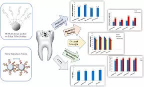
A study was done to evaluate the influence of the nature of silane coupling agents on the consistency of dental composites at various temperatures. Silanes SI 1–4 were synthesized in one single step. They were characterized by 1H and 13C NMR spectroscopy. SI 1–4, as well as 3-methacryloyloxypropyltrimethoxysilane (MPTS), 8-methacryloyloxyoctyltrimethoxysilane (MOTS) and n-dodecyltrimethoxysilane were then used to functionalize a barium aluminum borosilicate glass filler (d50 = 1.0 µm). Silanizations were carried out in cyclohexane in the presence of a catalytic amount of n-propylamine. Each silane was used in an equimolar amount. Composites containing 67 wt% of silanized fillers and packable composites exhibiting a similar consistency at room temperature were subsequently formulated. The consistency of the uncured composites was determined at various temperatures (23 °C, 30 °C, 50 °C and 60 °C) using a texture analyzer. The flexural strength and modulus of the cured composites were assessed according to ISO 4049. Results: The structure of the silane was shown to strongly influence the consistency of composites. The spacer length between silyl and methacrylate groups, as well as the presence of a urea or a urethane moiety, were demonstrated to be key parameters. Heating of each composite resulted in a drop of the consistency. The decrease was however significantly stronger if coupling agents with long spacers were selected. Especially, the use of SI 3 provided a packable composite which exhibited a packable consistency at 30 °C and flowable consistency at 60 °C. Regarding mechanical properties, it was shown that the coupling agent must be able to copolymerize with the monomers of the organic matrix to obtain high flexural strength and modulus values. The silanization of glass fillers using silanes bearing a long spacer was shown to have an additional advantage: packable composites having a higher filler content, and consequently improved flexural modulus, can be formulated. The use of filler particles functionalized with silanes containing long alkyl spacers between the silyl and methacrylate moiety is a promising strategy for the development of packable dental composites which exhibit good mechanical properties and a strong drop in consistency upon heating.

Reference:

Benjamin Grob, Nathan Wachter, Robert Liska, Yohann Catel,

Heating of dental composites: The crucial role of the silane coupling agent on the consistency change, Dental Materials, 2025, ISSN 0109-5641,

https://doi.org/10.1016/j.dental.2025.06.022.

(https://www.sciencedirect.com/science/article/pii/S0109564125006785)

Keywords:

Silane-Modified, Fillers, Enhance, Dental, Composite, Performance, suggest, study, Benjamin Grob, Nathan Wachter, Robert Liska, Yohann Catel, Heating of dental composites

Powered by WPeMatico
Matrix rhythm therapy represents promising advancement in treatment of tissue adhesions: study

Matrix rhythm therapy (MRT) is an innovative therapeutic approach that utilizes vibrational and rhythmic stimulation to restore physiological cellular function. Developed by Randoll et al, MRT targets the ECM and cellular rhythm, promoting tissue regeneration and healing. Unlike traditional manual or instrument-assisted techniques, MRT offers a non-invasive solution that acts on both mechanical and physiological levels.

By influencing the ECM directly, MRT helps to reverse the pathological changes that lead to adhesion formation, offering a novel avenue for improving patient outcomes.

MRT demonstrates promising results in breaking tissue adhesions, improving mobility, and reducing pain by restoring cell matrix dynamics and tissue elasticity.

MRT acts on adhesions through several mechanisms – Mechanical disruption, ECM remodeling, Improved microcirculation, Cellular Effects.

Studies and case reports highlight MRT’s efficacy in various conditions.

• Post-surgical adhesions

Patients undergoing MRT post-abdominal or orthopedic surgery report significant improvements in mobility and reductions in pain.

• Chronic pain conditions

In frozen shoulder and myofascial pain syndrome, MRT demonstrated significant pain relief and enhanced range of motion. Clinical trials indicate a 30–50% reduction in pain scores after 4–6 weeks of treatment.

• Athletic recovery

MRT has facilitated faster recovery in athletes by addressing scar tissue and restoring tissue dynamics. Athletes with hamstring or Achilles tendon injuries returned to activity sooner with MRT compared to standard physiotherapy

MRT offers several benefits compared to traditional therapies.

Non-invasive and painless – MRT provides a targeted approach to specific adhesion sites with minimal risk of complications or side effects.

Enhanced patient comfort – The gentle oscillatory mechanism ensures higher patient compliance, particularly in individuals sensitive to pain.

Comprehensive action – By addressing both mechanical and physiological factors, MRT delivers a more holistic approach to tissue healing.

The authors concluded that – ‘Matrix rhythm therapy (MRT) represents a promising advancement in the treatment of tissue adhesions. By addressing the underlying causes of adhesions and promoting ECM remodeling, MRT offers a non-invasive and effective solution for improving mobility and reducing pain. While preliminary evidence supports its efficacy, further research is essential to standardize protocols and establish its role in clinical practice. MRT has the potential to transform adhesion management, providing hope for patients and clinicians alike.’

Further reading:

The role of matrix rhythm therapy in managing and breaking tissue adhesions

International Journal of Research in Orthopaedics

Rizvi A et al. Int J Res Orthop. 2025 May;11(3):671-674 http://www.ijoro.org

Powered by WPeMatico

New Gene therapy trial shows restored hearing and speech in children born deaf, treated in both ears

A novel gene therapy designed to target a form of inherited deafness restored hearing function in five children who were treated in both ears. The children also experienced better speech perception and gained the ability to localize and determine the position of sound. The study, the world’s first clinical trial to administer a gene therapy to both ears (bilaterally), demonstrates additional benefits than what were observed in the first phase of this trial, published earlier this year, when children were treated in one ear. The research was led by investigators from Mass Eye and Ear (a member of the Mass General Brigham healthcare system) and Eye & ENT Hospital of Fudan University in Shanghai, and findings were published June 5th in Nature Medicine.

“The results from these studies are astounding,” said study co-senior author Zheng-Yi Chen, DPhil, an associate scientist in the Eaton-Peabody Laboratories at Mass Eye and Ear. “We continue to see the hearing ability of treated children dramatically progress and the new study shows added benefits of the gene therapy when administrated to both ears, including the ability for sound source localization and improvements in speech recognition in noisy environments.”

The researchers noted their team’s goal was always to treat children in both ears to achieve the ability to hear sound in three dimensions, a capability important for communication and common daily tasks such as driving.

“Restoring hearing in both ears of children who are born deaf can maximize the benefits of hearing recovery,” said lead study author Yilai Shu MD, PhD, professor, director of Diagnosis and Treatment Center of Genetic Hearing Loss affiliated with the Eye & ENT Hospital of Fudan University in Shanghai, “These new results show this approach holds great promise and warrant larger international trials.”

Over 430 million people around the world are affected by disabling hearing loss, of which congenital deafness constitutes about 26 million of them. Up to 60 percent of childhood deafness is caused by genetic factors. Children with DFNB9 are born with mutations in the OTOF gene that prevent the production of functioning otoferlin protein, which is necessary for the auditory and neural mechanisms underlying hearing.

This new study is the first clinical trial to use bilateral ear gene therapy for treating DFNB9. The new research presents an interim analysis of a single-arm trial of five children with DFNB9 who were observed over either a 13-week or 26-week period at the Eye & ENT Hospital of Fudan University in Shanghai, China. Shu injected functioning copies of the human OTOF transgene carried by adeno-associated virus (AAV) into the inner ears of patients through a specialized, minimally invasive surgery. The first case of bilateral treatment was conducted in July 2023. During follow-up, 36 adverse events were observed, but no dose-limiting toxicity or serious events occurred. All five children showed hearing recovery in both ears, with dramatic improvements in speech perception and sound localization. Two of the children gained an ability to appreciate music, a more complex auditory signal, and were observed dancing to music in videos captured for the study. The trial remains ongoing with participants continuing to be monitored.

In 2022, this research team delivered the first gene therapy in the world for DFNB9 as part of a trial of six patients in China treated in one ear. That trial, which had results published in The Lancet in January 2024, showed five of six children gained improvements in hearing and speech. Shu initially presented the data at the 30th annual congress of European Society of Gene and Cell Therapy (ESGCT) in Brussels, Belgium in October 2023, becoming the first in the world to report clinical data on using gene therapy to restore hearing.

“These results confirm the efficacy of the treatment that we previously reported on and represent a major step in gene therapy for genetic hearing loss,” said Shu. Shu trained under Chen for four years as a postdoctoral fellow at Mass Eye and Ear, with their collaboration continuing for more than a decade since he returned to Shanghai.

“Our study strongly supports treating children with DFNB9 in both ears, and our hope is this trial can expand and this approach can also be looked at for deafness caused by other genes or non-genetic causes,” added Chen, who is also an associate professor of Otolaryngology–Head and Neck Surgery at Harvard Medical School. “Our ultimate goal is to help people regain hearing no matter how their hearing loss was caused.”

Currently, there are no drugs available to treat hereditary deafness, which has made room for novel interventions like gene therapies.

Mass General Brigham’s Gene and Cell Therapy Institute is helping to translate scientific discoveries made by researchers into first-in-human clinical trials. Chen and his colleagues are working with the Institute to develop platforms and vectors with good manufacturing practice standards that would enable his team to more easily test this therapeutic approach with other genes in the future.

The authors note that more work is needed to further study and refine the therapy. The bilateral study requires more consideration compared to the unilateral (one-ear) study as operations in both ears, in the course of one surgery, doubles the surgical time. Furthermore, by injecting double doses of AAVs into the body, the immune response is likely to be stronger and the potential for adverse effects could be greater. Looking ahead, more patients as well as a longer follow-up duration are necessary, and continued analysis of gene therapies and cochlear implants in larger randomized trials will be valuable.

Reference:

Wang, H., Chen, Y., Lv, J. et al. Bilateral gene therapy in children with autosomal recessive deafness 9: single-arm trial results. Nat Med 30, 1898–1904 (2024). https://doi.org/10.1038/s41591-024-03023-5

Powered by WPeMatico

Blood Urea Nitrogen-to-Albumin Ratio Predicts 28-Day Mortality in ICU Patients with CKD: Study

Researchers have found in a new study that Blood Urea Nitrogen-to-Albumin Ratio (BAR) shows a significant association with 28-day mortality risk in ICU patients suffering from chronic kidney disease (CKD), highlighting its potential as a valuable tool for mortality risk stratification.

Chronic kidney disease (CKD) is prevalent worldwide, with patients facing significant mortality risk in intensive care units (ICUs). Early identification of high-risk CKD patients is crucial for improving clinical outcomes. The blood urea nitrogen to albumin ratio (BAR) is a simple and measurable indicator, but its relationship with 28-day mortality in CKD patients is not well established. This study aimed to investigate this association. They conducted a retrospective analysis of eligible CKD patients from the MIMIC IV database. The association between the BAR and 28-day mortality was assessed using Kaplan-Meier survival curves, multivariable Cox regression models, and restricted cubic spline models. Results: A total of 4,625 patients were included, with a 28-day mortality rate of 25.2%. Kaplan-Meier survival curve analysis indicated that patients in the high BAR tertile had significantly lower survival probabilities than those in the low BAR tertile. The adjusted Cox regression model showed that compared to low BAR patients (T1 ≤ 9.8 mg/g), those in T2 (10.0-17.4 mg/g) and T3 (≥ 17.5 mg/g) had increased risks of 28-day mortality, with HRs of 1.49 (95% CI: 1.26–1.76) and 2.04 (95% CI: 1.73–2.40), respectively. Restricted cubic spline analysis indicated a nonlinear association. The BAR is significantly associated with 28-day mortality risk in ICU patients with CKD and may serve as a valuable tool for mortality risk stratification.

Reference:

He, K., Zhu, Y., Wang, W. et al. Association between the blood urea nitrogen to serum albumin ratio and the risk of mortality in patients with chronic kidney disease: a cohort study. BMC Nephrol 26, 275 (2025). https://doi.org/10.1186/s12882-025-04214-z

Keywords:

Blood, Urea, Nitrogen-to-Albumin, Ratio, Predicts, 28-Day, Mortality, ICU Patients, CKD, Study, He, K., Zhu, Y., Wang, W.

Powered by WPeMatico