U.S. survey finds salt substitutes rarely used by people with high blood pressure
Powered by WPeMatico
Powered by WPeMatico
Powered by WPeMatico
Powered by WPeMatico
Powered by WPeMatico
Powered by WPeMatico
Powered by WPeMatico
Powered by WPeMatico
Powered by WPeMatico

Madrid: Routine coronary computed tomography (CCT)-based follow-up after percutaneous coronary intervention (PCI) of the left main coronary artery did not reduce death, myocardial infarction (MI), unstable angina or stent thrombosis compared with symptom-based follow-up, according to late-breaking research presented in a Hot Line session today at ESC Congress 2025
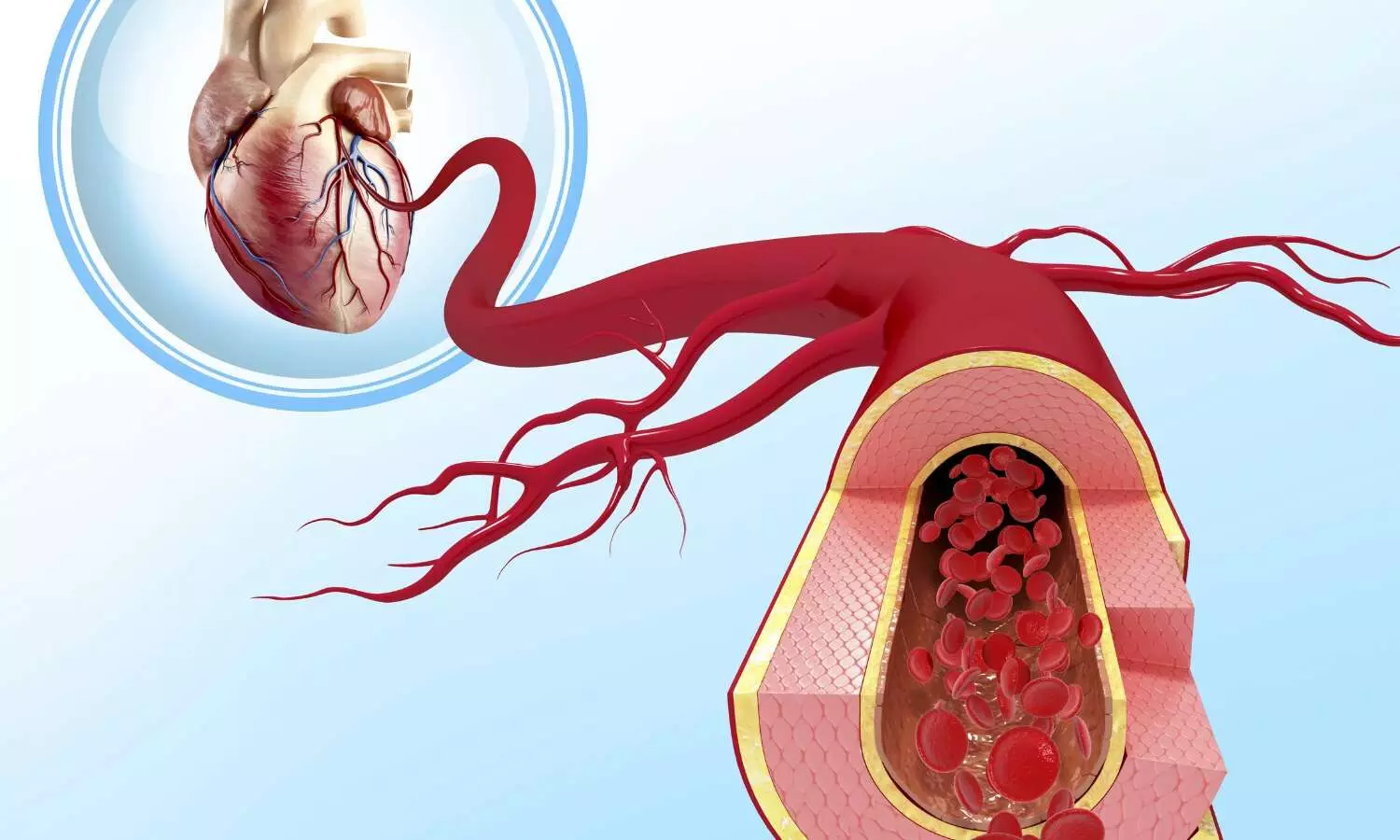
The left main coronary artery supplies a large proportion of the heart muscle and significant left main coronary artery disease is associated with high morbidity and mortality. The introduction of coronary stents along with the improvements in technology and pharmacological management has increased the use of PCI in these high-risk patients with similar results achieved compared with coronary artery bypass grafting.
“Detrimental complications, such as stent restenosis, and recurrent ischaemic events can occur after left main PCI; however, the optimal surveillance strategy remains a subject of debate,” explained trial presenter, Doctor Ovidio De Filippo from Hospital Citta Della Salute e della Scienza di Torino, Turin, Italy.
“In recent years, CCT has emerged as a valuable tool for diagnosis and monitoring, providing accuracy comparable to invasive angiography, while minimising procedural risks and reducing healthcare costs. We conducted the first randomised trial to evaluate the potential benefit of routine CCT-based follow-up at 6 months compared with standard symptom- and ischaemia-driven management in patients after PCI for left main disease.”
PULSE was an open-label, blinded-endpoint, investigator-initiated, randomised trial conducted at 15 sites in Europe and South America.
Participants were consecutive patients with critical stenosis undergoing PCI for left main coronary artery disease. Participants were randomised 1:1 to either a CCT-guided follow-up at 6 months (experimental arm) or standard symptom and ischaemia-driven management (control arm). Participants were followed for an additional 12 months (total follow-up 18 months).
In the CCT arm, if significant left main in-stent restenosis was detected, patients underwent invasive coronary angiography followed by target lesion revascularisation if in-stent restenosis was confirmed. If any significant stenosis was detected in a different site, management was conducted according to the current guidelines. In the standard-of-care arm, patients were managed per clinical guidelines and according to each centre’s standard practice. The primary endpoint was a composite of all-cause death, spontaneous MI, unstable angina or definite/probable stent thrombosis at 18 months.
A total of 606 patients were randomised who had a mean age of 69 years and 18% were female. CCT was performed in 89.8% of patients in the experimental arm at a median of 200 days.
A primary-endpoint event occurred in 11.9% of patients in the CCT arm and 12.5% of patients in the control arm at 18 months (hazard ratio [HR] 0.97; 95% confidence interval [CI] 0.76 to 1.23; p=0.80).
There was a reduced risk of spontaneous MI in the CCT arm vs. the control arm (0.9% vs. 4.9%; HR 0.26; 95% CI 0.07 to 0.91; p=0.004). An increase in imaging-triggered target-lesion revascularisation was observed in the CCT arm compared with the control arm (4.9% vs. 0.3%; HR 7.7; 95% CI 1.70 to 33.7; p=0.001); however, the incidence of clinically driven target-lesion revascularisation was similar between the arms (5.3% vs. 7.2%; HR 0.74; 95% CI 0.38 to 1.41; p=0.32).
Summing up the main findings, Principal Investigator, Professor Fabrizio D’Ascenzo, also from Hospital Citta Della Salute e della Scienza di Torino, said: “Systematic 6-month CCT-based follow-up did not result in a reduction in 18-month all-cause death, spontaneous MI, unstable angina and stent thrombosis. While universal CCT-based follow-up may not be useful, the marked reduction in spontaneous MI and identification of obstructive lesions requiring repeat PCI suggest this approach may be worth investigating further in selected patients with complex anatomies and over longer follow-up.”
Powered by WPeMatico

Pulsed field ablation did not have superior efficacy to radiofrequency ablation in patients with drug-resistant paroxysmal (intermittent) atrial fibrillation, according to results from a late-breaking trial presented in a Hot Line session today at ESC Congress 2025.
Atrial fibrillation (AF) is the most common sustained cardiac arrhythmia. Patients whose AF is not controlled by antiarrhythmic drugs may undergo catheter ablation to disrupt the abnormal electrical pathways that cause the arrhythmia.
Principal Investigator, Professor Pierre Jaïs from the IHU LIRYC (L’Institut de Rythmologie et Modélisation Cardiaque), Bordeaux, France, explained why the trial was carried out: “Pulmonary vein isolation using thermal radiofrequency-based ablation (RFA) is a widely accepted and established treatment for antiarrhythmic drug-resistant AF. However, pulmonary vein isolation has evolved with the introduction of pulsed field ablation (PFA), which is a faster, more straightforward nonthermal procedure that potentially offers more selective tissue targeting than thermal energy sources. Other trials have compared PFA with thermal energy sources with inconclusive results.2,3 We conducted the BEAT-PAROX-AF trial to directly compare PFA with advanced RFA in patients with antiarrhythmic drug-resistant symptomatic paroxysmal AF.”
BEAT-PAROX-AF was an open-label, randomised controlled superiority trial conducted at nine high-volume centres across France, Czechia, Germany, Austria and Belgium. Eligible patients were aged 18–80 years with symptomatic paroxysmal AF that was resistant to at least one antiarrhythmic drug, with a Class I or IIa indication for AF ablation according to ESC Guidelines and effective oral anticoagulation for >3 weeks prior to the planned procedure. Patients were randomised 1:1 to pulmonary vein isolation using either single-shot PFA or point-by-point RFA following the CLOSE protocol. The primary endpoint was the single-procedure success rate after 12 months, defined as the absence of ≥30-second atrial arrhythmia recurrence, cardioversion, Class I/III antiarrhythmic drug resumption after a 2-month blanking period or any repeat ablation. For follow-up, participants were instructed to perform weekly self-recorded single-lead ECGs and to capture recordings during symptomatic episodes using a mobile ECG system.
A total of 289 patients were analysed who had a mean age of 63.5 years and 42% were female. The mean duration of drug-resistant AF was 39 months.
The primary endpoint, single-procedure success at 12 months, was high and similar between the procedure types: 77.2% in the PFA group and 77.6% in the RFA group, with an adjusted difference of 0.9% (95% confidence interval [CI] –8.2 to 10.1; p=0.84).
The mean total procedure duration was significantly shorter for PFA (56 vs. 95 minutes), with an adjusted difference of −39 minutes (95% CI −44 to −34).
Overall, the safety profile was excellent in both groups. Procedure-related serious adverse events Including unplanned or prolonged hospitalisations occurred in 5 patients (3.4%) in the PFA group and 11 patients (7.6%) in the RFA group. Complications appeared more frequent with RFA. One transient ischaemic attack was observed with PFA, while two tamponade percutaneously drained and two cases of pulmonary vein stenosis >70% were observed with RFA. Pulmonary vein stenosis >50% occurred in 12 patients and 15 patients, respectively. No deaths, persistent phrenic palsy or stroke occurred.
Professor Jaïs concluded: “Both PFA and RFA using the CLOSE protocol showed excellent and similar efficacy. Single-procedure success rates were comparable, although there appeared to be fewer complications and a shorter procedure time with PFA.”
Powered by WPeMatico
