Developed by a broad expert collaboration led by Julie De Backer, Cardiology, Ghent University Hospital, Ghent, Belgium, and Center for Medical Genetics, Ghent University Hospital, Ghent, Belgium, these guidelines aim to optimize diagnostic and therapeutic approaches, significantly reducing maternal and fetal morbidity and mortality associated with CVD.
Here are 10 pivotal takeaways from the groundbreaking new guidelines:
1. Pregnancy Heart Team (PHT) is now central, mandated for women with mWHO 2.0 class II–III CVD and above, ensuring multidisciplinary, individualized care from pre-pregnancy through post-partum.
2. The mWHO 2.0 classification, refined with CARPREG II insights, offers nuanced maternal and fetal risk assessment, guiding care intensity and PHT involvement.
3. Pre-pregnancy genetic counseling and testing in specialized centers are recommended for heritable CVD, vital for assessing outcomes and discussing diagnosis options.
4. Essential contraception counseling for all women with CVD is highlighted. For mWHO 2.0 class IV, discussing the exceptionally high risks, including pregnancy termination and psychological support, is strongly recommended.
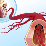
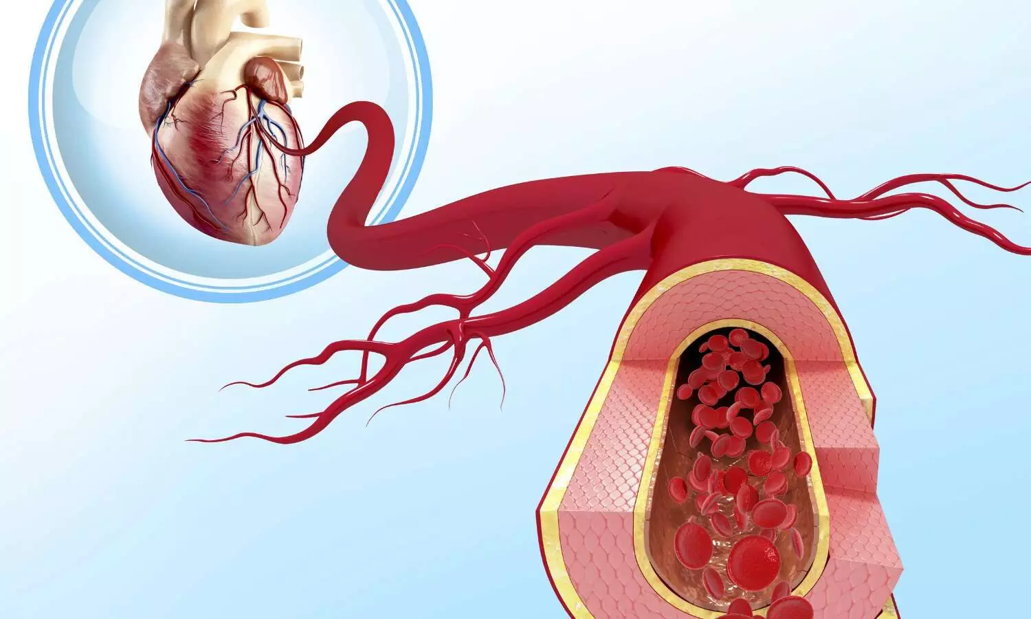
5. ACE-Is, ARBs, ARNIs, renin inhibitors, and SGLT2 inhibitors are contraindicated in pregnancy. DOACs are not recommended. Statins may be considered for established ASCVD or familial hypercholesterolemia. Nadolol/propranolol are recommended beta-blockers for LQTS/CPVT during pregnancy/lactation.
6. Women with PAH are strongly advised against pregnancy due to very high risks. Multidisciplinary counseling, including termination options in specialized centers if pregnancy occurs, is essential.
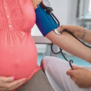
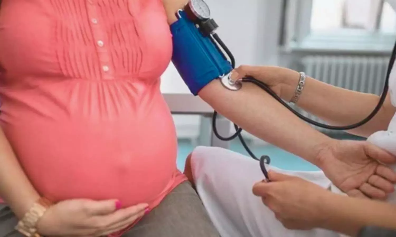
7. Target BP is <140/90 mmHg. Severe hypertension (≥160/110 mmHg) is a hospital emergency. Low-dose aspirin (75–150 mg daily) for pre-eclampsia risk (weeks 12–36/37) is recommended. Methyldopa, labetalol, and dihydropyridine CCBs are first-line treatments; IV hydralazine is second-line for severe cases. Avoid methyldopa post-partum.
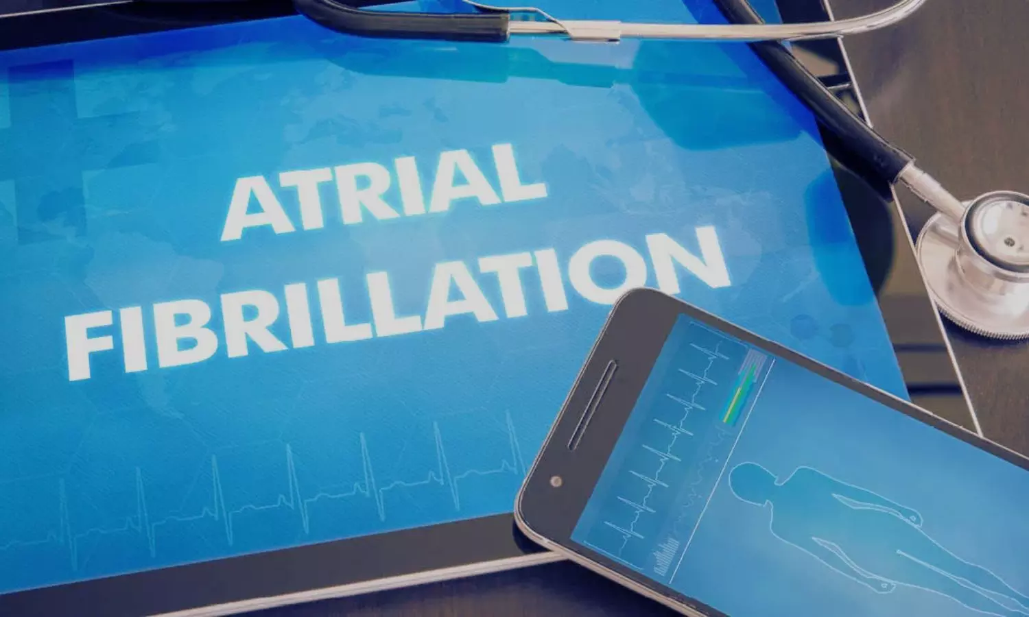
8. For cardiac arrest (≥20 weeks of gestation), continuous manual left uterine displacement, IV access above the diaphragm, and standard CPR/defibrillation are recommended. No drugs should be withheld for teratogenicity. Immediate Caesarean section is considered if ROSC is not achieved after 4 minutes and the fetus is viable.
9. Vaginal delivery is preferred for most women with CVD. Caesarean section for obstetric reasons or severe cardiac conditions (e.g., severe heart failure, uncontrolled arrhythmias, outflow obstruction, VKA use in labor).
10. A new focus highlights Adverse Pregnancy Outcomes (APOs) and their long-term CVD implications. Risk assessment and lifestyle counseling are recommended for women with APO history (e.g., gestational hypertension, pre-eclampsia, GDM). Breastfeeding may reduce future CVD risk in these women.
“These updated guidelines, a product of extensive expert collaboration and the endorsement of leading societies, signify a crucial step towards optimizing diagnostic and therapeutic strategies, ultimately striving to reduce maternal and fetal morbidity and mortality associated with cardiovascular disease, and enhancing long-term cardiovascular health for women,” De Backer and colleagues concluded.
Reference:
De Backer, J., Haugaa, K. H., Hasselberg, N. E., De Hosson, M., Brida, M., Castelletti, S., Cauldwell, M., Cerbai, E., Crotti, L., De Groot, N. M., Estensen, M., Goossens, E. S., Haring, B., Kurpas, D., McEniery, C. M., Peters, S. A., Rakisheva, A., Sambola, A., Schlager, O., . . . Zakirova, F. 2025 ESC Guidelines for the management of cardiovascular disease and pregnancy: Developed by the task force on the management of cardiovascular disease and pregnancy of the European Society of Cardiology (ESC)Endorsed by the European Society of Gynecology (ESG). European Heart Journal. https://doi.org/10.1093/eurheartj/ehaf193