Routine heart CT scanning not warranted after stenting for left main CAD: ESC Study

Madrid: Routine coronary computed tomography (CCT)-based follow-up after percutaneous coronary intervention (PCI) of the left main coronary artery did not reduce death, myocardial infarction (MI), unstable angina or stent thrombosis compared with symptom-based follow-up, according to late-breaking research presented in a Hot Line session today at ESC Congress 2025

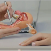
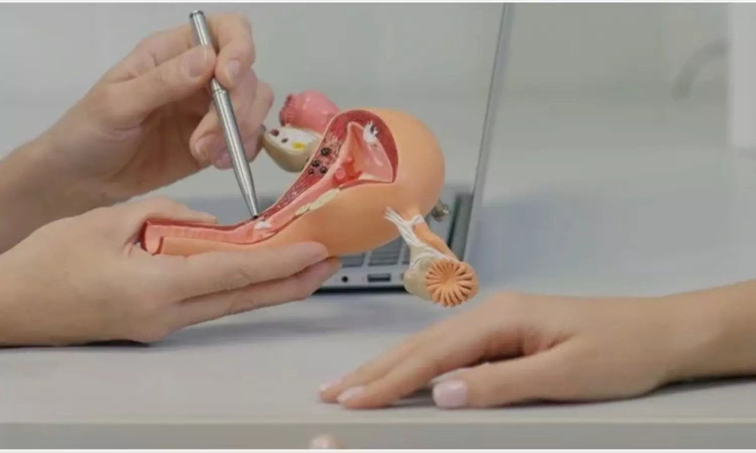
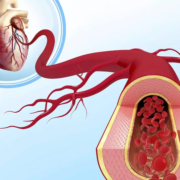
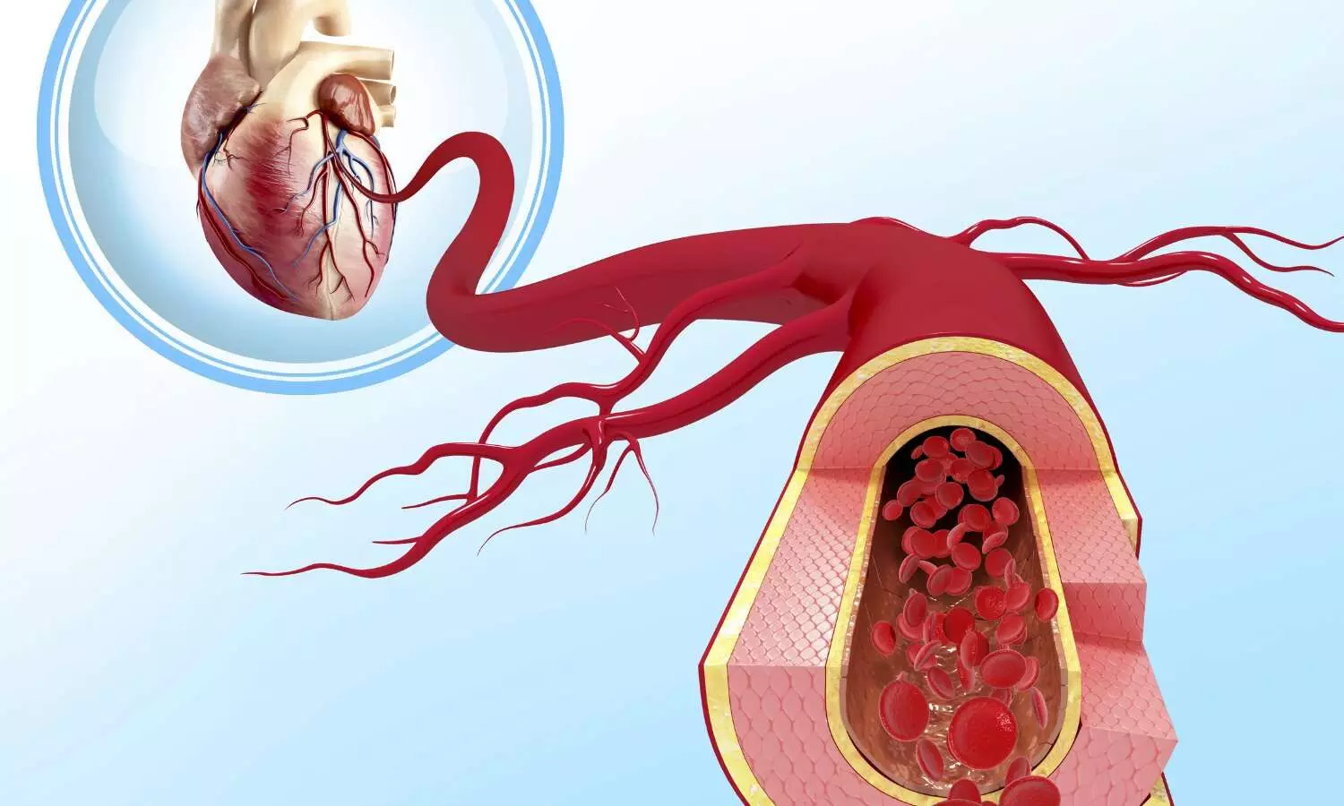
The left main coronary artery supplies a large proportion of the heart muscle and significant left main coronary artery disease is associated with high morbidity and mortality. The introduction of coronary stents along with the improvements in technology and pharmacological management has increased the use of PCI in these high-risk patients with similar results achieved compared with coronary artery bypass grafting.

“Detrimental complications, such as stent restenosis, and recurrent ischaemic events can occur after left main PCI; however, the optimal surveillance strategy remains a subject of debate,” explained trial presenter, Doctor Ovidio De Filippo from Hospital Citta Della Salute e della Scienza di Torino, Turin, Italy.

“In recent years, CCT has emerged as a valuable tool for diagnosis and monitoring, providing accuracy comparable to invasive angiography, while minimising procedural risks and reducing healthcare costs. We conducted the first randomised trial to evaluate the potential benefit of routine CCT-based follow-up at 6 months compared with standard symptom- and ischaemia-driven management in patients after PCI for left main disease.”

PULSE was an open-label, blinded-endpoint, investigator-initiated, randomised trial conducted at 15 sites in Europe and South America.

Participants were consecutive patients with critical stenosis undergoing PCI for left main coronary artery disease. Participants were randomised 1:1 to either a CCT-guided follow-up at 6 months (experimental arm) or standard symptom and ischaemia-driven management (control arm). Participants were followed for an additional 12 months (total follow-up 18 months).

In the CCT arm, if significant left main in-stent restenosis was detected, patients underwent invasive coronary angiography followed by target lesion revascularisation if in-stent restenosis was confirmed. If any significant stenosis was detected in a different site, management was conducted according to the current guidelines. In the standard-of-care arm, patients were managed per clinical guidelines and according to each centre’s standard practice. The primary endpoint was a composite of all-cause death, spontaneous MI, unstable angina or definite/probable stent thrombosis at 18 months.

A total of 606 patients were randomised who had a mean age of 69 years and 18% were female. CCT was performed in 89.8% of patients in the experimental arm at a median of 200 days.

A primary-endpoint event occurred in 11.9% of patients in the CCT arm and 12.5% of patients in the control arm at 18 months (hazard ratio [HR] 0.97; 95% confidence interval [CI] 0.76 to 1.23; p=0.80).

There was a reduced risk of spontaneous MI in the CCT arm vs. the control arm (0.9% vs. 4.9%; HR 0.26; 95% CI 0.07 to 0.91; p=0.004). An increase in imaging-triggered target-lesion revascularisation was observed in the CCT arm compared with the control arm (4.9% vs. 0.3%; HR 7.7; 95% CI 1.70 to 33.7; p=0.001); however, the incidence of clinically driven target-lesion revascularisation was similar between the arms (5.3% vs. 7.2%; HR 0.74; 95% CI 0.38 to 1.41; p=0.32).

Summing up the main findings, Principal Investigator, Professor Fabrizio D’Ascenzo, also from Hospital Citta Della Salute e della Scienza di Torino, said: “Systematic 6-month CCT-based follow-up did not result in a reduction in 18-month all-cause death, spontaneous MI, unstable angina and stent thrombosis. While universal CCT-based follow-up may not be useful, the marked reduction in spontaneous MI and identification of obstructive lesions requiring repeat PCI suggest this approach may be worth investigating further in selected patients with complex anatomies and over longer follow-up.”

Powered by WPeMatico

Pulsed field ablation not superior to radiofrequency ablation in paroxysmal atrial fibrillation: ESC Study

Pulsed field ablation did not have superior efficacy to radiofrequency ablation in patients with drug-resistant paroxysmal (intermittent) atrial fibrillation, according to results from a late-breaking trial presented in a Hot Line session today at ESC Congress 2025.

Atrial fibrillation (AF) is the most common sustained cardiac arrhythmia. Patients whose AF is not controlled by antiarrhythmic drugs may undergo catheter ablation to disrupt the abnormal electrical pathways that cause the arrhythmia.

Principal Investigator, Professor Pierre Jaïs from the IHU LIRYC (L’Institut de Rythmologie et Modélisation Cardiaque), Bordeaux, France, explained why the trial was carried out: “Pulmonary vein isolation using thermal radiofrequency-based ablation (RFA) is a widely accepted and established treatment for antiarrhythmic drug-resistant AF. However, pulmonary vein isolation has evolved with the introduction of pulsed field ablation (PFA), which is a faster, more straightforward nonthermal procedure that potentially offers more selective tissue targeting than thermal energy sources. Other trials have compared PFA with thermal energy sources with inconclusive results.2,3 We conducted the BEAT-PAROX-AF trial to directly compare PFA with advanced RFA in patients with antiarrhythmic drug-resistant symptomatic paroxysmal AF.”

BEAT-PAROX-AF was an open-label, randomised controlled superiority trial conducted at nine high-volume centres across France, Czechia, Germany, Austria and Belgium. Eligible patients were aged 18–80 years with symptomatic paroxysmal AF that was resistant to at least one antiarrhythmic drug, with a Class I or IIa indication for AF ablation according to ESC Guidelines and effective oral anticoagulation for >3 weeks prior to the planned procedure. Patients were randomised 1:1 to pulmonary vein isolation using either single-shot PFA or point-by-point RFA following the CLOSE protocol. The primary endpoint was the single-procedure success rate after 12 months, defined as the absence of ≥30-second atrial arrhythmia recurrence, cardioversion, Class I/III antiarrhythmic drug resumption after a 2-month blanking period or any repeat ablation. For follow-up, participants were instructed to perform weekly self-recorded single-lead ECGs and to capture recordings during symptomatic episodes using a mobile ECG system.

A total of 289 patients were analysed who had a mean age of 63.5 years and 42% were female. The mean duration of drug-resistant AF was 39 months.

The primary endpoint, single-procedure success at 12 months, was high and similar between the procedure types: 77.2% in the PFA group and 77.6% in the RFA group, with an adjusted difference of 0.9% (95% confidence interval [CI] –8.2 to 10.1; p=0.84).

The mean total procedure duration was significantly shorter for PFA (56 vs. 95 minutes), with an adjusted difference of −39 minutes (95% CI −44 to −34).

Overall, the safety profile was excellent in both groups. Procedure-related serious adverse events Including unplanned or prolonged hospitalisations occurred in 5 patients (3.4%) in the PFA group and 11 patients (7.6%) in the RFA group. Complications appeared more frequent with RFA. One transient ischaemic attack was observed with PFA, while two tamponade percutaneously drained and two cases of pulmonary vein stenosis >70% were observed with RFA. Pulmonary vein stenosis >50% occurred in 12 patients and 15 patients, respectively. No deaths, persistent phrenic palsy or stroke occurred.

Professor Jaïs concluded: “Both PFA and RFA using the CLOSE protocol showed excellent and similar efficacy. Single-procedure success rates were comparable, although there appeared to be fewer complications and a shorter procedure time with PFA.” 

Powered by WPeMatico

Sleep problems in early teens associated with future self-harm: Study

Self-harm in young people is a major public health concern, rates are rising, and the adolescent years presents a critical period of intervention. Another modern challenge facing adolescents is sleep deficiency, with global reductions in total sleep time and inconsistent sleep patterns, and as many as 70% of teenagers getting inadequate sleep.

Published today in the Journal of Child Psychology and Psychiatry, researchers at The University of Warwick and University of Birmingham have investigated this relationship between multiple measures of sleep problems and self-harm, using data from over 10,000 teenagers from the Millenium Cohort.

10,000 teenagers, aged 14, were asked about their sleep problems including how long they slept on school days, how long it took to get to sleep, and how often they awoke during the night. They were also asked whether they had self-harmed at 14, a question they were asked again three years later when surveyed at 17 years old.

Michaela Pawley, PhD Candidate, Department of Psychology, University of Warwick, and first author said: “Using large scale data like this really allows you to explore longitudinal relationships at a population level. In this analysis, we discovered that shorter sleep on school days, longer time to fall asleep and more frequent night awakenings at age 14 associated with self-harm concurrently and 3 years later at age 17.”

“While this is clearly an unfavourable relationship, one positive from this research is that sleep is a modifiable risk factor – we can actually do something about it. If the link between sleep and self-harm holds true and with well-placed interventions in schools and homes, there is a lot we can do to turn the tide.”

The researchers found that sleep problems at age 14 were directly associated with self-harm behaviour at age 14 and again at age 17, showing that teenage sleep can have long lasting impacts on self-harm, and could be an avenue to support teenagers at risk.

Sleep problems contributed to risk, even when accounting for other factors that have shown to influence self-harm such as age, sex, socio-economic status, previous instances of self-harm, self-esteem and, importantly, levels of depression. Importantly, only sleep was consistently significant when looking cross-sectionally (age 14) and longitudinally (age 17).

Senior author Professor Nicole Tang, Director of Warwick Sleep and Pain Lab at The University of Warwick added: “Self-harm is one of the leading causes of death among adolescents and young adults. It is a sobering topic. Knowing that poor and fragmented sleep is often a marker preceding or co-occurring with suicidal thoughts and behaviour, it gives us a useful focus for risk monitoring and early prevention.”

The researchers were interested in what could explain this relationship and tested the idea that poor sleep is linked with poorer decision making, which increases your risk of self-harm behaviour. This turned out to not be the case, leaving an open question as to how poor sleep is associated with risk of self-harm.

Regardless, because adolescence is a critical period of vulnerability and potential prevention for self-harm, this study emphasises that sleep health needs to be prioritised in adolescents. Doing so could have long lasting protective effects.  

Reference:

Michaela Pawley, Isabel Morales-Muñoz, Andrew P Bagshaw, Nicole K Y Tang, The longitudinal role of sleep on self-harm during adolescence: A birth cohort study, Journal of Child Psychology and Psychiatry, https://doi.org/10.1111/jcpp.70018.

Powered by WPeMatico

ESC Releases 2025 Guidelines on Heart Disease in Pregnancy: 10 Key Takeaways

Belgium: The European Society of Cardiology (ESC), endorsed by the European Society of Gynecology (ESG), has published its “2025 ESC Guidelines for the management of cardiovascular disease and pregnancy in the European Heart Journal.” Updating the 2018 version, these critical guidelines integrate new evidence to address cardiovascular disease (CVD) as a primary cause of maternal mortality and morbidity, offering refined strategies for pregnant women with cardiac conditions.

Developed by a broad expert collaboration led by Julie De Backer, Cardiology, Ghent University Hospital, Ghent, Belgium, and Center for Medical Genetics, Ghent University Hospital, Ghent, Belgium, these guidelines aim to optimize diagnostic and therapeutic approaches, significantly reducing maternal and fetal morbidity and mortality associated with CVD.
Here are 10 pivotal takeaways from the groundbreaking new guidelines:
1. Pregnancy Heart Team (PHT) is now central, mandated for women with mWHO 2.0 class II–III CVD and above, ensuring multidisciplinary, individualized care from pre-pregnancy through post-partum.
2. The mWHO 2.0 classification, refined with CARPREG II insights, offers nuanced maternal and fetal risk assessment, guiding care intensity and PHT involvement.
3. Pre-pregnancy genetic counseling and testing in specialized centers are recommended for heritable CVD, vital for assessing outcomes and discussing diagnosis options.
4. Essential contraception counseling for all women with CVD is highlighted. For mWHO 2.0 class IV, discussing the exceptionally high risks, including pregnancy termination and psychological support, is strongly recommended.
5. ACE-Is, ARBs, ARNIs, renin inhibitors, and SGLT2 inhibitors are contraindicated in pregnancy. DOACs are not recommended. Statins may be considered for established ASCVD or familial hypercholesterolemia. Nadolol/propranolol are recommended beta-blockers for LQTS/CPVT during pregnancy/lactation.
6. Women with PAH are strongly advised against pregnancy due to very high risks. Multidisciplinary counseling, including termination options in specialized centers if pregnancy occurs, is essential.
7. Target BP is <140/90 mmHg. Severe hypertension (≥160/110 mmHg) is a hospital emergency. Low-dose aspirin (75–150 mg daily) for pre-eclampsia risk (weeks 12–36/37) is recommended. Methyldopa, labetalol, and dihydropyridine CCBs are first-line treatments; IV hydralazine is second-line for severe cases. Avoid methyldopa post-partum.
8. For cardiac arrest (≥20 weeks of gestation), continuous manual left uterine displacement, IV access above the diaphragm, and standard CPR/defibrillation are recommended. No drugs should be withheld for teratogenicity. Immediate Caesarean section is considered if ROSC is not achieved after 4 minutes and the fetus is viable.
9. Vaginal delivery is preferred for most women with CVD. Caesarean section for obstetric reasons or severe cardiac conditions (e.g., severe heart failure, uncontrolled arrhythmias, outflow obstruction, VKA use in labor).
10. A new focus highlights Adverse Pregnancy Outcomes (APOs) and their long-term CVD implications. Risk assessment and lifestyle counseling are recommended for women with APO history (e.g., gestational hypertension, pre-eclampsia, GDM). Breastfeeding may reduce future CVD risk in these women.
“These updated guidelines, a product of extensive expert collaboration and the endorsement of leading societies, signify a crucial step towards optimizing diagnostic and therapeutic strategies, ultimately striving to reduce maternal and fetal morbidity and mortality associated with cardiovascular disease, and enhancing long-term cardiovascular health for women,” De Backer and colleagues concluded.
Reference:
De Backer, J., Haugaa, K. H., Hasselberg, N. E., De Hosson, M., Brida, M., Castelletti, S., Cauldwell, M., Cerbai, E., Crotti, L., De Groot, N. M., Estensen, M., Goossens, E. S., Haring, B., Kurpas, D., McEniery, C. M., Peters, S. A., Rakisheva, A., Sambola, A., Schlager, O., . . . Zakirova, F. 2025 ESC Guidelines for the management of cardiovascular disease and pregnancy: Developed by the task force on the management of cardiovascular disease and pregnancy of the European Society of Cardiology (ESC)Endorsed by the European Society of Gynecology (ESG). European Heart Journal. https://doi.org/10.1093/eurheartj/ehaf193

Powered by WPeMatico

Robot assisted pancreatoduodenectomy Safe and feasible for Uncinate Process Dissection : Study

Pancreatoduodenectomy, also known as the Whipple procedure, is a complex surgical treatment for malignant and benign tumors of the pancreatic head and periampullary region. One of the most technically challenging aspects of this operation is dissection of the uncinate process, particularly when tumors are large and located adjacent to major vessels such as the superior mesenteric artery (SMA). With the advent of minimally invasive and robotic approaches, optimizing surgical strategies to enhance safety and oncologic outcomes has become a key focus in hepatopancreatobiliary surgery.

A new study has found that a mesenteric route, SMA-first approach facilitates precise uncinate process dissection and may represent a safe, feasible option for selected patients undergoing robot-assisted pancreatoduodenectomy. The approach was particularly beneficial in nonobese individuals with large pancreatic head tumors in close proximity to the SMA and portal vein, where conventional dissection can be more technically demanding. In the study, patients undergoing the mesenteric route SMA-first approach demonstrated favorable intraoperative and postoperative outcomes. The technique allowed for early identification and control of the SMA, reducing intraoperative blood loss and providing a clearer operative field. Importantly, the approach enabled meticulous clearance of the uncinate margin, improving the potential for R0 resection in oncologic cases. Operative times were comparable to standard techniques, and postoperative complication rates, including pancreatic fistula and delayed gastric emptying, remained within expected ranges. The authors noted that patient selection is critical. The technique may be best suited for nonobese patients, as excessive visceral fat can obscure visualization in the mesenteric window. Additionally, surgeons require advanced robotic expertise and familiarity with vascular dissection to ensure safety. The findings suggest that this modified approach has the potential to expand the role of robotic surgery in complex pancreatic resections, offering a viable alternative for patients with tumors involving the uncinate process.

Reference
Zhang, Y., Patel, N., & Kim, D. H. (2025). Mesenteric route SMA-first approach in robot-assisted pancreatoduodenectomy: Safety and feasibility for uncinate process dissection. Annals of Surgical Oncology, 32(4), 511–520. https://doi.org/10.1245/s10434-025-1234-7

Keywords: SMA-first approach, mesenteric route, uncinate process dissection, robot-assisted pancreatoduodenectomy, pancreatic head tumors, minimally invasive surgery, vascular involvement, R0 resection, surgical oncology, Annals of Surgical Oncology

Powered by WPeMatico

Half of Lupus Patients Develop Chronic Kidney Disease, Even Without Nephritis: Study Suggests

Israel: An 18-year follow-up of adults living with systemic lupus erythematosus (SLE) has revealed that more than half develop chronic kidney disease (CKD) over time, including a substantial proportion without a history of lupus nephritis (LN).

The findings, published in The Journal of Rheumatology, highlight the serious burden of CKD in SLE and its link to cardiovascular complications, higher hospitalization rates, and increased mortality.
The research, conducted by Dr. Keren Cohen-Hagai from the Department of Nephrology and Hypertension, Meir Medical Center, Kfar Saba, and the Faculty of Medical and Health Sciences, Tel Aviv University, Israel, and colleagues, examined long-term outcomes in 175 adults diagnosed with SLE. Patients were followed for a mean of over 18 years, making this one of the more extensive assessments of kidney involvement in lupus.
The study revealed the following notable findings: 
  • During the study period, chronic kidney disease was diagnosed in 54.6% of patients, with nearly one in three demonstrating both reduced kidney function and albuminuria.
  • Importantly, 46.1% of patients with CKD had no prior history of lupus nephritis, underscoring that renal complications in lupus are not limited to those with overt nephritis.
  • Of the 175 patients included in the study, 12 eventually required kidney replacement therapy.
  • Statistical analyses revealed that lupus nephritis was the strongest predictor of CKD, with a hazard ratio of 5.4.
  • Other significant predictors of CKD included advancing age and lower estimated glomerular filtration rate (eGFR) at diagnosis.
  • These findings suggest that while lupus nephritis is a powerful driver of kidney damage, clinicians should also remain vigilant in monitoring lupus patients without nephritis for potential signs of renal decline.
  • The study further established that CKD in systemic lupus erythematosus carries broad systemic consequences, with patients who had CKD experiencing higher rates of cardiovascular morbidity.
  • Patients with CKD were also more frequently hospitalized for lupus flare-ups and infections, highlighting the broader health burden associated with renal impairment in lupus.
  • Mortality risk was markedly higher, with 19.1% of patients with CKD dying during follow-up compared to only 1.4% of those without CKD.
“Our findings highlight the critical need for early identification and close monitoring of kidney health in all lupus patients, not just those with lupus nephritis,” the authors noted. They emphasized that CKD in SLE should be considered a major determinant of long-term outcomes, requiring proactive management strategies to mitigate associated risks.
By revealing that nearly half of lupus patients with CKD had no nephritis history, the study challenges assumptions about renal involvement in SLE and broadens the scope of patients who may require closer nephrological evaluation. Given the rising recognition of CKD’s impact on quality of life and survival, the results stress the importance of timely diagnosis, routine kidney assessments, and targeted preventive measures in this high-risk population.
“The long-term investigation highlights CKD as a prevalent and clinically significant complication in lupus, with implications beyond renal health. Addressing kidney involvement early in the disease course may improve cardiovascular outcomes, reduce hospitalizations, and enhance survival in patients with SLE,” the authors concluded.
Reference:
Prevalence, Risk Factors, and Outcomes of Chronic Kidney Disease in Patients With Systemic Lupus Erythematosus With and Without Lupus Nephritis. Keren Cohen-Hagai, Mor Saban, Sydney Benchetrit, Dorin Bar-Ziv, Naomi Nacasch, Moshe Shashar, Yael Pri-Paz Basson, Ori Wand, Ayelet Grupper, Shaye Kivity, Oshrat E. Tayer-Shifman. The Journal of Rheumatology Jul 2025, jrheum.2024-1087; DOI: 10.3899/jrheum.2024-1087

Powered by WPeMatico

Elevated serum vitamin D levels are linked with higher skin cancer risk, finds study

Skin cancer, the most common global malignancy, is linked to ultraviolet (UV)-driven serum 25-hydroxyvitamin D (25(OH)D)synthesis, with its controversial role possibly reflecting cumulative UV exposure. This study aimed to assess the association and causality between 25(OH)D levels and skin cancer risk using the National Health and Nutrition Examination Survey (1999–2018) data and Mendelian randomization (MR) analyses, evaluating 25(OH)D as a screening biomarker.

We integrated data from the National Health and Nutrition Examination Survey (1999–2018; n = 21,357 U.S. adults, including 631 skin cancer cases) with MR analyses using genome-wide association study-derived genetic variants to assess the causal relationship between serum 25(OH)D levels and skin cancer risk.

Higher 25(OH)D levels were associated with increased risks of nonmelanoma skin cancer [odds ratio (OR) (95% confidence interval (CI)) = 2.94 (2.10, 4.20)], melanoma [OR (95% CI) = 2.94 (1.73, 5.28)], and other skin cancers [OR (95% CI) = 2.10 (1.36, 3.36)]. MR analyses supported a causal relationship for nonmelanoma skin cancer [OR (95% CI) = 1.01 (1.00, 1.02)] and melanoma [OR (95% CI) = 1.00 (1.00, 1.01)]. Risks were highest in males, older adults, and individuals with obesity.

People with higher vitamin D levels had nearly 3× the odds of getting skin cancer compared to those with lower levels. Genetic analysis supports a modest causal link—meaning vitamin D might contribute to risk, but the effect is small.Vitamin D is produced in your skin when you’re exposed to UV rays—the primary risk factor for skin cancer. Therefore, high vitamin D probably indicates more sun exposure, which drives the increased risk.

Higher serum 25(OH)D levels are associated with increased skin cancer risk, likely reflecting cumulative UV exposure. Routine monitoring of 25(OH)D, combined with UV exposure management, is recommended for risk stratification in skin cancer screening, particularly among high-risk groups. Validation in multiethnic cohorts is needed to confirm these findings.

Reference:

Meng J, Du R, Li P, Lyu J. Association between Serum 25-Hydroxyvitamin D Levels and Skin Cancer Risk: An Observational Study Based on NHANES and Mendelian Randomization Analysis. Cancer Screen Prev. 2025;4(2):89-97. doi: 10.14218/CSP.2025.00010.

Powered by WPeMatico

Study Finds Periodontitis Significantly Increases Glaucoma Risk

Researchers have found in a new study that periodontitis, a common inflammatory disease of the gums and tooth support structures, is strongly associated with open angle glaucoma (OAG), particularly in individuals aged over 40 years, men, and diabetics. The study was conducted by Jeong H. N. and colleagues published in the Journal of Glaucoma.

This cross-sectional population-based questionnaire utilized the Korean National Health and Nutrition Examination Survey (KNHANES) data for 2010–2011 to determine whether periodontitis subjects are at greater risk of OAG, a chronic ocular condition that causes irreversible vision impairment if not treated. The finding of this study highlights that oral health could be implicated in ocular disease prevention or attenuation, especially among susceptible populations.

The research started with the assessment of 17,478 participants from the KNHANES database and narrowed the sample to 3,681 adults aged ≥19 years who had complete and valid data from dental and ophthalmologic examinations. Exclusion factors were thorough, excluding participants with a history of ocular surgeries (e.g., cataract, retina, or refractive surgery), with age-related macular degeneration, pregnant women undergoing orthodontic therapy, or with missing clinic data. The final population was stringently evaluated for periodontitis and glaucoma according to standardized definitions.

Periodontal health was determined by the World Health Organization’s (WHO) Community Periodontal Index (CPI), and glaucoma was diagnosed according to International Society of Geographical and Epidemiological Ophthalmology (ISGEO) criteria.

Key Findings

  • 197 out of 3,681 participants (4.59%) were found to have glaucoma, while 3,484 (95.41%) did not have it.

  • Among the patients with glaucoma, 80 (39.48%) also had periodontitis.

  • However, among the individuals without glaucoma, 892 (22.20%) had periodontitis.

The presence of periodontitis was strongly related to glaucoma:

  • Odds Ratio (OR): 1.53

  • 95% Confidence Interval (CI): 1.06–2.22

  • P < 0.001 after age, sex, diabetes (DM), hypertension (HTN), smoking, and alcohol consumption adjustment.

Subgroup analyses indicated even greater associations in specific groups:

  • For those ≥40 years old, the OR was 1.75 (95% CI: 1.18–2.61).

  • In men, the OR was 1.65 (95% CI: 1.01–2.70).

  • In diabetic patients, the OR was 2.70 (95% CI: 1.46–5.02), indicating they were almost three times more likely to have glaucoma if also diagnosed with periodontitis.

These results reinforce the developing understanding of the mouth–eye relationship and necessitate more longitudinal studies investigating causal pathways and mechanisms. Incorporation of monitoring for oral health into general health evaluations has the potential to enhance the early detection and prevention of glaucoma and perhaps other systemic conditions.

Reference:

Noh, J. H., Lee, M. Y., Yoo, C., Sung, K. R., & Kim, J. M. (2025). Relationship Between Periodontitis and Open Angle Glaucoma: The Korea National Health and Nutrition Examination Survey. Journal of glaucoma, 34(8), 565–574. https://doi.org/10.1097/IJG.0000000000002584

Powered by WPeMatico

Hysterectomy with bilateral oophorectomy may increase risk of stroke, reveals research

Given the seriousness and increased frequency of strokes, many studies have been conducted to assess the relationship between hysterectomy and/or bilateral oophorectomy and the risk of stroke with varying results. A new study suggests women having a hysterectomy and/or bilateral oophorectomy have higher risks of stroke compared with those who did not have surgery. Results of the study are published online today in Menopause, the journal of The Menopause Society.

Stroke is the third dominant cause of death and the fourth dominant cause of disability around the world, representing a significant public health challenge. Therefore, ongoing prevention efforts that address modifiable risk factors are essential to reduce the burden of this disease.

Estrogen levels play a major role. Women of reproductive age have a lower stroke risk, whereas postmenopausal women are roughly two times more likely to have a stroke within a decade of menopause. Both hysterectomy and oophorectomy significantly affect estrogen levels. Hysterectomy may result in lower ovarian sex steroid levels, resulting in earlier menopause. An oophorectomy can reduce premenopausal serum estradiol by up to 80% and androgen levels by about 50% in both premenopausal and postmenopausal women.

Although multiple studies have previously been conducted around the relationship between surgery and stroke risk, results have been mixed. This latest study using data from the National Health and Nutrition Examination Survey (NHANES) included more than 21,000 women, with an average of 8.3 follow-up years, documenting 193 stroke-related deaths. The analysis of these results found an increased risk for hysterectomy with bilateral oophorectomy but not for hysterectomy alone or hysterectomy with unliteral oophorectomy. A pooling analysis of this study’s results with other cohorts, however, revealed an 18% higher risk of stroke for hysterectomy with bilateral oophorectomy, and a 5% higher risk of stroke for hysterectomy alone.

Although the new study lacked surgical indication data, meta-analysis studies show that there is no connection between a benign or malignant diagnosis when determining the associated risk of surgery. Similarly, current evidence does not differentiate the amount of risk based on specific indications (ie, endometriosis, adenomyosis, fibroids, abnormal uterine bleeding, prolapse, or other rare conditions).

Additional studies with a large sample size and longer follow-up period are needed to address the disparities of type of stroke, age at surgery, surgical techniques, and menopause status on the association between stroke risk and hysterectomy and/or bilateral oophorectomy.

Survey results are published in the article “Stroke risk in women with or without hysterectomy and/or bilateral oophorectomy: evidence from the NHANES 1999-2018 and meta-analysis.”

“The results of this study demonstrate increased stroke risk related to hysterectomy and/or bilateral oophorectomy, highlighting that these common procedures carry longer-term risks. They also call attention to an opportunity for more careful assessment of cardiovascular risk and implementation of risk reduction strategies in women who undergo these surgeries,” says Dr. Stephanie Faubion, medical director for The Menopause Society.

Reference:

Shao, C., et al. (2025) Stroke risk in women with or without hysterectomy and/or bilateral oophorectomy: evidence from the NHANES 1999-2018 and meta-analysis. Menopause. doi.org/10.1097/GME.0000000000002616

Powered by WPeMatico

Dehydration in elderly may increase risk of intracerebral hemorrhage and stroke: Study

A new study published in the Journal of Stroke and Cardiovascular Diseases showed that dehydration was associated with an increased risk of stroke in a large sample of persons aged 80 and over.

Stroke is still a major cause of morbidity and death globally, and early clinical status and modifiable risk factors have a significant impact on outcomes. It is becoming more widely acknowledged that dehydration, a prevalent but sometimes disregarded ailment in hospitalized and elderly patients, may have a role in the severity and recovery of stroke.

Dehydration can exacerbate ischemia damage by increasing blood viscosity, promoting thrombus formation, and impairing brain perfusion. This study looked through a sizable database to see whether dehydration and stroke are related. Adults 80 years of age and older who had medical visits prior to the pandemic, from January 1, 2018, to December 31, 2019, made up the research population.

Laboratory test data and ICD-10 diagnostic codes were used to identify the dehydration cohort. Based on ICD-10 codes, outcome measurements included one of 3 stroke types: transient ischemic attack, ischemic stroke, or intracerebral hemorrhage. A subanalysis was conducted on people with diabetes.

This study found a total of 563,476 of the 3,125,610 people aged 80 and above were dehydrated. There were 443,450 diabetics and 101,661 dehydrated people. In both groups, the dehydration cohorts were somewhat older (82.7 vs. 82.4, p <0.001), slightly more female, and non-Hispanic white.

Dehydrated people had 1.98–3.99 times the risk of stroke after adjusting for common covariates in propensity score matching: ischemic stroke (OR=1.98, 95% CI=1.9-2.07), TIA (OR=2.88, 95% CI=2.74-3.28), and intracerebral hemorrhage (OR=3.99, 95% CI=3.41-4.67). The diabetic group experienced ischemic stroke (OR=1.97, 95% CI=1.81-2.16), TIA (OR=2.81, 95% CI=2.33-3.39), and intracerebral hemorrhage (OR=6.76, 95% CI=4-11.42).

Overall, in this sizable sample of 3 million people, as well as in a subset with diabetes, a substantial correlation between dehydration and stroke was discovered. The risk of dehydration in older persons is increased by both the physiological changes associated with natural aging and drugs used to address cardiovascular stroke risk factors. Blood viscosity may account for the link between ischemic stroke and TIA, but the intracerebral hemorrhage correlation is unexpected. Future research ought to assess if better hydration results in fewer strokes.

Source:

Hamrick, I., Tuan, W.-J., Harker, P., Adogwa, O., & Hyacinth, H. I. (2025). Association between dehydration and stroke, a retrospective cohort study of a large database. Journal of Stroke and Cerebrovascular Diseases: The Official Journal of National Stroke Association, 108430, 108430. https://doi.org/10.1016/j.jstrokecerebrovasdis.2025.108430

Powered by WPeMatico