United Doctors Front announces National Core Committee for 2025-26

New Delhi: United Doctors Front (UDF), an organization dedicated to the rights and welfare of doctors and medical students, has announced its National Core Committee for the 2025-26 session.
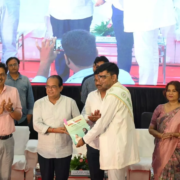
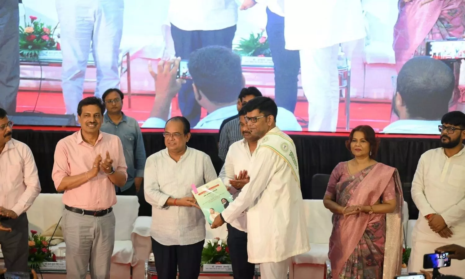
The 2025–26 term will place special emphasis on the safety and dignity of the medical community. The new team comprises both experienced experts and dedicated young doctors.
Dr Lakshya Mittal has been appointed as the Chairperson and National President. Dr Amit Vyas has been designated as the National Vice President, while Dr Arun K. Kumar takes over as the National General Secretary.
Also Read:Doctors Urge Union Health Minister to Implement Rotatory Headship at AIIMS, PGI Chandigarh
Dr Meer Wasim and Dr. Rakesh Beniwal have been given the role of State Joint Secretaries. Dr. Yagika Pareek and Dr. Bhanu Kumar have been appointed as State Spokespersons.
The responsibility of the National Finance Secretary has been entrusted to Dr. Bharat Rathore. Additionally, Dr. Aditi Singh (Social Media Secretary), Swami Das (Head, National RTI Cell), Dr. Charu Mathur and Satyam Singh Rajput (Legal Advisors), and Dr. Lay Paghadar (Divyangjan Secretary) have been announced. Dr. Akshat Gautam and Dr. Manoj Jat have been appointed as JR Secretaries.
Furthermore, Dr. Shubhapratap Solanki, Dr. Hanuman Bishnoi, Dr. Krishna Sharma, and Dr. Alok Singh have been appointed as North Zone Secretaries. Dr. Hariharan will serve as the South Zone Secretary. Dr. Anshuman Patra, Dr. Jugal Krishna Dole, and Dr. Amit Kumar Giri will hold the positions of East Zone Secretaries. Dr. Ajit Singh Shekhawat, Dr. Samyak Bansal, and Dr. Sukharam Gehlot will serve as West Zone Secretaries. Dr. Vikas Milky, Dr. Sasanpuri Sai Santosh Teja, and Dr. Rajat Khurana have been appointed as Central Zone Secretaries.
Dr. Harshit Naranival, Dr. Divjot Singh Kalra, and Dr. Akshay Sharma have been made Anti-Ragging Secretaries. The role of Mental Health Secretaries will be taken up by Dr. Prashant Sharma, Dr. Lalit Tanwar, and Dr. Anshita Chhabra. Dr. Kanchan Dochaniya and Rhythm will lead the Research Cell, while Dr. Abhinandana Tokas and Dr. Khushboo Vyas have been given charge of the Academic Cell. Dr. Yogendra Pal Yadav and Dr. Vansh Chopra have been appointed as FMG Secretaries. The IT Cell will be handled by Dr. Rakesh Beniwal and Dr. Gopal Singh.
In the Student Wing, Batul Fatima, Amrit Singh, Aryan Kansal, and Akshat Tiwari have been appointed as Secretaries. Dr. Jagpati Bhardwaj will lead the Dental Wing. The Social Media team includes Dr. Shashank Tiwari, Deependra, Pragya Chauhan, Tripti Yadav, Dr. Ayaan Bhati, I.V. Sabarish, and Sheikh Kaifuddin.
National President Dr. Lakshya Mittal stated that the new team will take concrete steps toward major reforms in medical education, ensuring defined working hours for resident doctors, and preventing violence against doctors. The goal is to protect the dignity, safety, and well-being of doctors while ensuring quality healthcare services for every citizen.
Also Read:United Doctors Front Association Elects New Leadership for 2024-2025
Powered by WPeMatico