Adipokines: A New Tool for Early Detection of Metabolic Syndrome in PCOS patients, Study Finds

India: A recent study published in Cureus has found that over 40.2% of women with polycystic ovary syndrome (PCOS) developed metabolic syndrome (MetS) and identified a strong link between altered adipokine levels and the early stages of MetS development.

“Adipokines such as adiponectin, leptin, and resistin showed significant variations between women with and without MetS, suggesting their potential as early biomarkers for detection and intervention,” the researchers reported. “In contrast, traditional indicators like fasting glucose, waist circumference, and triglyceride levels were more predictive in later stages, reinforcing the value of adipokines in the early diagnosis and management of MetS in PCOS patients.”

Women with polycystic ovary syndrome are more likely to develop metabolic syndrome, as both conditions share common underlying factors. Adipokines—hormones produced by fat tissue—play a key role in this link by affecting how fat is stored and used in the body. These changes in fat metabolism can contribute to the development of MetS.

To better understand this connection, Chaitali Maitra, Biochemistry, Dr. S.S. Tantia Medical College Hospital and Research Centre, Sri Ganganagar, IND, and colleagues looked at adipokine levels in women with PCOS, both with and without MetS, aiming to explore whether these molecules could serve as early warning signs for metabolic complications in PCOS patients.

For this purpose, the researchers conducted a monocentric, tertiary hospital-based study involving 144 women with PCOS, classified according to the Rotterdam criteria and screened for MetS. Based on the presence or absence of MetS, participants were divided into two groups. The study measured and compared adipokine levels—adiponectin, leptin, and resistin—along with various biochemical and anthropometric parameters. Data analysis was performed using Jamovi statistical software (version 2.3), employing independent samples t-tests and multinomial regression to assess the associations.

The study led to the following findings:

  • Out of 144 women with PCOS included in the study, 58 were diagnosed with metabolic syndrome (MetS).
  • The highest prevalence of MetS was observed in the non-PCO (O+HA) phenotype, accounting for 53 of the 58 MetS cases.
  • Statistically significant differences in adipokine levels were found between the MetS and non-MetS groups.
  • Adiponectin, leptin, and resistin emerged as significant early predictors of MetS in women with PCOS.
  • In the later stages of MetS, traditional markers such as fasting blood glucose, waist circumference, and triglyceride levels remained reliable indicators.

The study found a high prevalence of metabolic syndrome in women with PCOS, with altered adipokine levels serving as early predictors of its development.

“While adipokines were significant in the early stages, traditional markers like fasting blood sugar, triglycerides, and waist circumference became more predictive in later stages. Measuring adipokines early on could aid in the timely diagnosis and management of MetS in PCOS patients,” the authors wrote.

Reference:

Maitra C, Maitra A (April 16, 2025) Role of Adipokines in the Development of Metabolic Syndrome in Patients With Polycystic Ovary Syndrome. Cureus 17(4): e82355. doi:10.7759/cureus.82355

Powered by WPeMatico

Study Reveals Link Between Abdominal CT Use and Sarcopenia Diagnosis Among Older Adults

A new Harvey L. Neiman Health Policy Institute study found evidence that a new diagnosis of sarcopenia, a potentially reversible disease with low muscle mass and strength, often coincides with an abdominal CT study. Increasing opportunistic evaluation of abdominal CT scans could facilitate diagnosis of sarcopenia and ultimately improve patient care. The research, published today in the Journal of the American College of Radiology was based on nearly 3 million Medicare fee-for-service beneficiaries over the 2017-2022 period.

The term sarcopenia was first proposed in 1989 to describe the loss of muscle mass, but it did not receive an International Classification of Disease (ICD) code until 2016. Yet, “Since sarcopenia received an ICD code, its diagnosis has increased multifold from 0.01% of Medicare beneficiaries in 2017 to 0.07% in 2022,” stated Eric Christensen, PhD, Research Director at the Neiman Health Policy institute. “Despite this increase in sarcopenia’s diagnosis, it remains underdiagnosed compared to the estimated 10-27% of the elderly population with this disease.”

“Improving sarcopenia diagnosis has the potential to improve patient care through tailored interventions, such as exercise programs and nutritional support” said Robert Boutin, MD, Clinical Professor and Director of Musculoskeletal Imaging Fellowship at the Stanford University School of Medicine. “The challenge is that screening for sarcopenia, which typically starts with a patient questionnaire or gait speed assessment prior to imaging, is not routine as is screening for osteoporosis. Because CT is a reference standard for quantifying muscle mass, using CT scans patients are already receiving for the additional purpose of sarcopenia screening could further radiologists’ role in population health management.”

Unlike dual-energy X-ray absorptiometry, known as DXA scans, which are specifically for diagnosing osteoporosis, there is no procedure code for the express purpose of diagnosing sarcopenia. Given the inability to directly observe screening events, the researchers identified all abdominal CT scans received by Medicare beneficiaries diagnosed with sarcopenia. They then analyzed the temporal association of the CT examination and the sarcopenia diagnosis.

“We looked back from each patient’s sarcopenia diagnosis date and found that the abdominal CT rate climbed exponentially starting about 90 days beforehand and peaked on the diagnosis date,” said Dr. Christensen. “These findings show a clear temporal association between abdominal CT use and a sarcopenia diagnosis, but we cannot determine from these data whether this represents intentional or opportunistic use of CT for diagnosing sarcopenia.”

“Given the underdiagnosis of sarcopenia, the increasing number of CT scans and CT scanners, and the interest in implementing AI tools for sarcopenia diagnosis at the point of imaging care, opportunistic abdominal CT could play an important role in increasing the diagnosis of sarcopenia,” stated Dr. Boutin. “In this way, radiologists can increase their role in population health management and reduce disparities across socioeconomic groups while increasing the value to patients when abdominal CT scans are ordered.”

Reference:

Christensen, Eric W. et al., Sarcopenia Diagnosis Trends and Opportunistic Use of Abdominal CT Among Medicare Beneficiaries, Journal of the American College of Radiology.

Powered by WPeMatico

Image-Based Memory May Heighten Symptoms of PTSD, reports study

When you’re asked to remember something, does an image pop into your head? Many people’s memories are intricately tied to their use of imagery.

In an upcoming Clinical Psychological Science study, Ryan Yeung, a postdoctoral fellow at the Baycrest Academy for Research and Education, examined this trait in people with post-traumatic stress disorder (PTSD).

Using imagery can help people keep their memories vivid for longer, previous studies have shown (Anderson et al., 2017; Rasmussen & Berntsen, 2014). The idea is so popular that people who compete in memorization contests often use imagery as a technique for memorizing large chunks of information quickly.

However, in a condition like PTSD, a strong sense of imagery could be harmful (Clark & Mackay, 2015; Holmes et al., 2015). Individuals with the disorder often experience vivid flashbacks, wherein the traumatized person feels transported back to a stressful scenario through sense memory, said Yeung.

“It was really shocking that when we looked into [the literature], maybe half of the studies find the effect, half of them don’t,” he said in an interview.

Yeung and his colleagues hypothesized that this discrepancy could be due to how earlier research had measured imagery itself. So, for their study, the team investigated two different traits of imagery: object imagery, which includes physical details such as shape, color, and form; and spatial imagery, which includes details such as location, direction, and movement.

To measure a person’s object imagery, a researcher might ask a person to conjure up an apple in their mind’s eye, and then have the participant describe its color, size, and shape. To measure spatial imagery, a researcher might ask a person to draw a map of a floor plan they’ve visited.

Yeung and colleagues tested how these different traits of imagery relate to PTSD symptoms. In two separate studies, participants were given a series of surveys to measure their object and spatial imagery traits. In total, the studies comprised 3,203 participants, some of whom had experienced a traumatic event. In the first study, participants ranged in age between 18 and 83. In the second study, participants were all undergraduates.

In both, participants were asked to respond to questions about their PTSD symptoms, depression, anxiety, and object and spatial imagery.

Some of the survey items designed to measure object imagery may have, for example, asked participants if it was easy for them to close their eyes and easily picture a scene they had experienced (Blajenkova et al., 2006), whereas a question measuring spatial imagery may have asked participants if they could easily rotate three-dimensional geometric figures in their mind.

During analysis of these datasets, the researchers controlled for depression symptoms and gender, which previous research had found could skew studies of this sort (Holmes et al., 2016; Ji et al., 2019). After controlling the data, they found that people with strong object imagery were more likely to have severe PTSD symptoms than people with strong spatial imagery.

“We were definitely surprised, because there isn’t that much out there, especially in terms of the spatial aspect,” Yeung said.

It’s not clear why spatial imagery might be less linked to negative symptoms of PTSD than object imagery. However, Yeung said that people who use spatial imagery frequently are able to recontextualize and process their traumas better than those with strong object memory. Thinking about their trauma in different ways instead of essentially reliving it could help a person work through a painful event, Yeung explained.

On the opposite end, people with strong object memory might more easily relive painful memories. Essentially, Yeung said, “this so-called benefit of having imagery just naturally could actually harm these people.”

In the future, Yeung believes these findings may inform new therapies. Clinicians could design interventions that illicit and strengthen a person’s spatial imagery, which could, in theory, help patients work through their trauma and become less burdened by it.  

Powered by WPeMatico

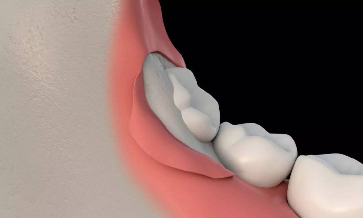
Low-Level Laser Therapy Shows No Significant Relief After Wisdom Tooth Removal: Study Finds

Switzerland: A new research, published in the Journal of Oral and Maxillofacial Surgery revealed that low-level laser therapy (LLLT), often promoted as a modern, non-invasive solution to reduce postoperative discomfort, may not offer the expected relief after wisdom tooth extraction.

Postoperative pain, swelling, and trismus are the most frequently encountered complications following the surgical removal of wisdom teeth. While various methods have been explored to manage these symptoms, the effectiveness of low-level laser therapy remains a topic of debate.

In this context, the present study aimed to assess whether LLLT could offer a significant reduction in pain, swelling, and trismus after the extraction of both upper and lower third molars. Esra Sekerci, Assistant Dentist in Further Training for Oral Surgery, Clinic of Cranio-Maxillofacial and Oral Surgery, Center of Dental Medicine, University of Zurich, Switzerland, and colleagues hypothesized that LLLT would result in greater postoperative relief compared to the side of the mouth treated with a placebo laser (PL), thereby providing a potential adjunctive tool for improving patient comfort and recovery.

For this purpose, the researchers conducted a double-blind, randomized, split-mouth study at the University of Zurich involving 20 healthy patients aged 18 to 24 with impacted wisdom teeth. Patients were randomly assigned to receive either low-level laser therapy (LLL) or placebo laser (PL) treatment. Pain, swelling, and trismus were assessed preoperatively and on days 3 and 7 after surgery using a visual analog scale, 3D scanner, and ruler, respectively. Age and sex were comparable across groups and included as covariates. Statistical analysis was performed using a two-sided Wilcoxon signed-rank test with a significance level of 0.05.

The key findings of the study were as follows:

  • The study included 20 subjects with a mean age of 20.4 years (±1.9), of whom 55% were female.
  • On day 3 post-surgery, the median pain score was 2.5 in the LLLT group and 2.0 in the placebo group.
  • Median swelling on day 3 was 10,697 mm³ in the LLLT group compared to 13,407 mm³ in the placebo group.
  • By day 7, median swelling was 2,379 mm³ in the LLLT group and 1,553 mm³ in the placebo group.
  • Median mouth opening (trismus) on day 3 was 35 mm in the LLLT group and 35.5 mm in the placebo group.
  • On day 7, the trismus measured 43.5 mm in the LLLT group and 48 mm in the placebo group.

The authors concluded that postoperative low-level laser therapy showed no statistically significant effect in reducing pain, swelling, or trismus following wisdom teeth removal. They acknowledged that a key limitation was the inability to assess the specific effect of the 670 nm wavelength, and noted that the laser was applied only once post-surgery.

“Future studies should explore varied LLLT protocols, including multiple irradiations, different wavelengths, and anatomical targets,” the authors wrote. They emphasized the importance of maintaining rigorous, bias-minimized study designs—such as their randomized, split-mouth, double-blind approach—and concluded that further research is needed to refine and optimize the clinical use of LLLT in oral surgery.

Reference:

Sekerci E, Schiefersteiner M, Wiedemeier D, Valdec S. Is Low-Level Laser Treatment Effective in Reducing Pain, Swelling, and Trismus After Removing Impacted Maxillary and Mandibular Third Molars? J Oral Maxillofac Surg. 2025 Apr 1:S0278-2391(25)00189-2. doi: 10.1016/j.joms.2025.03.017. Epub ahead of print. PMID: 40252691.

Powered by WPeMatico

Study identifies new method for improving lung growth and function in preterm infants

Adding two weeks of continuous positive airway pressure for preterm infants in the hospital substantially improves lung growth and function, according to new research from Oregon Health & Science University.

The study, published in the American Journal of Respiratory and Critical Care Medicine, found that extending nasal continuous positive airway pressure, or CPAP, in preterm infants beyond the usual time used in current clinical practice significantly improves lung growth and development.

OHSU physician-scientists say the extended use of a common, accessible tool in the neonatal intensive care unit, or NICU, may result in healthier respiratory development through childhood.

Preterm birth affects 10% of births in the United States — about 400,000 infants per year — and is the most common cause of altered lung development, which poses potential lifelong respiratory consequences. Following discharge from the NICU, infants born preterm have an increased risk for wheezing, asthma and respiratory illness hospitalizations compared with full-term infants. Additionally, reduced airway function during infancy persists into adulthood and is associated with increased respiratory complications.

Early application of CPAP after preterm delivery is the standard of care for infants experiencing respiratory distress, however, the optimal duration of this treatment remains unknown, with most NICUs stopping CPAP when the infant no longer shows signs of breathing problems.

This study was the first to describe new targets for this common therapy by investigating how a longer duration of treatment would affect infant lung growth.

“Lower lung function early in life often tracks and remains low into adulthood, so it’s critical we identify early interventions to address these risks,” said the study’s principal investigator Cindy McEvoy, M.D., professor of pediatrics in the OHSU School of Medicine, OHSU Doernbecher Children’s Hospital. “Our findings suggest that implementing slight adjustments to our current common practice of CPAP in the NICU may be a safe and non-pharmacologic intervention to improve a child’s lifelong trajectory of lung function and overall respiratory health. We hope extending CPAP is a simple intervention that can soon be applied globally.”

In a randomized controlled trial of 100 preterm infants, OHSU researchers found that among stable preterm infants using CPAP in the NICU, an extended two weeks of treatment with CPAP beyond usual care resulted in significantly larger lung volumes — a key indicator of lung growth and development — when the infants were assessed following NICU discharge at 6 months of age.

The infants’ lungs could more effectively diffuse oxygen and carbon dioxide and showed signs of better airway function. Importantly, through 12 months of age, there was evidence of less wheezing in the infants who received the extended CPAP in the NICU.

OHSU Doernbecher Children’s Hospital is now implementing this practice for preterm infants in its NICU and continues to see positive results. Many NICUs around the country also are evaluating the amount of time preterm infants are kept on CPAP based on the study findings.

Researchers say future studies need to further evaluate the optimal duration of CPAP treatment for preterm infants, especially for infants with very low birth weight. Additionally, longer-term follow up with patients is needed to determine whether this treatment will improve the longer-term respiratory health of children born preterm, including protecting against common lung conditions such as asthma and chronic obstructive pulmonary disease as adults.

McEvoy’s research team plans to continue to study the children from the randomized trial at OHSU through age 10, with yearly lung function tests to investigate the duration of the benefit of extended CPAP given in the NICU.

“CPAP treatment is something every NICU has in use, which makes this intervention very accessible and easy to implement,” said Dmitry Dukhovny, M.D., M.P.H., professor of pediatrics in the OHSU School of Medicine and medical director of the neonatal intensive care unit at OHSU Doernbecher Children’s Hospital. “Our goal is to give children the best possible start to a full and healthy life, so it’s exciting to see that we’re able to significantly improve lung growth and function in preterm infants by simply altering the duration of an already standard treatment.”

Reference:

Cindy T. McEvoy, Kelvin D. MacDonald, Extended Continuous Positive Airway Pressure in Preterm Infants Increases Lung Growth at 6 Months: A Randomized Controlled Trial, American Journal of Respiratory and Critical Care Medicine, https://doi.org/10.1164/rccm.202411-2169OC.

Powered by WPeMatico

Low Vitamin D Levels Linked to Higher BPPV Risk and Recurrence, Supplementation Offers Relief: Study Finds

China: A recent systematic review and meta-analysis published in Frontiers in Neurology has highlighted a significant association between vitamin D levels and benign paroxysmal positional vertigo (BPPV), a common inner ear disorder characterized by brief dizziness or vertigo episodes triggered by head movements.              

The analysis found that patients with BPPV had notably lower vitamin D levels (WMD −2.84), with recurrent cases showing even greater deficiency (WMD −5.01). Drawing from 60 studies, the researchers observed that vitamin D supplementation nearly halved the recurrence rate (RR 0.45). Moreover, higher vitamin D levels were linked to a reduced incidence of BPPV (RR 1.36), suggesting that vitamin D deficiency could be a modifiable risk factor in both the onset and recurrence of the condition.

Benign paroxysmal positional vertigo, a common peripheral vestibular disorder, presents with symptoms like dizziness and vertigo that can significantly disrupt daily functioning and diminish quality of life. Emerging evidence has suggested a potential link between BPPV, particularly its recurrent form, and vitamin D deficiency. Some studies propose that addressing severe vitamin D insufficiency may help lower the recurrence of BPPV, although findings across individual studies have varied and lacked consistency.

To explore this relationship more conclusively, researchers conducted the present meta-analysis to assess the association between vitamin D levels and the occurrence and recurrence of BPPV. Yanyan Li, Department of Neurology Ward, Chaoyang Central Hospital of China Medical University, Chaoyang, China, and colleagues aimed to evaluate the effectiveness of vitamin D supplementation in preventing BPPV relapses, offering a clearer understanding of whether vitamin D could serve as a modifiable factor in managing this condition.

For this purpose, the researchers conducted a comprehensive search of electronic databases, including PubMed, EMBASE, SCOPUS, and the Cochrane Library, to identify relevant studies on vitamin D or vitamin D supplementation and its association with the incidence or recurrence of benign paroxysmal positional vertigo. The search spanned from the inception of these databases to December 22, 2024. A total of 60 studies, encompassing 16,368 participants, were included in this meta-analysis.

The findings of the meta-analysis are as follows:

  • In the BPPV cohort, there was a significant reduction in vitamin D levels compared to the control group, with a weighted mean difference (WMD) of −2.84.
  • Recurrent BPPV groups showed notably lower vitamin D levels than non-recurrent groups, with a WMD of −5.01.
  • Vitamin D levels were significantly lower in the cupulolithiasis BPPV group compared to the canalolithiasis BPPV group, with a WMD of 5.09.
  • Increased vitamin D levels were inversely associated with BPPV incidence, as indicated by a multivariable-adjusted relative risk (RR) of 1.36. However, there was no significant relationship between vitamin D and recurrence (RR = 0.95).
  • The vitamin D supplementation group showed a significantly lower recurrence rate than the control group that did not receive vitamin D supplementation, with a relative risk (RR) of 0.45.

The meta-analysis highlights a significant association between lower vitamin D levels and the incidence and recurrence of benign paroxysmal positional vertigo, particularly in recurrent cases.

“The study indicates that vitamin D deficiency may play a role in the occurrence of BPPV episodes, with significantly lower vitamin D levels observed in patients with canalolithiasis compared to those with cupulolithiasis,” the authors noted. They further emphasized, “Additionally, vitamin D supplementation in patients with deficiency or insufficiency significantly reduced the recurrence of BPPV.”

“However, due to the limited quality and quantity of the studies included, further research with larger sample sizes is necessary to confirm these findings and validate the role of vitamin D in managing BPPV,” they concluded.

Reference:

Li Y, Gao P, Ding R, Xu Y, Wang Z, Pei X and Li L (2025) Association between vitamin D, vitamin D supplementation and benign paroxysmal positional vertigo: a systematic review and meta-analysis. Front. Neurol. 16:1560616. doi: 10.3389/fneur.2025.1560616

Powered by WPeMatico

Study Decodes Deescalation: Impact of BRCA Pathogenic Variants on Neoadjuvant Treatment in Triple-Negative Breast Cancer

Recent investigation focuses on the effects of BRCA1 and BRCA2 tumor pathogenic variants (tPVs) within the parameters of two deescalated neoadjuvant chemotherapy regimens—nab-paclitaxel combined with either carboplatin or gemcitabine—on key outcomes, specifically pathologic complete response (pCR), invasive disease-free survival (IDFS), and overall survival (OS) in early-stage triple-negative breast cancer (TNBC) patients. This analysis is centered on a phase 2 randomized clinical trial, ADAPT-TN, conducted across 45 sites in Germany.

DNA Sample Analysis

Out of 307 available DNA samples collected from pretreatment biopsies, successful sequencing was achieved for 266 patients due to low DNA amounts and quality control failures in some samples. The cohort comprised entirely of female patients with a median age of 51 years. Investigators found that the association between pCR rates and survival outcomes, particularly IDFS, was crucial. While TP53 tPVs did not markedly impact pCR, there was a trend indicating that PIK3CA tPVs could be deleterious, although results were not statistically significant.

Challenges in TNBC Treatment

The study underscores the ongoing challenges in treating TNBC due to its aggression and the lack of effective therapeutic targets. The prevalence of germline pathogenic variants in BRCA1/2 ranges from 9% to 18% among unselected TNBC patients, implying a significant link between hereditary factors and treatment response. Notably, previous studies noted superiority in pCR rates and disease-free survival when carboplatin is included in therapy.

Trial Methods and Outcomes

In the ADAPT-TN trial, patients were randomly assigned to receive either nab-paclitaxel with carboplatin or nab-paclitaxel with gemcitabine for 12 weeks. A statistically significant improvement in pCR rates was observed in the carboplatin group (45.9%) compared to the gemcitabine group (28.7%). Importantly, the enhanced pCR rates did not translate to improved 5-year IDFS or OS, suggesting that while pCR serves as a critical marker for response, it does not automatically guarantee better long-term outcomes.

BRCA1/2 tPV Status Insights

A significant aspect of the findings is the potential role of BRCA1/2 tPV status in determining which patients may benefit from carboplatin. Within the smaller subgroup of patients with BRCA1/2 tPVs treated with carboplatin, there was a notable pCR rate of 64.3%, which is promising compared to the BRCA1/2 wild-type group (24.6%). Despite these encouraging results, the impacts on long-term survival rates for this cohort were not statistically significant and require further investigation.

Survival Outcomes and Trends

Additionally, survival outcomes indicated a concerning trend for patients with PIK3CA tPVs showing poorer survival rates compared to those with wild-type status, aligning with existing literature linking PIK3CA mutations to chemotherapy resistance. Conversely, patients without TP53 tPVs exhibited better survival outcomes, a finding echoed in previous studies.

Analysis Limitations

The exploratory nature of this secondary analysis highlights several limitations. The small sample sizes within genetic subgroups resulted in insufficient statistical power, which hinders definitive conclusions. Furthermore, survival was a secondary endpoint of the primary trial, complicating the interpretation of the survival data. In conclusion, while deescalated neoadjuvant nab-paclitaxel plus carboplatin appears effective, the role of BRCA1/2 tPVs presents a promising avenue for further research. The study advocates for larger trials that can focus on survival endpoints to better delineate the potential benefits of chemotherapy deescalation based on genetic profiling. This research heralds the necessity of precision medicine in refining treatment strategies for TNBC.

Key Points

– **Investigation Focus**: The study evaluates the impact of BRCA1 and BRCA2 tumor pathogenic variants (tPVs) on outcomes of de-escalated neoadjuvant chemotherapy regimens, specifically comparing nab-paclitaxel combined with carboplatin versus gemcitabine, in early-stage triple-negative breast cancer (TNBC) patients. Key outcomes assessed include pathologic complete response (pCR), invasive disease-free survival (IDFS), and overall survival (OS).

– **DNA Sample Insights**: Out of 307 collected DNA samples from pretreatment biopsies, 266 were successfully sequenced. The patient cohort, consisting entirely of women with a median age of 51 years, showed a crucial relationship between pCR rates and survival outcomes, particularly IDFS. While TP53 tPVs had minimal effects on pCR, there was a suggestive trend toward deleterious implications of PIK3CA tPVs, although not statistically significant.

– **Challenges and Prevalence**: The investigation emphasizes the treatment challenges posed by TNBC’s aggressiveness and the dearth of effective therapeutic targets. It notes that 9% to 18% of unselected TNBC patients carry germline pathogenic variants in BRCA1/2, highlighting the hereditary link to treatment responses. Prior studies have suggested improved pCR rates and disease-free survival with carboplatin inclusion in therapy.

– **Trial Design and Results**: In the ADAPT-TN trial, patients were randomly assigned to nab-paclitaxel with either carboplatin or gemcitabine for 12 weeks. Results indicated a significant improvement in pCR rates within the carboplatin group (45.9%) compared to the gemcitabine group (28.7%). However, this increase did not correlate with improved 5-year IDFS or OS, indicating that pCR alone does not ensure better long-term outcomes.

– **BRCA1/2 tPV Impact**: The analysis suggests that the BRCA1/2 tPV status may identify patients who would particularly benefit from carboplatin, as evidenced by a pCR rate of 64.3% in the BRCA1/2 tPV subgroup compared to 24.6% in the wild-type group. However, differences in long-term survival rates for this group were not statistically significant and warranted further exploration.

– **Limitations and Future Directions**: The exploratory nature of the analysis identifies limitations such as small genetic subgroup sample sizes affecting statistical power and the secondary endpoint nature of survival data. It calls for larger trials focusing on survival outcomes to better understand the implications of chemotherapy deescalation based on genetic profiles, emphasizing the need for precision medicine in TNBC treatment strategies.

Reference –

Lisa Richters et al. (2025). Genetic Alterations, Therapy Response, And Survival Among Patients With Triple-Negative Breast Cancer. *JAMA Network Open*, 8. https://doi.org/10.1001/jamanetworkopen.2024.61639.

Powered by WPeMatico

Patient navigators improve colonoscopy rates after abnormal stool tests, study shows

 A University of Arizona Health Sciences-led study found that patients are more likely to get colonoscopies following abnormal stool test results if patient navigators assist them through the process.

The paper, published in the Annals of Internal Medicine, showed that 55% of patients who were assigned to a patient navigator received follow-up colonoscopies within a year compared with 42.5% of patients who received usual care without a navigator.

“It is important for patients with abnormal stool test results to get a colonoscopy as soon as possible to prevent an increased risk of colorectal cancer and later-stage cancer detection,” said first author Gloria Coronado, PhD, the U of A Cancer Center’s associate director of population science and a professor at the U of A Mel and Enid Zuckerman College of Public Health. “We found that using patient navigators improved rates of colonoscopy follow-up and reduced time to follow-up among patients who had an abnormal fecal immunochemical test, or FIT test. This guidance through the process can improve patient survival rates through earlier cancer detection.”

Patients ranging in age from 50 to 75 who had abnormal fecal test results in the prior month were enrolled in PRECISE, which stands for Predicting and Addressing Coloscopy Non-Adherence in Community Settings. All of the patients were seen at Sea Mar Community Health Centers, a federally qualified health center that operates 32 clinics in western Washington.

A total of 970 patients with abnormal FIT results were enrolled, and 479 were randomly selected and assigned to a patient navigator on staff at Sea Mar. Patients not assigned to a navigator received usual care consisting of up to two phone calls and a letter to schedule a colonoscopy appointment.

Patient navigators sent introductory letters, made phone calls and delivered text messages to patients addressing six topic areas at scheduled times: introduction and barrier assessment, barrier resolution, bowel preparation instructions, bowel preparation reminder, colonoscopy check-in and a final checkup following the completed procedure. Navigators made up to six attempts for each scheduled call, and patients not initially reached were called up to 12 more times and sent a follow-up letter.

Patient navigation resulted in a 12% improvement in one-year follow-up colonoscopy completion rates over usual care. Additionally, patients allocated to navigation obtained a colonoscopy in an average of 229 days, which was 27 days shorter than usual care patients.

Coronado said the colorectal cancer mortality rate could be substantially reduced if patients with abnormal stool test results received timely follow-up colonoscopies, as patients who delay are seven times more likely to die from colorectal cancer.

In the future, Coronado said her team hopes clinics will use navigators for patients who receive an abnormal stool test result.

“By standardizing navigation, clinics can establish procedures that include notifying patients of their results and assisting them in understanding the importance of a colonoscopy,” Coronado said.

According to the American Cancer Society, colorectal cancer is the third-leading cause of cancer-related deaths in U.S. men and the fourth-leading cause in U.S. women, but it’s the second most common cause of cancer deaths when numbers for men and women are combined.

Reference:

Gloria D. Coronado, Amanda F. Petrik, Jamie H. Thompson, et al. Patient Navigation to Improve Colonoscopy Completion After an Abnormal Stool Test Result: A Randomized Controlled Trial. Ann Intern Med. [Epub 1 April 2025]. doi:10.7326/ANNALS-24-01885

Powered by WPeMatico

Spironolactone Use among Young Women for acne treatment Growing despite Safety Concerns: JAMA

Researchers have found in a new study that use of spironolactone among young women and girls has increased since 2000, primarily for acne and androgen-related conditions. While the drug is well-established for cardiovascular issues, its safety for relatively healthy individuals using it off-label remains unproven. The FDA warns of potential risks, including hyperkalemia, hypotension, worsening renal function, electrolyte imbalances, metabolic abnormalities, and gynecomastia.

Spironolactone is FDA-approved for cardiovascular disease but is widely prescribed off-label for androgen-related conditions such as acne in young women and girls, often at higher doses (100-200 mg/d) than cardiovascular uses (25 mg/d). While clinical trials have primarily focused on older adults with heart failure, safety concerns related to androgen deprivation, including vulvar pain, have emerged in case reports. However, large-scale safety evaluations in younger populations remain limited. Given spironolactone’s increasing use for acne, particularly among adolescents, understanding prescribing trends can inform the need for further safety studies.This cohort study analyzed prescribing trends from 2000 to 2020 among 38 million insured females aged 12 to 40 years using deidentified claims data. Among 451,234 new spironolactone users (1.2% of the cohort), 11.5% were aged 12 to 18 years at initiation. Acne was the most common diagnosis (55.5%), followed by hirsutism (8.3%) and polycystic ovary syndrome (8.1%). Cardiovascular conditions were rare, with hypertension (4.0%) and congestive heart failure (0.8%). Notably, 29.4% of users lacked a recorded diagnosis for these conditions, possibly indicating alternative uses such as cancer-related edema management. New prescriptions increased from 17 per 100,000 covered individuals in 2000 to 88 per 100,000 in 2020, with the highest growth among those aged 19 to 25 years. Despite potential misclassification of indications due to insurance reimbursement factors, the study found a significant rise in spironolactone use among young women and girls, with most users requiring doses higher than those evaluated in cardiovascular trials. Given its widespread use and the potential for safety concerns, particularly regarding androgen deprivation effects, further targeted safety studies may be necessary to assess risks in this population.

Reference:

Soppe SE, Robinson WR, Lachiewicz MP, Wood ME. Trends in the Proportion of Young Women and Girls Prescribed Spironolactone. JAMA Netw Open. 2025;8(3):e250842. doi:10.1001/jamanetworkopen.2025.0842

Keywords:

Spironolactone, Use, among, Young, Women, acne, treatment, Growing, despite, Safety, Concern, JAMA, Soppe SE, Robinson WR, Lachiewicz MP, Wood ME

Powered by WPeMatico

Study shows how new antibody therapy works against ovarian cancer: Study

Research has shed light on how a new type of antibody treatment reactivates patients’ immune cells to fight ovarian cancer.

The research, from the group of Professor Sophia Karagiannis at King’s College London, could help to better understand the responses of patients who receive this type of therapy.

Antibody treatments are a type of immunotherapy, which work by helping the body’s immune system to recognise and kill cancer cells. Almost all antibodies currently used in cancer treatment are made from a type of antibody called IgG, however, IgGs have not been effective against ovarian cancer.

Researchers at King’s are the first in the world to develop a treatment from a different type of antibody, called IgE. IgE has important roles in triggering the immune response during an allergic reaction or by stimulating immune cells to fight parasite infections. Unlike IgG antibodies, which activate immune cells circulating in the blood, IgE antibodies bind very tightly to immune cells found in tissues. The team has been working to harness these immune-boosting activities of IgE against solid cancers.

The team investigated an IgE antibody called MOv18, exploring its ability to activate immune cells from patients with ovarian cancer and its influence on the tumour’s environment.

The research showed that MOv18 IgE works in a unique way, by reversing the suppression of the immune system imposed by the tumour, through activation of different groups of immune cells against the cancer.

MOv18 IgE treatment has already shown promising results in a phase Ia clinical trial designed and run by the King’s researchers in the National Institute for Health and Care Research (NIHR) Guy’s and St Thomas’ Clinical Research Facility and in collaboration with Cancer Research UK’s Centre for Drug Development. At low doses, MOv18 IgE shrank the tumour of a patient with ovarian cancer who had not responded to conventional therapy. In a new study, the team set out to understand exactly how the antibody works in the immune environment conditions of ovarian cancer.

The findings were published today in Nature Communications and the work was supported by Cancer Research UK, the Medical Research Council and Breast Cancer Now.

Understanding the biology

In the multidisciplinary study, conducted at King’s in collaboration with colleagues at Guy’s and St Thomas’ NHS Foundation Trust, the Medical University of Vienna, Fondazione IRCCS Instituto Nazionale dei Tumori, Milan, and SeromYx Systems, Inc, the team looked at how MOv18 IgE interacts with different groups of immune cells in ovarian cancer patients. They principally investigated macrophages, an immune cell which normally fights infection and kills microorganisms. However, cancer can corrupt macrophages – suppressing their ability to trigger an immune response and re-programming them to support tumour growth instead.

Previous research in animal models suggested that MOv18 IgE activates these corrupted macrophages to drive them towards fighting the cancer. To investigate this in the human context of ovarian cancer, the team first collected macrophages from healthy donors and then exposed them to cancerous fluid samples from the peritoneal cavity (the main site of ovarian cancer spread) of patients with ovarian cancer. The team then isolated macrophages directly from these patient-derived cancerous fluid samples. All patient samples were collected from Guy’s and St Thomas’ NHS Foundation Trust.

In both cases, they found that ovarian cancer suppressed the immune activity of macrophages. However, they discovered that MOv18 IgE could bind and activate these suppressed macrophages to kill ovarian cancer cells. Additionally, through this activation, MOv18 IgE reversed the suppressive effect of ovarian cancer macrophages on other immune cells called T cells, which are known to be key in maintaining long-term immune responses against cancer in patients.

Dr Gabriel Osborn, who conducted the research when he was a PhD student at King’s College London, said: “We found that in patients, ovarian cancer re-programmed macrophages away from normal immune activation. Instead, they formed an immunosuppressive web in association with T cells, that could restrict anti-cancer immunity in patients. MOv18 IgE however induced patient macrophages to kill cancer cells and undergo a highly inflammatory activation, which reversed their suppressive effects on T cells. This study adds important patient-level information to support what we previously observed for MOv18 IgE in the laboratory and reveals, for the first time, that IgE-driven macrophage stimulation can activate the wider tumour immune system.”

After seeing these results in the lab, the team looked at tumour biopsies from two patients who took part in the phase Ia clinical trial. They analysed one biopsy from each patient that had been collected before treatment with MOv18, and a second biopsy that was collected after treatment. They saw increased numbers of macrophages and T cells present in the post-treatment samples, indicating these two groups of immune cells play a key role in the anti-tumour activity of MOv18 IgE.

“Understanding the biology of how a treatment works is essential for bringing treatments closer to patients,” says Professor Sophia Karagiannis, Professor of Translational Cancer Immunology and Immunotherapy at King’s College London and senior author of the study. “We found that immune cells which are otherwise inhibited in the ‘microenvironment’ of the tumour, are directed by IgE to target the cancer cells. While we are still progressing with clinical testing in patients, it is imperative that we continue in our quest towards understanding how MOv18 IgE, and a wider panel of IgE-based antibodies we are studying, harness the immune system in different groups of patients and cancer types.”

Dr Debra Josephs, consultant medical oncologist at Guy’s and St Thomas’ NHS Foundation Trust and co-author of the study who developed pre-clinical research studies that guided MOv18 IgE to clinical testing said: “Our focus is to deepen our understanding of the immune system and its interaction with cancer, with the goal of discovering better treatments for patients. During the preclinical development of MOv18 IgE we demonstrated the important role of activation and migration of tumour-associated macrophages into cancer lesions for this antibody treatment to be effective. This research marks an important next step in the development of MOv18 IgE by advancing our understanding of macrophage-mediated mechanisms, thus supporting the therapeutic potential of this novel antibody.”

Professor James Spicer, Professor of Experimental Cancer Medicine at King’s College London, consultant in medical oncology at Guy’s and St Thomas’ NHS Foundation Trust and Chief Clinical Investigator of the MOv18 IgE Phase Ia trial, who is co-author of the study said: “We need to achieve better outcomes for our patients. Clear progress is being made by studying the immune system and the environment in which the cancer grows. In our ongoing research we are striving to understand how we can capitalise on the power of IgE to develop novel effective treatments, which will complement established IgG antibody drugs used in the clinic.”

Reference:

Osborn, G., López-Abente, J., Adams, R. et al. Hyperinflammatory repolarisation of ovarian cancer patient macrophages by anti-tumour IgE antibody, MOv18, restricts an immunosuppressive macrophage:Treg cell interaction. Nat Commun 16, 2903 (2025). https://doi.org/10.1038/s41467-025-57870-y

Powered by WPeMatico