Personalized brain stimulation shows benefit for depression
Powered by WPeMatico
Powered by WPeMatico

Researchers have determined in a new study that psychological distress is extremely common in adolescents and young adults presenting with hip pain and is strongly correlated with poorer patient-rated pain and dysfunction. Young adults were at risk of depression, and extreme psychological distress was prevalent in female patients, obese patients, and those who had undergone previous hip surgery. The study was published in The Journal of Bone and Joint Surgery by Michael C. and colleagues.
The research involved 500 patients between the ages of 10 and 24 years who had hip pain for the first time when attending their initial orthopaedic clinic. All the patients fulfilled three evidence-based screening instruments: Patient Health Questionnaire-9 (PHQ-9) for symptoms of depression, 17-item Optimal Screening for Prediction of Referral and Outcome-Yellow Flag (OSPRO-YF) for psychological distress, and International Hip Outcome Tool-12 (iHOT-12) for patient-reported function of the hip.
• Depression was classified as mild or less versus moderate or more by PHQ-9 score.
• Psychological distress was classified as none/mild, moderate, or severe by OSPRO-YF scores.
• Predictors including age, sex, BMI, previous surgery, and diagnosis were evaluated by logistic regression models.
• Functional outcomes (iHOT scores) were compared between groups.
Results
• The findings showed a significant prevalence of psychological distress and depression symptoms in the young population with hip pain.
• 10.6% of patients reported moderate or more depression symptoms.
• 26.9% of patients reported severe psychological distress.
• Young adults (20–24 years) were 2.09 times more likely to have moderate or more depression symptoms than adolescents aged 10–19 years (p = 0.016).
• Female patients were 1.86 times at increased risk of severe psychological distress (p = 0.026).
• Patients who underwent previous hip surgery were at 2.29 times increased risk (p = 0.025).
• Overweight patients were at 2.10 times greater risk of severe psychological distress (p = 0.008).
The research proved that psychological distress is prevalent among adolescents and young adults with hip pain and is associated with poorer pain and functional outcomes. Young adults are more vulnerable to depression symptoms, while substantial psychological distress occurs most frequently in females, obese patients, and those with previous hip surgery.
Reference:
Willey, M. C., Seffker, C. J., Jensen, J., Murray, T., Bender, N., Kochuyt, A., Lentz, T. A., Gao, Y., & Westermann, R. W. (2025). Psychological Distress Is Common and Associated with Greater Hip Dysfunction in Adolescents and Young Adults. The Journal of bone and joint surgery. American volume, 107(17), 1949–1956. https://doi.org/10.2106/JBJS.24.01219
Powered by WPeMatico

Sleep-disordered breathing (SDB) in children is a common but often underdiagnosed condition that can impact growth, behavior, and long-term health. Identifying modifiable risk factors is therefore critical.
A new study published in Frontiers in Pediatrics has highlighted a strong association between mouth breathing and sleep-disordered breathing in school-aged children, pointing toward the importance of early detection and intervention.
The study evaluated school-aged children for breathing patterns, sleep symptoms, and clinical indicators of airway obstruction. Researchers found that children who habitually breathed through the mouth had a markedly higher risk of developing Sleep-disordered breathing compared to those who primarily breathed nasally.
Mouth breathing was associated with increased rates of snoring, restless sleep, and observed breathing pauses during the night, which are hallmark features of Sleep-disordered breathing. Importantly, the link between mouth breathing and Sleep-disordered breathing persisted even after accounting for confounding variables such as age, sex, and body mass index.
This suggests that mouth breathing itself is an independent risk factor for sleep-related breathing disorders. Researchers emphasized that chronic mouth breathing may reflect underlying nasal obstruction, enlarged tonsils or adenoids, or allergic rhinitis, which in turn contribute to airway collapse during sleep.
The findings underscore the need for pediatricians, dentists, and caregivers to pay closer attention to children’s breathing habits. Simple screening for habitual mouth breathing in school-aged children could serve as an early marker for those at risk of Sleep-disordered breathing, facilitating timely referral for further evaluation and management. Researchers concluded that targeted interventions—such as managing nasal obstruction, orthodontic evaluation, or adenotonsillectomy in select cases—may help reduce the burden of Sleep-disordered breathing in children.
However, they also cautioned that longitudinal studies are required to better understand causality and the long-term outcomes of early intervention strategies. Overall, the study provides strong evidence that mouth breathing is not a benign habit but a clinically relevant marker of potential sleep-disordered breathing, highlighting the importance of early recognition in improving pediatric health outcomes.
Reference
Zheng, Y., Zhang, J., Li, Z., Li, Y., & Chen, R. (2025). Association between mouth breathing and sleep-disordered breathing in school-aged children: A cross-sectional study. Frontiers in Pediatrics, 13, 12412591. https://doi.org/10.3389/fped.2025.12412591
Powered by WPeMatico

China: Low vitamin D levels may contribute not only to the development of Meniere’s disease (MD) but also to the severity of associated hearing loss, a new study published in Frontiers in Neurology has revealed. Conducted by Dr. Yunqin Wu and colleagues from the Department of Neurology at Ningbo No. 2 Hospital, China, the research found that patients with MD had significantly lower serum 25-hydroxyvitamin D levels compared to healthy controls, with deficiency more than doubling the risk of MD.
Powered by WPeMatico

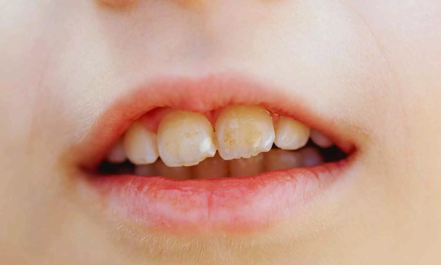
Researchers have found in a new study that white spot lesions (WSLs) on enamel allow greater penetration of hydrogen peroxide and yield less favorable optical outcomes—especially after in-office bleaching—suggesting that at-home bleaching may be a safer, more esthetically favorable option for managing these lesions. In this in-vitro study published in Clinical Oral Investigations, forty premolars were divided into four groups: sound and artificially demineralized (WSLs), each subjected to in-office (35 % hydrogen peroxide) or at-home (16 % carbamide peroxide) bleaching. Analysts measured HP permeability into the pulp chamber and tracked color change using spectrophotometry.
The results confirmed that WSLs significantly increased HP diffusion with in-office bleaching (p < .05), indicating heightened sensitivity risk. Moreover, the optical outcomes were consistently poorer: WSL-teeth showed lower lightness (L*) and whitening index (WI_D) values compared to sound teeth across treatments. Notably, differences remained regardless of bleaching method, although at-home bleaching avoided excessive HP penetration and produced marginally better esthetic results.
These findings raise concern about the safety and effectiveness of aggressive bleaching in teeth with WSLs, which are already structurally compromised. The authors propose that gentler, home-based bleaching protocols combined with remineralization strategies may preserve enamel integrity while improving appearance safely. For clinicians, this means evaluating lesion type before choosing a bleaching method and advising conservative options when WSLs are present. Further clinical trials are needed to verify long-term outcomes and guide tailored bleaching protocols for patients with enamel hypomineralization.
Barbosa, L.M.M., Baracco, B., Carneiro, T.S. et al. Impact of bleaching on white spot lesions: hydrogen peroxide permeability and color alteration. Clin Oral Invest 29, 401 (2025). https://doi.org/10.1007/s00784-025-06490-3
Powered by WPeMatico

A recent study published in the journal of BMC Nephrology revealed that no significant relationship between high-sensitivity cardiac troponin T (h-cTnT) and IV iron, but IV iron therapy remains important for hemodialysis patients.
The cross-sectional investigation from February 2023 to October 2024 in a dialysis unit, involved 244 patients who had been on hemodialysis for at least 3 months and were receiving IV iron therapy. All participants were aged 18 years or older. High-sensitivity cardiac troponin T levels were measured prior to dialysis, with patients grouped according to whether their h-cTnT values were at or below 60 ng/L or above that threshold.
Of the 224 patients who completed the study (137 male and 87 female, with a mean age of 59.9 years), the average IV iron dose was 255.5 mg per month. The overall mean h-cTnT was 90.5 ng/L. More than half the patients (58.5%) had h-cTnT levels exceeding 60 ng/L.
Despite initial concerns, this research found no statistically significant relationship between IV iron dose and troponin levels. However, several other factors did correlate with higher troponin values. Male patients, older individuals, and those with ischemic heart disease or cerebrovascular events showed significantly elevated h-cTnT. The patients on statins and doxazosin were more likely to fall into the high troponin group.
Clinical parameters tied to dialysis performance and nutritional status also showed noteworthy differences. The patients with h-cTnT levels above 60 ng/L had a lower processed blood volume, shorter effective dialysis time, and reduced KT/V urea values, suggesting less efficient treatment. Albumin levels were also lower in the high-troponin group, indicating potential systemic vulnerability.
These findings suggest that while IV iron therapy does not directly influence cardiac troponin T levels, other clinical and patient-related factors play a substantial role. Monitoring troponin remains critical, not for iron management alone, but for identifying patients at higher cardiovascular risk.
Overall, the results highlighted that IV iron appears to be safe in terms of its effect on troponin and is effective in managing anemia in dialysis patients. However, elevated h-cTnT still flags patients with poorer overall health, comorbidities, and less effective dialysis sessions.
Reference:
Hunjul, M., Owda, D., Sawaid, B., Saadeh, H., Enaya, A., Hamdan, Z., & Nazzal, Z. (2025). Exploring the relationship between intravenous iron therapy and troponin T levels in hemodialysis patients: a cross-sectional study. BMC Nephrology, 26(1), 510. https://doi.org/10.1186/s12882-025-04436-1
Powered by WPeMatico

USA: A study found that female veterans with systemic lupus erythematosus (SLE) or rheumatoid arthritis (RA) had significantly higher rates of pregnancy loss and severe maternal morbidity compared to other veterans. These risks were elevated beyond those seen in the general population.
Powered by WPeMatico

For patients with amputations affecting the hand, toe transfer surgery provides an alternative to replanting the amputated digits and may lead to greater improvement in hand function and other key outcomes, reports a study in the August issue of Plastic and Reconstructive Surgery®, the official medical journal of the American Society of Plastic Surgeons (ASPS). The journal is published in the Lippincott portfolio by Wolters Kluwer.
“Our study provides the first evidence that toe transfer surgery provides better long-term hand function compared to attempted replantation of the amputated fingers,” comments Fu-Chan Wei, MD, of Chang Gung Memorial Hospital, Taipei, Taiwan. “The findings challenge current approaches to emergency replantation surgery after digital amputations.”
Amputations of the fingers and thumb are common injuries, affecting about 45,000 people per year in the United States alone. Digit amputations can leave patients with years of disability, especially if the thumb is lost. Emergency replantation of the amputated digit is the current standard of treatment but is sometimes impossible or unsuccessful.
Toe transfer surgery – using one or more of the patients’ toes to replace the amputated digits – is a potential alternative to replantation. While toe transfer surgery is not a new procedure, few studies have evaluated its impact on hand function and other patient-reported outcomes.
Dr. Wei and coauthor Steven Lo, MD, of Canniesburn Plastic Surgery Unit, Glasgow, Scotland, analyzed long-term outcomes of 126 toe transfer procedures in 75 patients after digital amputation. Hand function and other patient-reported outcomes were compared to those of 96 replantation procedures in 52 patients. All patients were treated at Dr. Wei’s hospital; outcomes were assessed at least five years after surgery.
On the validated Michigan Hand Questionnaire, hand function was significantly better for patients undergoing toe transfer, compared to replantation. The difference in favor of toe transfer was substantial, with hand function scores about three times higher than the benefit considered clinically important. The more severe the injury, the greater the magnitude of improvement after toe transfer surgery.
Patients in the toe transfer group also had greater improvement in physical health-related quality of life, assessed using the standard SF-36 score. Foot function after toe transfer surgery was comparable to that in the general population.
The researchers performed physical assessments to evaluate the most important factors affecting hand function outcomes. Hand range of motion, tripod pinch (three-finger grip, as in holding a pencil), and moving two-point discrimination (a measure of nerve sensation) were predictors of better hand function after toe transfer. Higher overall physical and mental health scores were also associated with improved hand function.
Previous studies of toe transfer surgery have consistently yielded high surgical success rates. However, these studies have not included validated assessments of hand function. No evidence-based guidelines have been developed to help guide the decision to perform toe transfer versus attempted replantation for patients with amputations of the fingers and/or thumbs.
“These data provide the first evidence for the potential functional superiority of toe transfers over replantation in digital amputation using one of the largest validated outcome datasets of toe transfers to date,” Drs. Wei and Lo write. They believe their findings challenge the assumption that emergency replantation should always be the “gold standard” after digital amputation, and suggest that toe transfer can be considered “a viable alternative” for some patients. Furthermore, this study suggests that integrating toe transfers into national healthcare frameworks has the potential to positively address one of the single largest causes of disability worldwide.
Reference:
Lo, S., & Wei, F. C. (2025). Toe transfers outperform replantation after digit amputations: outcomes of 126 toe transfers. Plastic & Reconstructive Surgery. doi.org/10.1097/prs.0000000000012053.
Powered by WPeMatico

A new study published in the journal of PloS One found that people who used smartphones while sitting on the toilet had a 46% higher risk of developing hemorrhoids when compared to non-users. One key factor was prolonged toilet time, with 37.3% of phone users reporting spending more than 5 minutes per trip. This research suggest that physicians should ask patients with bowel issues about their smartphone use in the bathroom, as it may be an overlooked contributor to hemorrhoid risk.
This study investigated the link between smartphone use during toilet time and the prevalence of hemorrhoids. The study surveyed 125 adults undergoing screening colonoscopies, provides one of the first multivariate analyses exploring this modern habit. The participants completed detailed surveys about their bathroom behaviors, smartphone usage, and lifestyle factors like fiber intake, physical activity, and frequency of straining.
Of the 125 participants, 43% were found to have hemorrhoids. Smartphone use on the toilet was common—66% admitted to bringing their devices into the bathroom. Interestingly, smartphone users were younger on average than non-users (mean ages 55.4 vs. 62.1).
The analysis showed that time spent on the toilet was notably longer among smartphone users. While only 7.1 percent of non-users reported sitting for more than 5 minutes per visit, a substantial 37.3% of smartphone users did so. The study pointed out that prolonged sitting can increase rectal pressure, which is a known risk factor for hemorrhoids.
After accounting for potential confounding factors like age, sex, body mass index, fiber intake, straining, and physical activity, this association held. Smartphone use on the toilet was linked to a 46% increased risk of hemorrhoids, a finding that reached statistical significance (p = 0.044).
Beyond the numbers, the study also explored what people actually do on their phones while seated. The most common activity was reading news, reported by 54.3% of users, followed by scrolling through social media (44.4%).
Taken together, the findings suggest that the seemingly harmless habit of checking phones in the bathroom may be contributing to a quiet rise in hemorrhoid cases. While the study does not prove causation, the research found that the data adds weight to long-standing anecdotal advice to limit the time spent sitting on the toilet.
Overall, the study recommend that physicians consider asking about smartphone use when evaluating patients with hemorrhoids and that public health messaging address the potential consequences of prolonged toilet sitting.
Source:
Ramprasad, C., Wu, C., Chang, J., Rangan, V., Iturrino, J., Ballou, S., Singh, P., Lembo, A., Nee, J., & Pasricha, T. (2025). Smartphone use on the toilet and the risk of hemorrhoids. PloS One, 20(9),. https://doi.org/10.1371/journal.pone.0329983
Powered by WPeMatico

Breast milk is the first ‘super food’ for many babies. Full of vitamins, minerals, and other bioactive compounds, it helps build the young immune system and is widely considered the optimal source of infant nutrition. Not all mothers, however, have the opportunity to directly breastfeed multiple times during the day and night, and might use expressed milk stored for later.
Breast milk delivers a variety of cues from the mother to the infant, including signals that are thought to influence babies’ circadian rhythms. The hormones and proteins involved in circadian signaling, however, may vary in breast milk concentration over 24 hours. To learn more about these fluctuations, researchers in the US investigated expressed breast milk samples taken during different times of the day. They published their findings in Frontiers in Nutrition.
“We noted differences in the concentrations of bioactive components in breast milk based on time of day, reinforcing that breast milk is a dynamic food. Consideration should be given to the time it is fed to the infant when expressed breast milk is used,” said first author Dr Melissa Woortman, a recent PhD graduate from the Department of Nutritional Sciences at Rutgers University.
“The timing of these cues would be particularly critical in early life, when the infant’s internal circadian clock is still maturing,” added senior author Prof Maria Gloria Dominguez-Bello, a researcher at the Department of Biochemistry and Microbiology at Rutgers University.
The researchers took 10 milliliter breast milk samples from 21 participants at 6am, 12pm, 6pm, and 12am on two different days, which were about a month apart. A further 17 participants provided samples taken at the same times once, resulting in 236 samples included in the analysis. The samples were examined for levels of melatonin, cortisol, and oxytocin – all hormones – as well as immunoglobulin A (IgA), an antibody protein part of the immune system, and lactoferrin, a milk protein. Melatonin and cortisol are involved in the regulation of the circadian rhythm, whereas the other examined components influence intestinal development and gut microbiome dynamics.
They found that some breast milk components, especially melatonin and cortisol, varied over the course of the day. Melatonin peaked at midnight, whereas cortisol was at the highest level in the early morning. “We all have circadian rhythms in our blood, and in lactating mothers, these are often reflected in breast milk,” explained Woortman. “Hormones like melatonin and cortisol follow these rhythms and enter milk from maternal circulation.” The other examined components were mostly stable throughout the day. This might be because they may not be as strongly influenced by signals dictating circadian rhythms.
The team also found that as infants got older, the levels of different compounds in breast milk varied. For example, the levels of cortisol, IgA, and lactoferrin were highest when babies were less than one month old. Higher levels of these compounds likely support immune defense and gut colonization in very young babies.
“When it comes to differences in day/night variations by infant age, this could reflect the stabilizing of the maternal circadian clock that occurs with time after giving birth, as well as the maturing and stabilization of the infant’s circadian rhythm,” Woortman pointed out.
The researchers said their study was not able to account for all potentially relevant demographic factors, including delivery mode and maternal diet, due to sample size. Larger and more diverse cohorts will be needed in the future to ensure the generalizability of these results. In addition, future research should examine how infants respond to the variations observed here.
Still, the findings suggest that feeding expressed milk could be timed to maximize natural biological alignment. This way, circadian signals that support infant sleep, metabolism, and immune development – adaptations shaped through evolution – could be maintained.
“Labeling expressed milk as ‘morning,’ ‘afternoon,’ or ‘evening’ and feeding it correspondingly could help align expressing and feeding times and preserve the natural hormonal and microbial composition of the milk, as well as circadian signals,” Dominguez-Bello pointed out.
“In modern societies where it may not be feasible for mothers to stay with their infants throughout the day, aligning feeding times with the time of milk expression is a simple, practical step that maximizes the benefits of breast milk when feeding expressed milk,” Woortman concluded.
Reference:
Melissa A. Woortman, Day/night fluctuations of breast milk bioactive factors and microbiome, Frontiers in Nutrition, DOI:10.3389/fnut.2025.1618784
Powered by WPeMatico
