Minimally Invasive tissue-selecting therapy Effective and Safe for Severe Hemorrhoids, reveals research

Researchers have found in a new study that minimally invasive tissue-selecting therapy (TST) is effective in managing severe hemorrhoids, significantly enhances anorectal motility, and demonstrates a low rate of postoperative complications, indicating a high level of safety.

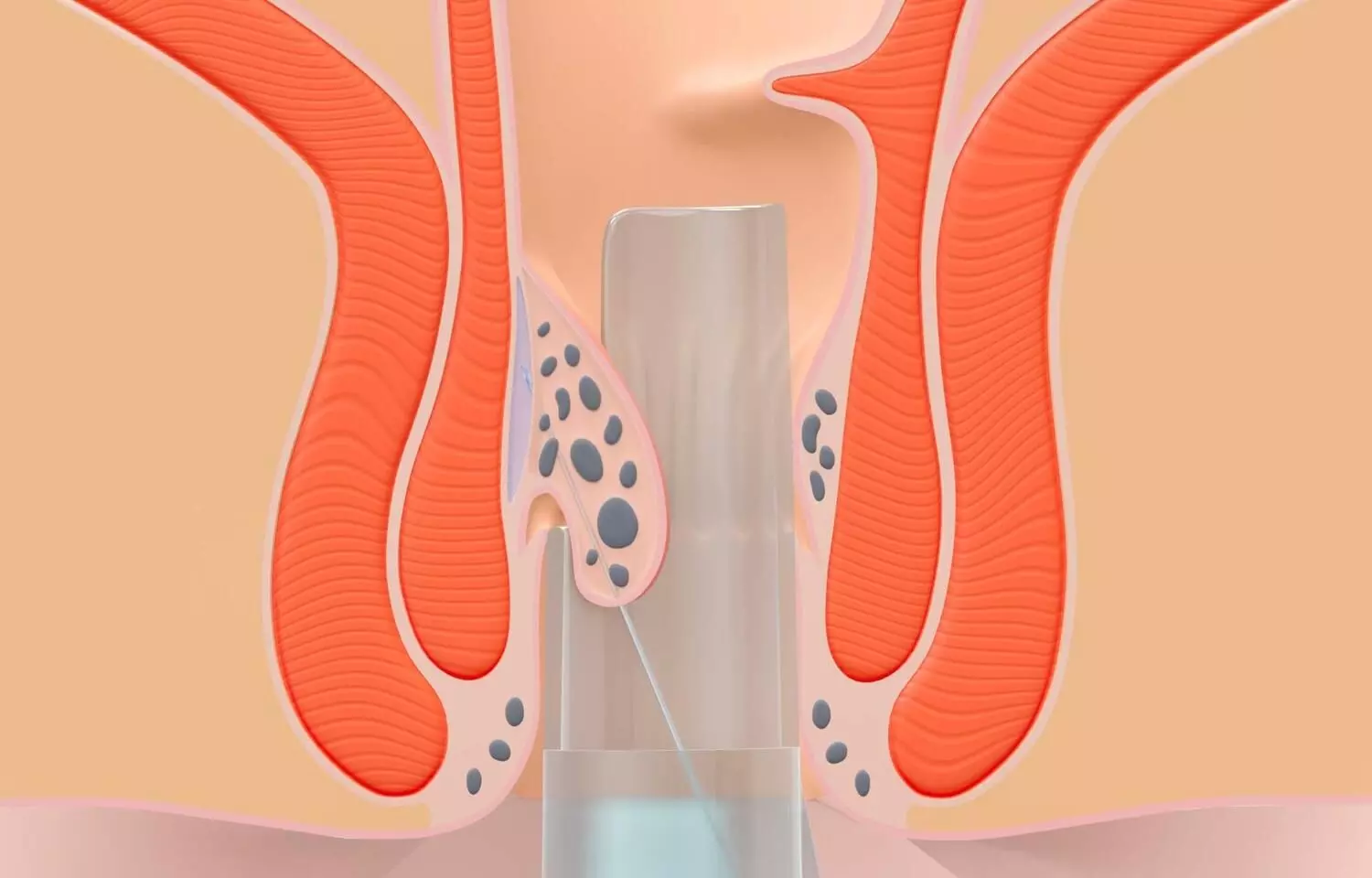
Severe hemorrhoids have a serious impact on patients’ quality of life. At present, the main method for treating severe hemorrhoids is surgery, but surgical treatment causes greater trauma to patients and has a high degree of complications. There is an urgent need to find new surgical methods to solve these problems. To assess the ability of tissue-selective therapy stapler (TST) minimally invasive surgery to treat severe hemorrhoids by studying the clinical efficacy, postoperative complications, and anal dynamics of selective supramedullary hemorrhoid stomy. In the hospital, between March 2015 and February 2018, 141 patients with severe hemorrhoids were included into three groups according to the block randomization method: the TST group, the pieces per hour the PPH group, and the eastern rubber band ligation and coincident of dislocation for prolapse and hemorrhoid (EPH)group. There were 47 patients in each group. The clinical efficacy of the three groups of patients, changes in hospital stay time, operation time, intraoperative blood loss, postoperative complications, and anal dynamic indicators (rectal resting pressure, anal resting pressure, anorectal pressure difference, rectal tolerance, rectal perception threshold and length of the anal high-pressure zone) were observed and compared. Results: The total effective rate in the TST group was 93.62%, which was higher than the 74.47% and 72.34% in the PPH and EPH groups, respectively, after treatment. Hospitalization time, operation time, and intraoperative blood loss were lower in the TST group than in the EPH and PPH groups. Three months after surgery, the rectal resting pressure, rectal tolerance, and rectal sensing thresholds of patients in the TST group were lower than those in the PPH and EPH groups, and the anal canal resting pressure, the anorectal pressure difference and the length of the anal high-pressure zone were greater than those in the PPH and EPH groups. After surgery, the complication rate in the TST group was 10.64%, which was lower than the 27.66% and 31.91% reported in the PPH and EPH groups, respectively. Minimally invasive TST is effective in treating severe hemorrhoids, can significantly improve anorectal motility, has a low incidence of postoperative complications and is highly safe.

Reference:

Huang, R., Fan, M., Lin, H. et al. Tissue selecting therapy stapler minimally invasive surgery to treat severe hemorrhoids. BMC Surg 25, 293 (2025). https://doi.org/10.1186/s12893-025-02918-5

Keywords:

1500X 600 Minimally, Invasive, tissue-selecting, therapy, Effective, Safe, Severe, Hemorrhoids, reveals, research, Huang, R., Fan, M., Lin, H, Selective suprahemorrhoidal, mucosa anastomosis, Severe hemorrhoids, Clinical efficacy, Complications, Anal dynamics

Powered by WPeMatico

Remote chronic behavioral therapy improves chronic pain symptoms and quality-of-life: JAMA

A new study published in the Journal of American Medical Association showed that when compared to standard therapy, remote, scalable cognitive behavioral therapy skills training programs for chronic pain (CBT-CP) therapies (either through telehealth or self-completed online courses) produced moderate reductions in pain and associated functional/quality-of-life outcomes in people with high-impact chronic pain.

Although they are not generally available, cognitive behavioral therapy (CBT) skills training programs are advised as the initial nonpharmacologic treatment for chronic pain. Thus, this study was set to evaluate the efficacy of telehealth and self-completed online remote, scalable CBT-CP therapies with standard care for those with high-impact chronic pain.

From January 2021 to February 2023, 2331 eligible individuals with high-impact chronic musculoskeletal pain from four geographically disparate US health care systems participated in this comparative efficacy, 3-group, phase 3 randomized clinical research. The period of follow-up ended in April 2024.

The participants were randomly assigned 1:1:1 to one of two remote, 8-session CBT-based skills training treatments: normal care with a resource guide (n = 777) or a health coach-led program via videoconference or phone (health coach; n = 778) or an online self-completed program (painTRAINER; n = 776).

Achieving or surpassing the minimum clinically meaningful difference (MCID) in pain intensity score (≥30% drop; score range, 0-10) on the 11-item Brief Pain Inventory–Short Form between baseline and 3 months was the main goal. Pain severity, pain-related interference, PROMIS (Patient-Reported Outcomes Measurement Information System) social role and physical functioning, and the patient’s overall perception of change were secondary outcomes at 3, 6, and 12 months.

A total of 2,210 (94.8%) of the 2,331 eligible randomized participants finished the trial. The adjusted proportion of individuals who experienced a 30% or more reduction in their pain severity score at 3 months was 26.6 (95% CI, 23.4-30.2) in the painTRAINER group, 20.8 (95% CI, 18.0-24.0) in the usual care group, and 32.0 (95% CI, 29.3-35.0) in the health coach group.

The relative risk [RR] for both intervention groups was 1.54 [95% CI, 1.30-1.82] when compared to the control group. The RR for painTRAINER versus usual care was 1.28 [95% CI, 1.06-1.55]; and the RR for the health coach program was 1.20 [95% CI, 1.03-1.40] when compared to the online self-completed painTRAINER program.

At 6 and 12 months following randomization, both intervention groups showed statistically significant improvements in pain severity outcomes as well as various secondary pain and functional outcomes as compared to conventional treatment. Overall, the results indicate that centralizing the delivery of CBT-CP-based programs using online interventions and telephone/videoconferencing is successful.

Reference:

DeBar, L. L., Mayhew, M., Wellman, R. D., Balderson, B. H., Dickerson, J. F., Elder, C. R., Justice, M., Keefe, F. J., McMullen, C. K., Owen-Smith, A. A., Rini, C., Von Korff, M., Waring, S., Yarava, A., Shen, Z., Thompson, R. E., Clark, A. E., Casper, T. C., & Cook, A. J. (2025). Telehealth and online cognitive behavioral therapy-based treatments for high-impact chronic pain: A randomized clinical trial: A randomized clinical trial. JAMA: The Journal of the American Medical Association. https://doi.org/10.1001/jama.2025.11178

Powered by WPeMatico

Rizatriptan Ineffective for Vestibular Migraine Treatment, reveals study

Researchers have discovered in a new study that rizatriptan was ineffective at relieving vestibular migraine attacks within 1 hour and showed only limited symptom improvement at 24 hours. These results do not support the use of rizatriptan for treatment of vestibular migraines. The study was published in JAMA Network by Jeffrey P. and colleagues.

This double-blind, placebo-controlled randomized clinical trial was conducted in two tertiary neurotologic centers. 222 patients (mean age 42.3 years; 70.7% female) were originally enrolled. After prospective observation to establish diagnosis and illness activity over a period, 134 patients (60.4%) with active disease were randomized in a 2:1 ratio to receive either rizatriptan 10 mg or placebo. Each patient was asked to treat up to three discrete attacks of vestibular migraine.

The major results were whether rizatriptan was able to decrease vertigo and unsteadiness/dizziness from moderate or severe to absent or mild at 1 hour after taking medication. Secondary outcomes were total resolution of the symptoms, decrease in headache and related symptoms, rescue medication use, 24-hour symptom status, treatment satisfaction, and quality of life at 48 hours.

Key Findings

  • The last efficacy analysis comprised 240 attacks with vestibular symptoms rated moderate or severe at treatment.

At 1 hour, rizatriptan and placebo had no statistically significant differences in relief of symptoms:

  • Vertigo relief: 48.3% (73/151 attacks) with rizatriptan vs. 56.8% (50/88 attacks) with placebo (Odds Ratio [OR], 0.71; 95% CI, 0.42–1.21)

  • Relief of unsteadiness/dizziness: 19.2% (29/151) on rizatriptan vs. 12.4% (11/89) on placebo (OR, 1.69; 95% CI, 0.80–3.57)

  • Use of rescue medication at 1 hour: Same for both groups at 26.4%

  • Total relief of symptoms at 1 hour: Not different

  • At 24 hours, rizatriptan had modest advantages:

  • Unsteadiness/dizziness: OR 2.65 favoring rizatriptan

  • Motion sensitivity: OR 3.58 favoring rizatriptan

  • Medium effects on headache, photophobia, and phonophobia were found by post hoc analysis

  • But treatment satisfaction and overall quality of life measures were inconsistent, and there were no severe adverse effects or side effect discontinuations.

The usage of rizatriptan is not recommended for the management of vestibular migraine attack, particularly when immediate control of the symptoms is warranted. These results highlight the urgent necessity of the discovery of targeted vestibular migraine therapies since the disease still has no standardized treatment.

Reference:

Staab JP, Eggers SDZ, Jen JC, et al. Rizatriptan vs Placebo for Attacks of Vestibular Migraine: A Randomized Clinical Trial. JAMA Neurol. 2025;82(7):676–686. doi:10.1001/jamaneurol.2025.1006

Powered by WPeMatico

New MRI approach maps brain metabolism, revealing disease signatures: Study

A new technology that uses clinical MRI machines to image metabolic activity in the brain could give researchers and clinicians unique insight into brain function and disease, researchers at the University of Illinois Urbana-Champaign report. The non-invasive, high-resolution metabolic imaging of the whole brain revealed differences in metabolic activity and neurotransmitter levels among brain regions; found metabolic alterations in brain tumors; and mapped and characterized multiple sclerosis lesions — with patients only spending minutes in an MRI scanner.

Led by Zhi-Pei Liang, a professor of electrical and computer engineering and a member of the Beckman Institute for Advanced Science and Technology at the U. of I., the team reported its findings in the journal Nature Biomedical Engineering.

“Understanding the brain, how it works and what goes wrong when it is injured or diseased is considered one of the most exciting and challenging scientific endeavors of our time,” Liang said. “MRI has played major roles in unlocking the mysteries of the brain over the past four decades. Our new technology adds another dimension to MRI’s capability for brain imaging: visualization of brain metabolism and detection of metabolic alterations associated with brain diseases.”

Conventional MRI provides high-resolution, detailed imaging of brain structures. Functional MRI maps brain activity by detecting changes in blood flow and blood oxygenation level, which are closely linked to neural activity. However, they cannot provide information on the metabolic activity in the brain, which is important for understanding function and disease, said postdoctoral researcher Yibo Zhao, the first author of the paper.

“Metabolic and physiological changes often occur before structural and functional abnormalities are visible on conventional MRI and fMRI images,” Zhao said. “Metabolic imaging, therefore, can lead to early diagnosis and intervention of brain diseases.”

Both MRI and fMRI techniques are based on magnetic resonance signals from water molecules. The new technology measures signals from brain metabolites and neurotransmitters as well as water molecules, a technique known as magnetic resonance spectroscopic imaging. These MRSI images can provide significant new insights into brain function and disease processes, and could improve sensitivity and specificity for the detection and diagnosis of brain diseases, Zhao said.

Other attempts at MRSI have been bogged down by the lengthy times required to capture the images and high levels of noise obscuring the signals from neurotransmitters. The new technique addresses both challenges.

“Our technology overcomes several long-standing technical barriers to fast high-resolution metabolic imaging by synergistically integrating ultrafast data acquisition with physics-based machine learning methods for data processing,” Liang said. With the new MRSI technology, the Illinois team cut the time required for a whole brain scan to 12 and a half minutes.

The researchers tested their MRSI technique on several populations. In healthy subjects, the researchers found and mapped varying metabolic and neurotransmitter activity across different brain regions, indicating that such activity is not universal. In patients with brain tumors, the researchers found metabolic alterations, such as elevated choline and lactate, in tumors of different grades — even when the tumors appeared identical on clinical MRI images. In subjects with multiple sclerosis, the technique detected molecular changes associated with neuroinflammatory response and reduced neuronal activity up to 70 days before changes become visible on clinical MRI images, the researchers report.

The researchers foresee potential for broad clinical use of their technique: By tracking metabolic changes over time, clinicians can assess the effectiveness of treatments for neurological conditions, Liang said. Metabolic information also can be used to tailor treatments to individual patients based on their unique metabolic profiles.

“High-resolution whole-brain metabolic imaging has significant clinical potential,” said Liang, who began his career in the lab of the late Illinois professor Paul Lauterbur, recipient of the Nobel Prize for developing MRI technology. “Paul envisioned this exciting possibility and the general approach, but it has been very difficult to achieve his dream of fast high-resolution metabolic imaging in the clinical setting.

“As healthcare is moving towards personalized, predictive and precision medicine, this high-speed, high-resolution technology can provide a timely and effective tool to address an urgent unmet need for noninvasive metabolic imaging in clinical applications.”

Reference:

Zhao, Y., Li, Y., Jin, W. et al. Ultrafast J-resolved magnetic resonance spectroscopic imaging for high-resolution metabolic brain imaging. Nat. Biomed. Eng (2025). https://doi.org/10.1038/s41551-025-01418-4

Powered by WPeMatico

Dementia takes 3.5 years to diagnose after symptoms begin: Study

People with dementia are diagnosed an average of 3.5 years after symptoms are first noticed, or even longer (4.1 years) for those with early-onset dementia, finds a new study led by UCL researchers.

The study, published in the International Journal of Geriatric Psychiatry, is the first systematic review and meta-analysis of global evidence examining time to diagnosis in dementia.

The researchers reviewed data from 13 previously published studies which took place in Europe, US, Australia and China, reporting data on 30,257 participants.

The research team was investigating the average interval between symptom onset (rated by patients or family carers using interviews or medical records) to the final diagnosis of dementia.

Lead author Dr Vasiliki Orgeta (UCL Division of Psychiatry) said: “Timely diagnosis of dementia remains a major global challenge, shaped by a complex set of factors, and specific healthcare strategies are urgently needed to improve it. Other studies estimate that only 50-65% of cases are ever diagnosed in high-income countries, with many countries having even lower diagnostic rates.

“Timely diagnosis can improve access to treatments and for some people prolong the time living with mild dementia before symptoms worsen.”

In a pooled meta-analysis of 10 of the included studies, the researchers found that it typically takes 3.5 years from the first alert of symptoms to a patient receiving a diagnosis of dementia, or 4.1 years for those with early-onset dementia, with some groups more likely to experience longer delays.

They found that younger age at onset and having frontotemporal dementia were both linked to longer time to diagnosis. While data on racial disparities was limited, one of the studies reviewed found that black patients tended to experience a longer delay before diagnosis.

Dr Orgeta said: “Our work highlights the need for a clear conceptual framework on time to diagnosis in dementia, developed in collaboration with people with dementia, their carers, and supporters.”

Dr Phuong Leung (UCL Division of Psychiatry) said: “Symptoms of dementia are often mistaken for normal ageing, while fear, stigma, and low public awareness can discourage people from seeking help.”

Professor Rafael Del-Pino-Casado, of the University of Jaén, Spain, said: “Within healthcare systems, inconsistent referral pathways, limited access to specialists, and under-resourced memory clinics can create further delays. For some, language differences or a lack of culturally appropriate assessment tools can make access to timely diagnosis even harder.”

Dr Orgeta added: “To speed up dementia diagnosis, we need action on multiple fronts. Public awareness campaigns can help improve understanding of early symptoms and reduce stigma, encouraging people to seek help sooner. Clinician training is critical to improve early recognition and referral, along with access to early intervention and individualised support so that people with dementia and their families can get the help they need.”

Reference:

Olubunmi Kusoro, Moïse Roche, Time to Diagnosis in Dementia: A Systematic Review With Meta-Analysis, International Journal of Geriatric Psychiatry, https://doi.org/10.1002/gps.70129 

Powered by WPeMatico

Flapless Immediate Implants with Bone Graft May Aid Bone Preservation, reveals research

A recent study published in Clinical and Experimental Dental Research suggests that flapless immediate implant placement combined with bone grafting may help preserve alveolar bone more effectively than traditional approaches. Authored by Xiang et al., the research highlights that this minimally invasive technique—where implants are placed directly into the extraction socket without raising a flap—could reduce postoperative bone loss, improve esthetic outcomes, and potentially shorten recovery time. The study analyzed clinical outcomes from patients undergoing flapless immediate implant placement with graft material added to fill the peri-implant gap, observing promising early results in bone volume retention. However, the authors emphasize that while these findings are encouraging, long-term randomized trials are needed to validate the technique’s consistency and compare it against conventional implant protocols. Preservation of the alveolar ridge following tooth extraction is critical for both function and esthetics, especially in anterior zones, and bone grafting at the time of implant placement may offer structural support and enhance soft tissue contour. The study contributes to the growing interest in flapless surgical techniques in implant dentistry, which aim to reduce trauma, maintain blood supply, and promote faster healing. As implantology continues to evolve toward patient-centered, minimally invasive care, this approach offers a potential pathway for balancing surgical efficiency with biological preservation. Clinical decision-making, however, should consider individual bone quality, implant site morphology, and operator experience.

Keywords: flapless implant, immediate implant placement, bone grafting, alveolar bone preservation, Xiang et al., Clinical and Experimental Dental Research, dental implant surgery, ridge preservation, peri-implant bone, minimally invasive dentistry

Reference:
Xiang, Y., et al. (2024). Flapless immediate implant placement combined with bone grafting for alveolar ridge preservation: A clinical evaluation. Clinical and Experimental Dental Research. https://doi.org/10.1002/cre2.70182

Powered by WPeMatico

Polygenic Risk Score Outperforms PSA and MRI in Detecting Clinically Significant Prostate Cancer: NEJM

Researchers have found in a new BARCODE1 study that a polygenic risk score identified a higher proportion of clinically significant prostate cancer compared to PSA or MRI screening, which would have missed 71.8% of cases. Further Overdiagnosis rates were comparable to those seen in previous PSA-based studies. The study was published in The New England Journal of Medicine by Jana K. and fellow researchers. This method might provide a more accurate and focused way of detecting high-risk individuals who would be indicated for more extensive diagnostic assessment.

The UK trial sought to investigate whether germline genetic information could enhance prostate cancer detection earlier, by targeting those with the greatest genetic risk with screening. Through the use of genome-wide association data to determine a polygenic risk score based on 130 established variants, researchers were able to target those in the top 10% of genetic risk and invite them for screening with multiparametric MRI and transperineal biopsy, irrespective of PSA.

Men aged 55 to 69 years were recruited from primary care centers in the UK. Saliva was sampled to yield germline DNA that was then used to compute a polygenic risk score for each participant based on 130 known prostate cancer-associated genetic variants. Of 40,292 men invited to join, 8953 (22.2%) were interested, and 6393 received polygenic risk score testing. The 745 men with a score at or above the 90th percentile (11.7%) were offered intensive screening. This included multiparametric MRI followed by transperineal biopsy, regardless of the participants’ PSA levels.

Key Findings

  • Of 40,292 invited men, 6393 had polygenic risk scoring.

  • 745 men (11.7%) were classified in the top 10% of genetic risk.

  • 468 of these men (62.8%) underwent MRI and biopsy.

  • 187 (40.0%) were found to have prostate cancer.

  • 103 (55.1%) had cancer of intermediate or higher risk that needed treatment.

  • 74 (71.8%) of these high cases would not be detected with existing PSA-based methods.

  • 40 men (21.4%) had high-risk or very high-risk prostate cancer.

This British study offers strong evidence that prostate cancer screening using polygenic risk scores can identify more clinically relevant cancers than conventional PSA-based methods. The approach has the potential to be a revolutionary step towards more personalized and efficient cancer screening. Applying genetic information to select men most likely to gain from more sophisticated diagnostic equipment can contribute to alleviating the population burden of undetected, high-risk prostate cancer.

Reference:

McHugh, J. K., Bancroft, E. K., Saunders, E., Brook, M. N., McGrowder, E., Wakerell, S., James, D., Rageevakumar, R., Benton, B., Taylor, N., Myhill, K., Hogben, M., Kinsella, N., Sohaib, A. A., Cahill, D., Hazell, S., Withey, S. J., Mcaddy, N., Page, E. C., … Eeles, R. A. (2025). Assessment of a polygenic risk score in screening for prostate cancer. The New England Journal of Medicine, 392(14), 1406–1417. https://doi.org/10.1056/nejmoa2407934

Powered by WPeMatico

Rising FIB-4 Scores Signal Higher Risk of Liver Stiffness and Mortality in MASLD Patients: Study Shows

China: A new study published in Clinical Gastroenterology and Hepatology suggests that tracking dynamic changes in the Fibrosis-4 Index (FIB-4) can provide valuable insights into liver stiffness progression, fibrosis advancement, and future clinical outcomes in patients with metabolic dysfunction-associated steatotic liver disease (MASLD). The research, led by Ming-Hua Zheng and colleagues from the Institute of Hepatology, Wenzhou Medical University, emphasizes the prognostic value of monitoring FIB-4 over time rather than relying solely on baseline measurements.

The study assessed three large cohorts to evaluate the relationship between longitudinal FIB-4 changes and disease outcomes. The VCTE-Prognosis cohort (n=10,203) was analyzed for liver stiffness progression, the Paired Liver Biopsy cohort (n=1,145) for histological fibrosis progression, and the Wenzhou Real-World (WRW) cohort (n=41,105) for clinical outcomes of mortality, cardiovascular events, and liver-related complications.

Patients were divided into low (<1.3) and high (≥1.3) FIB-4 groups. An increase in FIB-4 was defined as a ≥20% rise, with additional thresholds for patients moving from low to high categories.

The study led to the following findings:

  • In the VCTE-Prognosis cohort, patients with low baseline FIB-4 who showed rising scores had over twice the risk of liver stiffness progression compared to those with stable levels (adjusted OR=2.36).
  • In the high FIB-4 group, rising scores were linked to more than a threefold increase in liver stiffness progression risk (adjusted OR=3.42).
  • Data from the Paired Liver Biopsy cohort indicated that increasing FIB-4 scores significantly raised the likelihood of a one-stage fibrosis progression (adjusted OR=2.20 in low FIB-4; adjusted OR=3.68 in high FIB-4).
  • In the WRW real-world cohort, dynamic increases in FIB-4 were strongly associated with higher risks of all-cause mortality, cardiovascular events, and liver-related complications.

“These findings indicate that longitudinal changes in FIB-4 serve as a predictive marker not only for structural liver deterioration but also for overall prognosis in MASLD patients,” the researchers wrote.

According to the researchers, this approach addresses a critical gap in current clinical practice, where single-time FIB-4 values are often used for risk stratification. By incorporating serial monitoring, clinicians can identify patients at higher risk earlier and intervene proactively to prevent disease progression and adverse outcomes.

However, the authors note that despite the robust sample size across cohorts, further validation in diverse populations is warranted. They suggest integrating dynamic FIB-4 tracking into routine MASLD management protocols alongside other non-invasive assessments to improve patient monitoring and treatment strategies.

“The study highlights the clinical significance of monitoring FIB-4 trends over time. A ≥20% rise in FIB-4 levels was consistently linked with increased risks of worsening liver stiffness, fibrosis progression, and adverse events, highlighting its potential as a practical tool for early risk detection and personalized care in MASLD management,” the authors concluded.

Reference:

Zhou, X., Li, Y., Kim, S. U., Cheuk-Fung Yip, T., Petta, S., Nakajima, A., Tsochatzis, E., Shi, J., Chan, W., Boursier, J., Bugianesi, E., Yilmaz, Y., Hagström, H., Romero-Gomez, M., Alswat, K., Calleja, J. L., Takahashi, H., De Lédinghen, V., Castéra, L.,… Zheng, M. (2025). Longitudinal Changes in Fibrosis Markers: Monitoring Stiffness/Fibrosis Progression and Prognostic Outcomes in MASLD. Clinical Gastroenterology and Hepatology. https://doi.org/10.1016/j.cgh.2025.07.011

Powered by WPeMatico

Older paternal age linked to higher miscarriage risk and lower live birth rates in donor egg IVF cycles, new study finds

A new international study presented today at the 41st Annual Meeting of the European Society of Human Reproduction and Embryology (ESHRE) reveals that IVF cycles involving male partners over the age of 45 carry significantly increased miscarriage risks and lower live birth rates – even when young donor eggs are used.

By isolating paternal age from female reproductive factors through the exclusive use of donor eggs from young women, the study provides robust evidence that male age plays a critical role in reproductive success, challenging the common assumption that sperm age has little impact once fertilisation occurs.

The retrospective study analysed 1,712 first oocyte donation cycles conducted between 2019 and 2023 across six IVF centres in Italy and Spain. All cycles used fresh donor oocytes (mean donor age 26.1 years) and frozen sperm from male partners, with only the first single blastocyst transfer included. Female recipients had a mean age of 43.3 years.

Participants were divided into two groups: men aged 45 or younger (n=1,066) and those over 45 (n=646). While fertilisation rates and embryo development were comparable between groups, significant differences emerged in clinical outcomes.

Miscarriage rates were notably higher among couples where the male partner was over 45, reaching 23.8% compared to 16.3% in the younger paternal age group. Similarly, live birth rates were significantly lower in the older paternal age group, at 35.1% versus 41% for men aged 45 or younger.

Discussing the findings, Dr. Maria Cristina Guglielmo, Embryologist at Eugin Italy, said, “Traditionally, maternal age has been the central focus in reproductive medicine, but our results show that the age of the male partner also plays a crucial and independent role. Even when using eggs from young, healthy donors and transferring only a single, high-quality embryo, we observed poorer outcomes in men over 45.”

Explaining the potential underlying mechanisms, Dr. Guglielmo elaborated, “As men age, the continuous division of spermatogonial stem cells increases the chance of DNA replication errors. This results in a greater number of new genetic mutations and a higher rate of sperm aneuploidy, where sperm carry abnormal chromosomes. Older paternal age is also linked to increased sperm DNA fragmentation and changes in the epigenetic profile of sperms, such as DNA methylation.”

She continued, “Together, these factors affect both the genetic integrity and the functional quality of sperm, which can impair embryo development and contribute to a higher risk of miscarriage.”

Highlighting the clinical implications, Dr. Guglielmo stated, “Our findings underscore the need for fertility clinics to adopt a more balanced approach that recognises the role of paternal age, even in donor egg cycles where maternal factors are controlled”, she said. “Clinics should ensure that male patients are fully informed about how advancing paternal age can impact fertility potential, pregnancy success, and miscarriage risk.”

Looking ahead, Dr. Guglielmo outlined the research priorities: “Our future studies will aim to deepen understanding of the biological mechanisms behind the impact of paternal age, focusing on sperm DNA damage, oxidative stress, and epigenetic changes.”

She also emphasised the importance of examining how paternal age affects the health of offspring. “There is growing evidence linking advanced paternal age to an increased risk of neurodevelopment disorders in children. Our future work will investigate the long-term health and developmental outcomes of children conceived through donor egg cycles with older fathers, where maternal factors are minimised, to isolate paternal effects more clearly.”

Professor Dr. Carlos Calhaz-Jorge, Immediate Past Chair of ESHRE, said, “This is an important paper that draws attention to an often-overlooked factor in the IVF field. Although it might be interesting to further subdivide the ‘older paternal age’ group (for instance, would men over 55 show even poorer outcomes?), the results presented should be seriously considered during the counselling process for couples in which the male partner is over 45.”

Reference:

Older paternal age linked to higher miscarriage risk and lower live birth rates in donor egg IVF cycles, new study finds, European Society of Human Reproduction and Embryology, Meeting: ESHRE 41st Annual Meeting.

Powered by WPeMatico

Sulfhydryl-Containing Foods and Drugs Linked to Pemphigus Vulgaris Risk: Study

Turkey: New research has revealed that foods and medications containing sulfhydryl groups may trigger the onset of pemphigus vulgaris or worsen existing disease symptoms.

The study, published in Postepy Dermatologii i Alergologii (Advances in Dermatology and Allergology), was conducted by Dr. Goknur Ozaydın Yavuz and colleagues from the Department of Dermatology, Health Sciences University, Istanbul, Turkey. It explored the potential link between serum total sulfhydryl (TSH) levels and the autoimmune skin condition pemphigus vulgaris (PV). 

Pemphigus vulgaris is a rare and potentially life-threatening autoimmune blistering disease characterized by the production of autoantibodies against desmoglein-1 and desmoglein-3, which are essential components in maintaining skin cell adhesion. The disruption of these proteins leads to acantholysis—the loss of cell-to-cell adhesion in the skin—and the formation of painful blisters and erosions.

Sulfhydryl groups, also known as thiol groups, play a crucial role in maintaining cellular function. They are involved in antioxidant defense, detoxification, cellular proliferation, and programmed cell death. Previous research has suggested that chemical exposure, including that containing sulfhydryl compounds, may contribute to acantholysis and subsequent skin damage.

To investigate this further, the research team conducted a case-control study involving 177 participants, including 86 patients diagnosed with pemphigus vulgaris and 91 healthy individuals as controls. Detailed demographic and lifestyle data—including age, gender, smoking and alcohol habits, and dietary intake of foods known to trigger pemphigus—were recorded for each participant.

The diagnosis of PV was established through clinical evaluation, histopathological findings, and confirmation via direct and indirect immunofluorescence tests. Blood samples were collected, and serum total sulfhydryl levels were measured using a spectrophotometric method.

The findings revealed the following:

  • The average age of patients in the pemphigus vulgaris (PV) group was 41.94 years.
  • The average age in the control group was 39.86 years.
  • Smoking was in 62.8% of the PV patients.
  • Alcohol consumption was observed in 32.6% of the PV patients.
  • There was a statistically significant difference in serum total sulfhydryl levels between the PV and the control group.

Based on these results, the authors concluded that compounds containing sulfhydryl groups—commonly found in certain medications and foods—may play a role in triggering or exacerbating pemphigus vulgaris. The exact mechanisms remain to be fully understood, but the study emphasizes the potential importance of monitoring sulfhydryl exposure in individuals diagnosed with PV or at risk for the disease.

The research adds to growing evidence that environmental and dietary factors may influence the course of autoimmune skin diseases. Further studies are needed to clarify the causal relationships and to establish dietary guidelines that may help in managing or preventing PV flare-ups.

Reference:

Ozaydın Yavuz, Goknur, et al. “Evaluation of Total Sulfhydryl Levels and Dietary Habits in Pemphigus Vulgaris.” Postepy Dermatologii I Alergologii, vol. 42, no. 3, 2025, pp. 255-258.

Powered by WPeMatico