AI analysis of colonoscopy improves assessment of Crohn’s disease

In a new study, artificial intelligence matched and potentially exceeded the performance of gastroenterologists and conventional scoring in evaluating endoscopies of Crohn’s disease patients.

The results, published in Clinical Gastroenterology and Hepatology, show a computer vision model identified mucosal ulceration as accurately as physician reviewing videos, while also being strongly correlated with the most common endoscopic scoring system.

“A clinician has a gestalt of whether someone’s Crohn’s is mild or severe, if it’s improving or worsening,” said Ryan W. Stidham, M.D., M.S., associate professor of internal medicine at the University of Michigan Medical School and senior author of the paper.

“But the manual tools to quantify that expert opinion with objectivity and reproducibly are poor. AI image analysis can provide better solutions for more precise measurements and descriptions of what we are seeing on the screen during colonoscopy.”

In the study, two gastroenterologists annotated ulcer areas in 4,487 still images from endoscopic videos. The computer vision AI model reviewed the same images, from previous clinical trials.

The AI model’s results had a higher overall DICE similarity score with ground truth (.591) than when comparing the agreement between two gastroenterologists (.462).

Computer vision assessments of the colonoscopy videos were also strongly correlated with Simple Endoscopic Score for Crohn’s Disease (SES-CD) values. SES-CD is a metric that attempts to manually quantify various characteristics of a patient’s ulceration.

While study authors note the importance of SES-CD for Crohn’s disease research and assessing the effectiveness of treatment, they also highlight its limitations, including variability among observers and inability to capture certain ulcer features.

Researchers hope that AI-powered metrics will not only improve consistency between providers but more importantly may offer more quantitative detail to describe disease.

“It can be hard to justify decisions, particularly if you’re talking about expensive drugs or riskier therapies,” Stidham said.

“One of the reasons for this research is to one day provide more objective evidence for an experienced clinician’s impression.”

Researchers identify several other potential advantages to more consistent endoscopy metrics. In areas without IBD specialists AI endoscopy interpretation may be used to guide treatment change based on expert decision making. For more experienced providers, the information can help them understand their own decision-making process.

They also highlight possible implications for education and drug development.

“This paper is really the first step, and I hope that it gets the entire field thinking of better ways to quantify IBD that more completely describe an individual patient,” Stidham said.

“It’s very early, but computer vision tools like this may provide an important component for the future of automated care, where AI and experts work together in treating patients.” 

Reference:

Cai, Lingrui et al., Artificial Intelligence for Quantifying Endoscopic Mucosal Ulceration in Crohn’s Disease, Clinical Gastroenterology and Hepatology, DOI:10.1016/j.cgh.2025.05.026 

Powered by WPeMatico

Pregnancy Hypertension Linked to Lower Childhood IQ, Large Meta-Analysis Finds

Australia: Children born to mothers with hypertensive disorders of pregnancy face a heightened risk of cognitive and developmental challenges, according to a large systematic review and meta-analysis published in PLOS Medicine. The research shows that while links to autism and global developmental delay diminish when factors such as birthweight and gestational age are considered, a measurable reduction in average IQ persists, underscoring the importance of early monitoring and support for these children.

The study, led by Roxanne M. Hastie from the University of Melbourne’s Perinatal Epidemiology Group and Mercy Perinatal at Mercy Hospital for Women in Victoria, Australia, brings together data from 121 studies encompassing nearly 30 million children worldwide. Investigators pooled evidence from cohort, case–control, and cross-sectional studies to explore how maternal hypertensive disorders—including preeclampsia and gestational hypertension—might influence offspring neurodevelopment, intelligence, and educational outcomes.
The following were the key findings:
  • Children exposed in utero to hypertensive pregnancy disorders had a 65% higher odds of developing autism spectrum disorder.
  • The odds of attention-deficit/hyperactivity disorder (ADHD) increased by 27% in these children.
  • The likelihood of intellectual disability and global developmental delay each rose by 77%.
  • Mean intelligence scores were lower by an average of 2.2 points compared with children from unaffected pregnancies.
  • After adjusting for gestational age and birthweight, the elevated risks for autism and global developmental delay were no longer statistically significant.
  • The reduction in IQ remained evident after adjusting for birthweight but weakened when gestational age was considered, indicating overlapping and complex pathways.
The review team emphasized that despite these nuances, the findings point to a vulnerable population of children who could benefit from proactive developmental assessments and early interventions. Early identification of cognitive or behavioral difficulties can enable timely educational and therapeutic support, potentially improving long-term outcomes.
Researchers also noted limitations within the available evidence. Many of the included studies lacked comprehensive adjustment for confounding factors, and there was considerable variation in study design and quality. Nonetheless, sensitivity analyses excluding high-risk studies did not change the overall conclusions.
While the exact biological mechanisms remain unclear, hypertensive disorders of pregnancy are known to affect placental function and fetal growth, which may influence brain development. Dr. Hastie and colleagues call for further research to disentangle the roles of gestational age, birthweight, and other mediating factors.
The large-scale analysis highlights that children born following hypertensive pregnancies, even those without obvious complications at birth, may carry a subtle but significant risk of lower cognitive performance. The authors advocate early developmental surveillance and targeted support as a prudent strategy to help these children achieve their full potential.
Reference:
Atkinson JA, Gordon HG, Tong S, Walker SP, Mehdipour P, Lindquist AC, et al. (2025) Hypertensive disorders of pregnancy and childhood neurodevelopment: A systematic review and meta-analysis. PLoS Med 22(9): e1004558. https://doi.org/10.1371/journal.pmed.1004558

Powered by WPeMatico

New bioactive dressing promises faster diabetic wound healing, reveals research

Chronic diabetic wounds, including diabetic foot ulcers, are a significant burden for patients, as impaired blood vessel growth hinders the healing process. A recent breakthrough offers hope by combining small extracellular vesicles (sEVs) loaded with miR-221-3p and a GelMA hydrogel to target thrombospondin-1 (TSP-1), a protein that suppresses angiogenesis. This new bioactive wound dressing not only accelerates healing but also promotes blood vessel formation, offering a promising new approach to treating one of the most challenging complications of diabetes.

Diabetic wounds, particularly foot ulcers, are notorious for their slow and often incomplete healing due to reduced blood flow and endothelial cell dysfunction. One of the major contributors to this issue is thrombospondin-1 (TSP-1), which inhibits the growth of new blood vessels, a process crucial for tissue repair. Despite various existing treatments, the challenge of addressing this barrier to healing remains unmet. With the global rise in diabetes cases, new treatments targeting the underlying causes of delayed wound healing have become a critical area of research. In light of these ongoing challenges, this study explores a new approach to stimulate angiogenesis and speed up the healing process.

In a new study (DOI: 10.1093/burnst/tkaf036) published in Burns & Trauma, a team of researchers from leading Chinese institutions has unveiled a novel therapeutic solution for diabetic wound healing. The study introduces an innovative wound dressing that combines miR-221OE-sEVs—engineered extracellular vesicles that target and reduce TSP-1 levels—with a GelMA hydrogel to create a sustained-release system. This cutting-edge approach has shown to significantly enhance wound healing and blood vessel formation in diabetic mice, offering hope for more effective treatments in the future.

In their study, the researchers discovered that high glucose conditions commonly found in diabetic wounds lead to increased levels of TSP-1 in endothelial cells, impairing their ability to proliferate and migrate—key processes for angiogenesis. By utilizing miR-221-3p, a microRNA that targets and downregulates TSP-1 expression, they restored endothelial cell function. The engineered miR-221OE-sEVs were encapsulated within a GelMA hydrogel, ensuring a controlled release at the wound site, mimicking the extracellular matrix. In animal trials, this composite dressing dramatically accelerated wound healing, with a notable increase in vascularization and a 90% wound closure rate within just 12 days, compared to slower healing in control groups.

Dr. Chuan’an Shen, a key researcher in the study, shared his excitement about the potential impact of this innovation: “Our results demonstrate the power of combining advanced tissue engineering with molecular biology. By targeting TSP-1 with miR-221OE-sEVs encapsulated in GelMA, we’ve not only improved endothelial cell function but also ensured a sustained and localized therapeutic effect. This breakthrough could revolutionize how we approach diabetic wound care, with the potential to improve patients’ quality of life significantly.”

The success of this engineered hydrogel in diabetic wound healing opens up several exciting possibilities. Beyond diabetic foot ulcers, the technology could be adapted for use in treating other chronic wounds, such as those caused by vascular diseases, or even in regenerating tissues like bone and cartilage. As further research and clinical trials progress, the promise of combining miRNA-based therapies with biocompatible hydrogels could become a cornerstone in regenerative medicine, offering patients more efficient and lasting wound healing solutions.

Reference:

Yan Cong, Sheng Meng, Xiaoye Xie, Yiqi Chen, Yucong Li, Yingqian Zhou, Wandi Li, Lipeng Zhang, Guoqing Yang, Qian Wei, Chuan’an Shen, Engineered sEVs encapsulated in GelMA facilitated diabetic wound healing by promoting angiogenesis via targeting thrombospondin-1, Burns & Trauma, 2025;, tkaf036, https://doi.org/10.1093/burnst/tkaf036

Powered by WPeMatico

Researchers find brain region that fuels compulsive drinking

What compels someone to keep engaging in alcohol use, even if it damages their health, relationships and wellbeing? A new study from Scripps Research offers an important clue: a small midline brain region plays a key role in how animals learn to continue drinking to avoid the stress and misery of withdrawal.

In a new study, published in Biological Psychiatry: Global Open Science on August 5, 2025, the Scripps Research team zeroed in on a set of brain cells in the paraventricular nucleus of the thalamus (PVT) in rats. They found that this region becomes more active, driving strong relapse behavior, when rats learn to associate environmental stimuli with the easing of withdrawal symptoms by alcohol. By illuminating this brain pathway, the research sheds light on one of the most stubborn features of addiction-drinking not for pleasure, but to escape pain-and could eventually lead to new treatments for substance use disorders (SUDs) as well as other maladaptive behaviors including anxiety.

“What makes addiction so hard to break is that people aren’t simply chasing a high,” says Friedbert Weiss, professor of neuroscience at Scripps Research and senior author of the study. “They’re also trying to get rid of powerful negative states, like the stress and anxiety of withdrawal. This work shows us which brain systems are responsible for locking in that kind of learning, and why it can make relapse so persistent.”

“This brain region just lit up in every rat that had gone through withdrawal-related learning,” says co-senior author Hermina Nedelescu of Scripps Research. “It shows us which circuits are recruited when the brain links alcohol with relief from stress0and that could be a game-changer in how we think about relapse.”

From behavior to brain maps

An estimated 14.5 million people in the United States have alcohol use disorder, which encompasses a range of unhealthy drinking behaviors. Like other drug addictions, alcohol addiction is characterized by cycles of withdrawal, abstinence and relapse.

In 2022, Weiss and Nedelescu used rats to study the types of learning that happen in the brain throughout this cycle. When rats initially begin drinking, they learn to associate pleasure with alcohol and seek more. However, that conditioning becomes far stronger during multiple cycles of withdrawal and relapse. After learning that alcohol eased the unpleasant feelings of withdrawal—what scientists call negative reinforcement or a relief of ‘negative hedonic state’—the animals sought out more alcohol and would remain persistent even when uncomfortable.

“When rats learn to associate environmental stimuli or contexts with the experience of relief, they end up with an incredibly powerful urge to seek alcohol in the presence of that stimuli –even if conditions are introduced that require great effort to engage in alcohol seeking,” says Weiss. “That is, these rats seek alcohol even if that behavior is punished.”

In the new work, the team wanted to pin down exactly what networks of cells in the brain were responsible for learning to associate environmental cues with the relief of this negative hedonic state.

The researchers used advanced imaging tools to scan entire rat brains, cell by cell, and pinpoint areas that became more active in response to alcohol-related cues. They compared four groups of rats: those that had gone through withdrawal and learned that alcohol relieves a negative hedonic state, and three different control groups that had not.

While several brain areas showed increased activity in the withdrawal-learned rats, one stood out: the PVT, which is known for its role in stress and anxiety.

“In retrospect, this makes a lot of sense,” says Nedelescu. “The unpleasant effects of alcohol withdrawal are strongly associated with stress, and alcohol is providing relief from the agony of that stressful state.”

The researchers hypothesize that this negative hedonic state, and the activation of the PVT in the brain as a response, is critical for how the brain learns and perpetuates addiction.

A better understanding of addiction

The implications of the new study extend well beyond alcohol, the researchers say. Environmental stimuli conditioned to negative reinforcement—the drive to act in order to escape pain or stress-is a universal feature of the brain, and can drive human behavior beyond substance use disorders such as anxiety disorders, fear-conditioning and traumatic avoidance learning.

“This work has potential applications not only for alcohol addiction, but also other disorders where people get trapped in harmful cycles,” says Nedelescu.

Reference:

Hermina Nedelescu, Elias Meamari, Nami Rajaei, Alexus Grey, Ryan Bullard, Nathan O’Connor, Nobuyoshi Suto, Friedbert Weiss, Recruitment of Neuronal Populations in the Paraventricular Thalamus of Alcohol Seeking Rats with Withdrawal-related Learning Experience, Biological Psychiatry Global Open Science, https://doi.org/10.1016/j.bpsgos.2025.100578.

Powered by WPeMatico

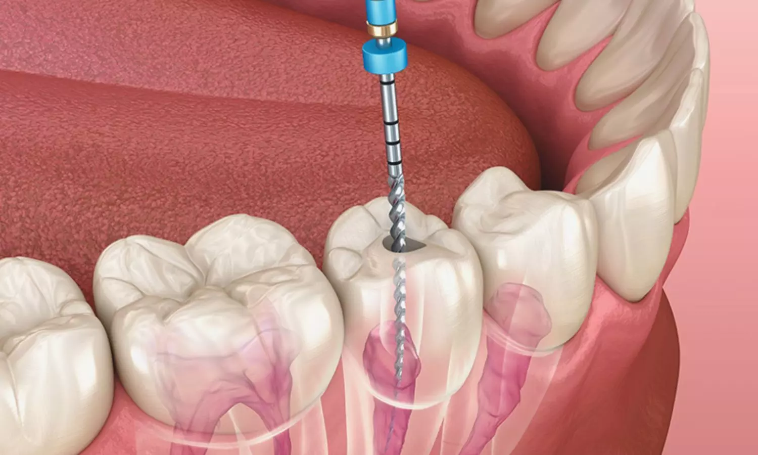
Nanoparticles Could Revolutionize Endodontics — Promise and Practical Hurdles: Study

Belgium: Researchers have found in a new study that nanoparticles offer significant potential in endodontics by enhancing antibacterial and biocompatible properties compared to conventional materials. However, concerns about long-term biocompatibility, manufacturing scalability, and clinical integration remain; the review calls for focused translational research to safely and practically integrate these laboratory advances into everyday dental practice.

The scoping review, published in Discover Nano by Una Ivković and colleagues from the OMFS-IMPATH Research Group, Department of Imaging and Pathology, Faculty of Medicine, University of Leuven, Belgium, highlights the expanding role of multifunctional nanoparticles in endodontics. These nanoscale materials are being investigated for their potential to regenerate the dentin–pulp complex, enhance the mechanical and biological performance of dental materials, and act as carriers for targeted therapeutic delivery.

Following PRISMA-Scoping Review guidelines, the team conducted an extensive literature search across Web of Science, PubMed, and Scopus. Of 490 initially identified articles, 70 met the inclusion criteria after full-text review. Data from both in vitro and in vivo studies were summarized, revealing that inorganic nanomaterials dominated the research landscape, featuring in 77% of studies, while organic nanomaterials accounted for 23%.

The study revealed the following findings:

  • Organic nanomaterials such as chitosan (29% of carrier applications) and extracellular vesicles (22% of therapeutic uses) showed considerable potential despite being less frequently studied.
  • Among inorganic nanomaterials, bioactive glass was a leading candidate, appearing in 31% of enhancer-related studies, 26% of therapeutic studies, and 13% of carrier-focused research.
  • Most applications (69% of studies) were directed toward pulp capping or root canal filling, reflecting the need for advanced materials in these procedures.
  • Research models included 75% in vitro experiments using relevant cell lines and 25% animal model studies, with 18% of the latter also incorporating parallel in vitro testing.
  • Nanoparticles consistently enhanced antibacterial activity and bioactivity compared with conventional endodontic materials, offering solutions to issues like reinfection and poor biological integration.

However, translating these benefits to clinical settings poses significant challenges. Current evidence is heavily based on controlled laboratory conditions that may not fully replicate the complexity of the oral environment. The authors highlight the need for more physiologically relevant models, including 3D culture systems, microfluidic tissue-on-chips, and organ-on-chips, to bridge this gap.

Future research priorities include optimizing nanoparticle formulations for specific therapeutic objectives, establishing standardized testing protocols to improve reproducibility, and expanding antibacterial evaluations to reflect the diverse microbial environment of the oral cavity. Additionally, robust clinical trials are essential to confirm real-world safety, effectiveness, and cost-efficiency.

The authors conclude that incorporating nanoparticles into endodontic practice could shift the field from a reliance on bio-inert materials to bioactive, regenerative solutions. By enabling tissue preservation, promoting healing, and facilitating targeted therapies, these innovative materials hold the potential to significantly enhance long-term outcomes for patients undergoing endodontic treatment.

Reference:

Ivković, U., Moreno-Rabié, C., Mignon, A. et al. Multifunctional nanoparticles in endodontics: applications, challenges, and future directions. Discover Nano 20, 130 (2025). https://doi.org/10.1186/s11671-025-04314-7

Powered by WPeMatico

Frailty Significantly Reduces Life Expectancy After Stroke or TIA in Elderly: Study

A recent study published in the Journal of Stroke and Cerebrovascular Diseases revealed that frailty significantly worsens outcomes for older adults who undergo ischemic stroke or transient ischemic attack (TIA), shortening life expectancy and increasing long-term mortality.

This research analyzed health data from the Vektis health insurance claims database, focusing on patients aged 70 and older who suffered a stroke or TIA in 2018. Frailty was identified using the U-PRIM frailty index, that evaluates vulnerability in older adults, while a non-frail control group was matched by age, sex, and socioeconomic status. The mortality data were tracked through May 2024, providing follow-up of roughly 5 to 6 years.

Among 16,778 frail and 10,069 non-frail participants, frail individuals underwent markedly higher mortality and shorter life expectancy after cerebrovascular events. Also, 66% of frail patients died following ischemic stroke when compared with 55% of non-frail patients. For TIA, the gap was even wider, with 54% of frail patients dying versus 36% of non-frail counterparts.

When examining life expectancy, the study found that frail patients who suffered a stroke lived an average of 3.8 years afterward, when compared with 5.2 years for non-frail individuals. TIA survivors demonstrated a similar pattern where frail patients lived about 5.9 years on average, while non-frail patients had life expectancy exceeding 6.4 years.

Using Cox regression analysis to adjust for potential confounders, frailty increased the risk of death by 30% after ischemic stroke and by 72% following TIA. The study examined secondary outcomes, finding that frail patients were more likely to experience additional cardiovascular events. After ischemic stroke, 40% of frail patients had further cardiovascular complications when compared with 38% of non-frail patients. Following TIA, rates were 18% versus 13%.

These results highlight the significant impact of frailty on survival in older adults after stroke or TIA. The study observed that recognizing frailty in clinical practice can inform treatment decisions, guide monitoring for recurrent cardiovascular events, and support discussions about advanced care planning.

The study highlights that age alone does not fully capture risk where frailty plays a critical role in predicting outcomes after cerebrovascular events. Overall, this research provides important evidence that frailty is a key factor of both mortality and life expectancy following ischemic stroke and TIA.

Reference:

Prins, S. R., Damoiseaux-Volman, B. A., van Erkelens, J. A., Vermeer, S. E., Van der Velde, N., & Van den Berg-Vos, R. M. (2025). Life expectancy after an ischemic stroke or transient ischemic attack in older adults – the role of frailty. Journal of Stroke and Cerebrovascular Diseases: The Official Journal of National Stroke Association, 108448. https://doi.org/10.1016/j.jstrokecerebrovasdis.2025.108448

Powered by WPeMatico

Over Half of Indian Patients With Nasal Symptoms Have Allergic Rhinitis: Study Finds

India: Allergic rhinitis (AR) is highly prevalent among Indian patients presenting with nasal complaints, with more than half of such individuals testing positive using the Score for Allergic Rhinitis (SFAR) questionnaire. The findings highlight the importance of early detection and targeted management to address this widespread health problem.         

The study, led by Dr. Carlton David Periera from the Department of ENT, Wadia Children’s Hospital, Parel, Mumbai, and colleagues, was published in the Journal of Asthma and Allergy. It represents one of the largest in-clinic evaluations of AR prevalence in India, involving patients aged 11 years and above across 19 states and union territories. Conducted between September 2022 and April 2023, the research enrolled 40,001 patients who visited doctors’ clinics and hospitals with nasal symptoms.

To determine AR, the researchers employed the SFAR questionnaire, with scores ≥7 indicating a positive diagnosis. A total of 3358 doctors, including ENT specialists and chest physicians, participated in the multicenter cross-sectional investigation.

The key findings were as follows:

  • 53.7% (21,480 patients) met the criteria for allergic rhinitis, reflecting a high prevalence in this clinical population.
  • Sneezing was the most common symptom, reported by 69.1% of all patients and nearly 87% of those with allergic rhinitis.
  • House dust mite exposure was identified as the leading trigger, affecting 73.8% of patients.
  • The burden of nasal symptoms peaked between October and January, indicating a seasonal pattern.
  • Allergic rhinitis was more frequent in females and individuals aged 11–40 years.
  • Around 50% of patients reported a family history of asthma, eczema, or allergic rhinitis.
  • Only 46% of allergic rhinitis–positive patients had been formally diagnosed by a doctor, and just 31.5% had undergone allergy testing, underscoring underdiagnosis.
  • After adjusting for confounders, allergic rhinitis was strongly associated with nasal complaints accompanied by watery, itchy eyes and with a history of physician-diagnosed allergies.
  • The authors noted that limited recognition may stem from a lack of standardized in-clinic diagnostic tools, which the SFAR questionnaire aims to address.

The researchers concluded that AR is a significant public health challenge in India. The high in-clinic prevalence suggests a pressing need for increased awareness, early screening, and proactive management. According to the team, the SFAR questionnaire is a reliable, easy-to-use screening instrument that can support timely diagnosis of AR in Indian clinical practice.

The authors emphasized that this dataset provides opportunities for further research into regional variations and risk factors. Expanding the use of SFAR beyond specialty clinics to general practice and community settings could give a clearer picture of the true burden of AR nationwide. Such efforts would not only improve early detection but also guide effective public health planning and strategies for managing allergic rhinitis in India.

Reference:

Periera CD, Warad V, Rodrigues JP, Pandit GS, Gupte V, Thakur GK, Upadhyaya A, Gogtay J. An Indian Cross-Sectional Study to Evaluate In-Clinic Allergic Rhinitis Prevalence in Patients Having Nasal Symptoms Using the SFAR Questionnaire. J Asthma Allergy. 2025;18:1239-1251. https://doi.org/10.2147/JAA.S516448

Powered by WPeMatico

ESIC Medical College Noida notifies MBBS fee structure, Check details

Uttar Pradesh- Through a notification, the Employees’ State Insurance Corporation (ESIC) Medical College Noida, UP has informed the complete fee structure for MBBS admission for the academic year 2025-26. The fee structure has been released for the IP Quota, State Quota and All India Quota (AIQ) students.

As per the fee structure, the IP Quota students have to pay a total of Rs 49,000/- and the State Quota students and AIQ students have to pay a total of Rs 1,25,000/- for MBBS admission.

Hostel Fee & Hostel Security Deposit are payable for those who want to opt for Hostel facilities. Those who do not want to opt for a Hostel may exclude both prescribed fees from the total fee structure to prepare a Demand Draft. Below is the complete fee structure-

FEE STRUCTURE

1 FOR ‘IP QUOTA’ STUDENT

S.NO

FEE HEAD (ANNUAL)

FEE TO PAY (IN RUPEES)

1

Tuition Fee

24,000/-

2

Caution Deposit (Refundable)

5,000/-

3

Hostel Fee

10,000/-

4

Hostel Security Deposit (Refundable)

10,000/-

TOTAL

49,000/-

2 FOR ‘STATE QUOTA’ STUDENTS

S.NO

FEE HEAD (ANNUAL)

FEE TO PAY (IN RUPEES)

1

Tuition Fee

1,00,000/-

2

Caution Deposit (Refundable)

5,000/-

3

Hostel Fee

10,000/-

4

Hostel Security Deposit (Refundable)

10,000/-

TOTAL

1,25,000/-

3 FOR ‘ALL INDIA QUOTA’ STUDENTS

S.NO

FEE HEAD (ANNUAL)

FEE TO PAY (IN RUPEES)

1

Tuition Fee

1,00,000/-

2

Caution Deposit (Refundable)

5,000/-

3

Hostel Fee

10,000/-

4

Hostel Security Deposit (Refundable)

10,000/-

TOTAL

1,25,000/-

To view the notification, click the link below

Prior to this, UPDGME has released the MD/MS fee structure for the academic session 2025–26 for all private medical colleges in the state. Medical Dialogues earlier reported that the fee structure includes the complete details of tuition fees (Clinical, Pathology, Non-Clinical), hostel charges, refundable security deposit, and annual miscellaneous charges applicable for each college. The fees for MD and MS programs in Uttar Pradesh vary considerably depending on the college and the specific course.

ADDITIONAL CHARGES

1 Security Deposit (Refundable): ₹5,00,000 (one-time) — includes hospital, lab, uniform, etc.

2 Miscellaneous Fees: ₹1,41,240 per year — covers university registration, development fee, library, student association, gym/sports, admission, examination, and all other charges.

Powered by WPeMatico

AI can spot which patients need treatment to prevent vision loss in young adults

Researchers have successfully used artificial intelligence (AI) to predict which patients need treatment to stabilize their corneas and preserve their eyesight, in a study presented at the 43rd Congress of the European Society of Cataract and Refractive Surgeons (ESCRS).

Powered by WPeMatico