Tetracyclines Reduce Fever and Hospital Stay in Mycoplasma pneumoniae Patients: Study

A new study published in the journal of Open Forum Infectious Diseases showed that tetracyclines are associated with shorter hospital stays and fever durations than macrolides and fluoroquinolones in patients with Mycoplasma pneumoniae pneumonia.

Knowing the epidemiological features of Mycoplasma pneumoniae pneumonia is essential for making well-informed diagnostic choices and developing the best possible treatment plans, especially in light of the disease’s recent pandemic resurgence following a period of decline. Infections with Mycoplasma pneumoniae occur both epidemically and endemically, peaking around every four years.

Due to their low minimum inhibitory concentrations and high tolerability, macrolide antibiotics are often used as the first line of therapy for M pneumoniae infections. Thus, this study was set to characterize the incidence rate, patient attributes, therapies, and results of adult patients who were admitted to the hospital due to Mycoplasma pneumoniae pneumonia.

Adults with M pneumoniae pneumonia who were referred to emergency rooms in Stockholm County, Sweden, between 2013 and 2017 were included in this retrospective cohort research. ICD-10 code J15.7 (M pneumoniae pneumonia) and a positive M pneumoniae polymerase chain reaction were used to identify the patients. Population data was taken from statistics sources and medical records were examined by hand. 

Nearly, 55% (385/747) of the 747 adults who were hospitalized with M pneumoniae pneumonia had a median age of 42 (interquartile range [IQR], 33–55) years. 8.5 instances per 100,000 person-years was the incidence rate, which peaked in 2016 at 14.1. 71% were hypoxemic at admission, and the most prevalent symptoms were fever (92%) and cough (95%).

The duration of symptoms at admission was prolonged for patients with severe illness. Almost, 0.4% of patients died in the hospital, and 6% needed to be admitted to an intensive care unit. those treated with fluoroquinolones (+0.8 [IQR, 0.1–1.4] days; P =.03) and macrolides (+1.0 [IQR, 0.9–1.2] days; P <.001) had a higher median duration of stay (4 [IQR, 2–6] days) than those treated with tetracyclines.

The patients treated with fluoroquinolones had a substantially longer median fever duration (+0.3 [IQR, 0.1–0.6] days; P =.02) than patients treated with tetracyclines. Overall, the results emphasize the value of prompt and precise therapy as well as the possible advantages of using doxycycline as a first-line medication. 

Source:

Hagman, K., Nilsson, A. C., Hedenstierna, M., & Ursing, J. (2025). Epidemiology, characteristics, and treatment outcomes of Mycoplasma pneumoniae pneumonia in hospitalized adults: A 5-year retrospective cohort study. Open Forum Infectious Diseases, 12(7), ofaf380. https://doi.org/10.1093/ofid/ofaf380

Powered by WPeMatico

Patients’ own autoantibodies may hold key to boosting cancer immunotherapy response, reveals research

A landmark study has revealed that autoantibodies-immune proteins traditionally associated with autoimmune disease-may profoundly influence how cancer patients respond to immunotherapy.

The study, published  in Nature, offers a potential breakthrough in solving one of modern-day oncology’s most frustrating mysteries: why checkpoint inhibitors work for some patients but not others-and how we can extend their benefits to more people.

“Our analysis shows that certain naturally occurring autoantibodies can tilt the odds dramatically toward shrinking tumors,” said senior author Aaron Ring, MD, PhD, an associate professor at Fred Hutch Cancer Center. “We saw some cases where autoantibodies boosted a patient’s likelihood of responding to checkpoint blockade by as much as five- to ten-fold.”

The Nature study suggests that autoantibodies could help reveal cancer’s weak spots and point to new targets for treatment.

Autoantibodies are proteins produced by the immune system that recognize the body’s own tissues. They are most associated with their harmful role in driving autoimmune diseases like lupus or rheumatoid arthritis. However, emerging evidence indicates that in some cases, autoantibodies can surprisingly exert health benefits.

“For years, autoantibodies were viewed mainly as bad actors in autoimmune disease, but we’re discovering they can also act as potent, built-in therapeutics,” said Ring, who holds the Anderson Family Endowed Chair for Immunotherapy at Fred Hutch. “My lab is mapping this hidden pharmacology so we can turn these natural molecules into new treatments for cancer and other illnesses.”

In the Nature study, Ring and his collaborators used a high-throughput assay he developed — called REAP (Rapid Extracellular Antigen Profiling) — to screen for over 6,000 types of autoantibodies in blood samples from 374 cancer patients receiving checkpoint inhibitors and 131 healthy individuals.

Checkpoint inhibitors have transformed treatment for a wide range of cancers including melanoma and non-small cell lung cancer by unleashing the immune system to see and attack cancer. But not all patients respond to these treatments and, in many cases, their anti-tumor effects are incomplete and do not result in a cure.

Using blood samples collected from patients and healthy individuals, the REAP analyses revealed that cancer patients had substantially higher levels of autoantibodies compared to healthy controls.

Importantly, certain autoantibodies were strongly linked to better clinical outcomes, indicating their potential role in enhancing the effectiveness of immunotherapy.

For example, autoantibodies that blocked an immune signal called interferon were linked to better anti-tumor effects from checkpoint inhibitors. This finding mirrors other studies showing how too much interferon can exhaust the immune system and then curtail the effects of immunotherapy.

“In some patients, their immune system essentially brewed its own companion drug,” Ring explained. “Their autoantibodies neutralized interferon and that amplified the effect of checkpoint blockade. This finding gives us a clear blueprint for combination therapies that intentionally modulate the interferon pathway for everyone else.”

Not all autoantibodies were beneficial. The team discovered several that were associated with worse outcomes from checkpoint inhibitors, likely because they disrupted critical immune pathways necessary for anti-tumor responses. Finding ways to eliminate or counteract these detrimental autoantibodies could open another promising avenue for enhancing the effectiveness of immunotherapy.

“This is only the beginning,” Ring said. “We’re now extending the search to other cancers and treatments so we can harness — or bypass — autoantibodies to make immunotherapy work for far more patients.” 

Reference:

Dai, Y., Aizenbud, L., Qin, K. et al. Humoral determinants of checkpoint immunotherapy. Nature (2025). https://doi.org/10.1038/s41586-025-09188-4

Powered by WPeMatico

Total Thyroidectomy Safe in Obese Patients Despite Longer Operative Times: Study

Researchers have found in a new study that Total thyroidectomy can be safely performed in obese patients without an increased risk of surgery-related complications, despite the longer operative durations associated with higher BMI.

Obesity is associated with an increased risk of postoperative morbidity. We aimed to analyze the impact of BMI on surgical complications in patients undergoing thyroidectomy. This retrospective study was conducted in a single academic center. A total of 484 patients with open total thyroidectomy were considered eligible. These patients were divided in the non-obese (BMI < 30 kg/m2) and obese (BMI ≥ 30 kg/m2) groups. A 1:2 case matching based on demographic (age and gender) and clinical (benign/malignant disease) variables was performed to generate homogenous study groups. A comparative analysis was carried out to show the differences between the two groups in terms of the occurrence of surgery-related outcomes. Results: After case matching, 193 non-obese and 98 obese patients were included in the final analysis. There was no statistically significant difference in the rate of primary outcomes in the non-obese and obese groups: hypoparathyroidism (transient: 29% versus 21.4%, p = 0.166; permanent: 11.4% versus 15.3%, p = 0.344, respectively) and recurrent laryngeal nerve palsy (transient: 13.9% versus 11.2%, p = 0.498; permanent: 3.1% versus 2.0%, p = 0.594, respectively). A BMI ≥ 30 kg/m2 was associated with a significantly longer operative time (p = 0.018), while other secondary outcomes were not significantly affected by BMI. Despite prolonged operative times in obese patients, total thyroidectomy could be performed safely and without increased risk of surgery-related morbidity, regardless of BMI.

Reference:

Vaghiri, S., Mirheli, J., Prassas, D. et al. The BMI impact on thyroidectomy-related morbidity; a case-matched single institutional analysis. BMC Surg 25, 286 (2025). https://doi.org/10.1186/s12893-025-03018-0

Keywords:

Total, Thyroidectomy, Safe, Obese, Patients, Despite, Longer, Operative, Times, Study, Vaghiri, S., Mirheli, J., Prassas, D, Obesity, Thyroid surgeryBMI, Postoperative complications

Powered by WPeMatico

During pregnancy, are newer antiseizure medications safer than older drugs?

A new study that examined older and newer medications to treat seizures has found that using some medications during pregnancy is linked to an increased risk of malformations at birth, or birth defects. The study is published July 16, 2025, in Neurology®, the medical journal of the American Academy of Neurology.

“Seizures can lead to falls and other complications during pregnancy, so seizure control for those with epilepsy is crucial to protect the health of both the mother and child,” said study author Sonia Hernandez-Diaz, MD, DrPH, of Harvard T.H. Chan School of Public Health in Boston, Massachusetts. “While some older drugs are known to increase the risk of major malformations, less is known about the safety of newer ‘second generation’ medications. Our study looked at a number of drugs and provides valuable information for health care providers and people who may become pregnant to make more informed decisions about the use of these medications during pregnancy.”

For the study, researchers looked at 7,311 women who were taking an antiseizure medication during the first trimester of pregnancy. They were compared to 1,311 women who did not take antiseizure medications.

Participants completed phone interviews at the start of the study, at seven months pregnant, and within three months after delivery.

Researchers confirmed birth defects through medical records. These included cleft lip, larger-than-normal holes in the heart, neural tube defects where the spinal cord does not develop properly such as spina bifida, missing or underdeveloped limbs and other issues where parts of the body did not form correctly.

They then looked at the risk of these birth defects among infants exposed to specific antiseizure medications taken during the first trimester of pregnancy and compared them to infants exposed to lamotrigine, an antiseizure medication used as the reference group.

Researchers found that the risk of major birth defects varied among medications. Valproate posed the highest risk, with major birth defects occurring in 9% of infants exposed. Phenobarbital followed at 6%. Among newer drugs, birth defects occurred in 5% of those exposed to topiramate, while the group exposed to lamotrigine had malformations in 2% of births.

When compared to those who took lamotrigine, those who took valproate had more than five times the risk of birth defects, those who took phenobarbital had nearly three times the risk, and those taking topiramate had over twice the risk.

“Our results confirm that using valproate, phenobarbital or topiramate during early pregnancy is linked to a higher chance of major birth defects in the infants when compared to lamotrigine,” Hernandez-Diaz said. “On the other hand, our results did not show an increased risk with medications like levetiracetam, oxcarbazepine, gabapentin and zonisamide. For lacosamide and pregabalin, the data wasn’t clear enough to make solid conclusions, so more research is needed.”

A limitation of the study was that participants were enrolled months after conception and in most cases early losses of pregnancy were not assessed for malformations, so the number of malformations overall may have been underestimated.

Reference:

Sonia Hernandez-Diaz, Use of Antiseizure Medications Early in Pregnancy and the Risk of Major Malformations in the Newborn,Neurology, doi/10.1212/WNL.0000000000213959.

Powered by WPeMatico

Blood test may rule out future dementia risk: Study

A new study, published in Nature Medicine, has investigated the potential of specific biomarkers such as tau217, Neurofilament Light (NfL), and Glial Fibrillary Acidic Protein (GFAP) to predict the occurrence of dementia, including Alzheimer’s disease, up to ten years before an actual diagnosis in cognitively healthy older adults living in the community.

Previous research has suggested that these biomarkers could be useful in early dementia diagnostics, but most studies involved individuals who have already sought medical care for cognitive issues, due to cognitive concerns or cognitive symptoms, such as memory difficulties.

A larger, community-based study, was necessary to determine the predictive value of biomarkers in the general population.

Led by researchers from the Aging Research Center of Karolinska Institutet in collaboration with SciLifeLab and KTH Royal Institute of Technology in Stockholm, the study analysed blood biomarkers in more than 2,100 adults aged 60+, who were followed over time to determine if they developed dementia.

At a follow-up ten years later, 17 percent of participants had developed dementia. The accuracy of the biomarkers used in the study was found to be up to 83 percent.

“This is an encouraging result, especially considering the 10-year predictive window between testing and diagnosis. It shows that it is possible to reliably identify individuals who develop dementia and those who will remain healthy,” says Giulia Grande, assistant professor at the Department of Neurobiology, Care Sciences and Society, Karolinska Institutet, and first author of the study.

“Our findings imply that if an individual has low levels of these biomarkers, their risk of developing dementia over the next decade is minimal”, explains Davide Vetrano, associate professor at the same department and the study’s senior author. “This information could offer reassurance to individuals worried about their cognitive health, as it potentially rules out the future development of dementia.”

However, the researchers also observed that these biomarkers had low positive predictive values, meaning elevated biomarker levels alone could not reliably identify individuals who would surely develop dementia within the next ten years. Therefore, the study authors advise against widespread use of these biomarkers as screening tools in the population at this stage.

“These biomarkers are promising, but they are currently not suitable as standalone screening tests to identify dementia risk in the general population,” says Davide Vetrano.

The researchers also noted that a combination of the three most relevant biomarkers – p-tau217 with NfL or GFAP – could improve predictive accuracy.

“Further research is needed to determine how these biomarkers can be effectively used in real-world settings, especially for elderly living in the community or in primary health care services,” says Grande.

“We need to move a step further and see whether the combination of these biomarkers with other clinical, biological or functional information could improve the possibility of these biomarkers to be used as screening tools for the general population”, Grande continues.

Reference:

Grande, G., Valletta, M., Rizzuto, D. et al. Blood-based biomarkers of Alzheimer’s disease and incident dementia in the community. Nat Med (2025). https://doi.org/10.1038/s41591-025-03605-x

Powered by WPeMatico

Brief Maternal Oxygen Inhalation in Third Trimester may Alter Fetal Hemodynamics, suggest study

A new study published in the BMC European Journal of Medical Research showed that brief maternal oxygen inhalation in third trimester leads to higher pulsatility index for the pulmonary artery (PPI) and lower cerebroplacental ratio (CPR). These changes suggest a potential impact on fetal circulation, warranting further investigation into its implications.

Newborns have not been demonstrated to benefit much from the maternal oxygen intake during birth. Its effect on fetal hemodynamics in the latter stages of pregnancy is yet unknown, though. Wu XQ and colleagues did this study to look into the relationship between fetal hemodynamic alterations and the maternal late-trimester oxygen inhalation. In particular, they analyzed if there are any possible advantages or disadvantages for the fetus from this approach and examined the short-term effects of maternal oxygen supplementation on fetal Doppler measurements.

Between January 2022 and December 2022, singleton pregnancies that had a prenatal ultrasound examination after 32[+0] weeks were the source of this retrospective data. The participants were divided into groups who inhaled oxygen and those that did not. Despite the lack of a therapeutic basis, oxygen inhalation was given at the mother’s request, mostly because of worries about hypoxia from extended mask usage during the COVID-19 epidemic.

The pregnant women in the oxygen inhalation group were given 3 L/min of oxygen via nasal cannula for 30 minutes, and then they proceeded to the ultrasound department for a sonographic evaluation within an hour. Before analysis, the CPR and PPI were predetermined as the main outcomes. The Doppler index, placental pulsatility index (PPI), and cerebroplacental ratio (CPR) were computed for every woman. Additionally, fetal heart function was evaluated in M-mode or pulsed Doppler. In comparison to the non-oxygen inhalation group, the exposed maternal oxygen inhalation group showed lower birth weight, lower CPR, and greater PPI as the primary result.

The final analysis comprised a total of 104 singleton pregnancies (oxygen inhalation group: n = 48). The resistance indices of the middle cerebral arteries, ductus venosus, descending aorta, umbilical vein, uterine arteries, and umbilical arteries did not change significantly. Variations were seen in the oxygen inhalation group, though.

Significant differences were seen between the groups for indices that have a higher sensitivity for predicting negative outcomes: PPI was higher in the oxygen inhalation group than in the non-oxygen inhalation group, and CPR was similarly lower in the oxygen inhalation group. Furthermore, the group that inhaled oxygen had a considerably lower birth weight than the group that did not. Overall, a short oxygen intake by the mother during the third trimester was linked to notable alterations in the fetal hemodynamics, including a decrease in CPR and an increase in PPI.

Source:

Wu, X.-Q., Yang, X.-F., Ye, L., Zhang, X.-B., Hong, Y.-Q., & Chiu, W.-H. (2025). Maternal oxygen inhalation affects the fetal hemodynamic in low-risk with uncomplicated late pregnancy. European Journal of Medical Research, 30(1), 222. https://doi.org/10.1186/s40001-025-02456-z

Powered by WPeMatico

RECK effective opioid-sparing local anesthetic for patients undergoing posterior spinal fusion: study

Ropivacaine-epinephrine-clonidine-ketorolac is an effective opioid-sparing local anesthetic for patients undergoing posterior spinal fusion: study

Ropivacaine-Epinephrine-Clonidine-Ketorolac (RECK) cocktail can improve pain control in patients undergoing lumbar decompression. Given the aging population, rising healthcare costs, the opioid epidemic, and associations of acute pain control with long-term opioid use, effective opioid-sparing analgesia following spinal fusion surgery may impart societal benefits.

Anthony V. Nguyen et al conducted a study to investigate whether RECK was an effective local anesthetic for patients undergoing posterior spinal fusion.

Primary outcomes were post operative pain levels as determined by Visual Analog Scale, in-hospital opioid consumption, length-of-stay<4days, and long-term opioid utilization at three months postoperatively. Secondary outcomes were rates of discharge to home, complication rates, readmissions within 90days.

The authors analyzed whether opioid exposure, patient-specific or surgery-specific factors, and administration of RECK (versus another local anesthetic) were associated with post operative pain levels, in hospital opioid consumption, length-of-stay, home discharge, long-term opioid utilization, complications, and readmissions within 90 days using multivariable regression.

KEY FINDINGS OF THE STUDY WERE:

• Of the162 patients meeting study criteria, 49(30.2%) received RECK.

• RECK was significantly associated with decreased pain levels at 2-,4-,6-, and 12-hours postoperatively (p≤.001−.01).

• RECK was associated with decreased total and daily inpatient opioid utilization (as measured by oral morphine equivalents) in multivariable linear regression (B=-159.6, 95% CI :-255.5−-63.6, p=.002andB=-27.9,95% CI :-48.9−-7.0, p=.01, respectively).

• Length-of stay duration of < 4days was associated with RECK administration (OR4.1,95%CI:1.4−13.2, p=.01) and was negatively associated with levels fused (OR0.4,95%CI:0.2−0.7, p=.005) and durotomy (OR0.02,95%CI:0.0009−0.1, p<.001).

• Prolonged post operative opioid utilization was associated with pre operative opioid prescription (OR3.6,95%CI:1.7−7.8,p=.001) and was negatively associated with RECK (OR0.4,95%CI:0.2−0.9,p=.04).

• RECK was not associated with readmissions, complications, or home discharge.

The authors concluded – ‘RECK administration during spinal fusion surgery was associated with decreased postoperative pain levels, inpatient opioid utilization, LOS, and chronic opioid use. It was not associated with increased complications or readmissions. Thus, RECK is a safe local anesthetic with opioid-sparing benefits for patients undergoing spinal fusion surgery and may reduce healthcare costs.’

Further reading:

Ropivacaine-epinephrine-clonidine-ketorolacisaneffectiveopioid-sparing local anesthetic for patients undergoing posterior spinal fusion

A.V. Nguyen et al.

The Spine Journal 25 (2025) 974−982

Powered by WPeMatico

From Womb to Wellness: The Impact of Prenatal and Perinatal Factors on Cardiovascular Health in Childhood, study finds

Associations between prenatal and perinatal factors and cardiovascular health (CVH) trajectories in children from childhood through adolescence were investigated, emphasizing the significance of the American Heart Association’s Life’s Essential 8 (LE8) framework. This framework includes four behavioral (diet, physical activity, sleep duration, nicotine exposure) and four biological factors (BMI, blood pressure, glucose, cholesterol) assessed on a 0-100 scale. Recent research highlights the crucial role that early life stages play in forming health behaviors and disease precursors.

Importance of Early Precursors in CVD

CVD is a leading cause of death in the U.S., with early precursors such as elevated BMI, blood pressure, and cholesterol levels observed by age three. Understanding how prenatal exposures impact long-term CVH is vital for primordial prevention strategies. Prior studies indicated that factors such as maternal obesity, gestational weight gain, and smoking during pregnancy correlate with subsequent increases in child CVD risk factors.

Methodology: Project Viva Cohort Study

Utilizing data from the Project Viva cohort study in eastern Massachusetts, the research examined how specific prenatal and perinatal factors, including maternal BMI, gestational weight gain, and breastfeeding practices, correlate with CVH. With 1,333 participants, the study analyzed CVH measures between early childhood (3 years) and late adolescence (18 years), employing mixed-effect models to assess CVH trajectories across sex.

Key Findings on Maternal Factors and CVH

The findings demonstrated that children born to mothers with prepregnancy obesity had significantly lower CVH scores throughout childhood and adolescence compared to those from healthy-weight mothers. Maternal gestational hypertension was linked to an earlier inflection point in CVH scores, indicating a faster decline after initial improvements, while smoking during pregnancy correlated with consistently lower CVH scores. Additionally, children exclusively formula-fed in their first six months exhibited poorer CVH outcomes.

Impact of Behavioral Factors on Health Trajectories

Behavioral factors accounted for most of the observed decline in CVH, highlighting the impact of early dietary habits on long-term health trajectories. The study reaffirmed previous observations regarding the connections between maternal health factors and child CVH trajectories, extending the knowledge on how prenatal environments influence long-term cardiovascular health.

Conclusion: Addressing Modifiable Prenatal Risks

Overall, the study underscores the importance of addressing modifiable prenatal and perinatal risks, such as maternal obesity and smoking, to improve childhood cardiovascular outcomes. This information is critical for developing early interventions aimed at fostering better health from infancy onward, ultimately reducing the risk of CVD in later life.

Key Points

– The study examines the associations between prenatal and perinatal factors and cardiovascular health (CVH) trajectories in children, utilizing the American Heart Association’s Life’s Essential 8 (LE8) framework, which assesses both behavioral and biological factors on a 0-100 scale to understand early life influences on health.

– Cardiovascular disease (CVD) is a principal cause of death in the U.S., with early risk indicators such as elevated BMI, blood pressure, and cholesterol emerging as early as three years of age, highlighting the importance of understanding prenatal exposure effects for effective prevention strategies.

– Data from the Project Viva cohort study (1,333 participants) in eastern Massachusetts was analyzed to investigate the impact of maternal factors (BMI, gestational weight gain, breastfeeding practices) on children’s CVH from early childhood (3 years) into adolescence (18 years), using mixed-effect models for trajectory assessments.

– Key findings indicate that children born to mothers with obesity pre-pregnancy exhibited consistently lower CVH scores, while maternal gestational hypertension and smoking during pregnancy were associated with negative shifts in CVH trajectories, including earlier declines in health measures.

– Behavioral factors heavily influenced CVH declines, particularly early dietary habits, reinforcing established links between maternal health conditions and their children’s cardiovascular outcomes and extending the understanding of prenatal influences on long-term health.

– The study highlights the critical need to address modifiable prenatal risks, such as maternal obesity and smoking, to enhance cardiovascular health in children and suggests that early interventions can mitigate CVD risks later in life.

Reference –

I. Aris et al. (2025). Prenatal And Perinatal Factors Of Life’S Essential 8 Cardiovascular Health Trajectories. *JAMA Network Open*, 8. https://doi.org/10.1001/jamanetworkopen.2025.7774.

Powered by WPeMatico

University of Houston optometry researcher provides new hope for injured corneas: Study

Consider the cornea: the eye’s transparent, dome-shaped surface that provides most of its focusing power and serves as a protective barrier. It directs light onto the retina for sharp vision and shields the eye from dust, debris and harmful UV rays.

That’s a lot to ask from a structure made of cells and proteins that’s about two-thirds the size of a dime, yet it does its job brilliantly.

Until it is damaged. Then vision can blur, distort—or be lost entirely. And the cornea is definitely prone to damage.

“When the cornea gets injured, it often heals with scar tissue and abnormal blood vessels, both of which can cause permanent vision loss,” said Tarsis G. Ferreira, assistant professor at the UH College of Optometry. “Unfortunately, there are few treatments for this, and in severe cases, patients need a corneal transplant.”

That dilemma led Ferreira to begin developing a treatment for corneal scarring. He is supported in his work with a five-year grant for $2.2 million from the National Institutes of Health. The treatment focuses on a natural protein in the cornea, named decorin, which plays an essential role in keeping the cornea clear and healthy. It does this by blocking another protein called TGF-β1 (transforming growth factor beta 1), which triggers the scarring process after injury.

“Our goal is to engineer a better version of decorin that more strongly blocks TGF-β1 and another protein receptor that promotes blood vessel growth (VEGFR2), to prevent both scarring and unwanted blood vessels from forming,” said Ferreira.

Natural decorin does that, but the protein is hard to produce. Ferreira has built a version of decorin, which he named mini-dec, that binds better to the two targets and is easy to produce and deliver as eyedrops.

“We hope this new therapy could soon be used to treat eye injuries and surgeries, helping people heal without losing vision,” said Ferreira. The decorin-based drugs will be simple to use and have the potential for treating corneal injuries in the clinic.”

Powered by WPeMatico

New discovery reveals the role of spinal cord in bladder control

Urinary incontinence is a devastating condition affecting over 33 million Americans, according to the National Association for Continence, leading to significant adverse impacts on patients’ mental health and quality of life. Disorders of urination are also a key feature of all neurological disorders.

A USC research team has now made major progress in understanding how the human spinal cord triggers the bladder emptying process. The discovery could lead to exciting new therapies to help patients regain control of this essential function.

In the pioneering study, a team from USC Viterbi School of Engineering and Keck School of Medicine of USC has harnessed functional ultrasound imaging to observe real-time changes in blood flow dynamics in the human spinal cord during bladder filling and emptying. The work was published in Nature Communications and was led by Charles Liu, the USC Neurorestoration Center director at Keck School of Medicine of USC and professor of biomedical engineering at USC Viterbi and Vasileios Christopoulos, assistant professor in the Alfred E. Mann Department of Biomedical Engineering. The study’s co-first authors are biomedical engineering postdoctoral scholar Kofi Agyeman and Darrin Lee, associate director of the USC Neurorestoration Center and also affiliated with biomedical engineering at USC. Another key contributor was Evgeniy Kreydin from the Rancho Los Amigos National Rehabilitation Center and the USC Institute of Urology.

The spinal cord regulates many essential human functions, including autonomic processes like bladder, bowel, and sexual function. These processes can break down when the spinal cord is damaged or degenerated due to injury, disease, stroke, or aging. However, the spinal cord’s small size and intricate bony enclosure have made it notoriously challenging to study directly in humans. Unlike in the brain, routine clinical care does not involve invasive electrodes and biopsies in the spinal cord due to the obvious risks of paralysis. Furthermore, fMRI imaging, which comprises most of human functional neuroimaging, does not exist in practical reality for the spinal cord, especially in the thoracic and lumbar regions where much of the critical function localizes.

“The spinal cord is a very undiscovered area,” Christopoulos said. “It’s very surprising to me because when I started doing neuroscience, everybody was talking about the brain. And Dr. Liu and I asked, ‘What about the spinal cord?’ For many, it was just a cable that transfers information from the brain to the peripheral system. The truth was that we didn’t know how to go there – how to study the spinal cord in action, visualize its dynamics and truly grasp its role in physiological functions.”

Functional Ultrasound Imaging: A New Window into the Spinal Cord

To overcome these barriers, the USC team employed functional ultrasound imaging (fUSI), an emerging neuroimaging technology that is minimally invasive. The fUSI process allowed the team to measure where changes in blood volume occur on the spinal cord during the cycle of urination.

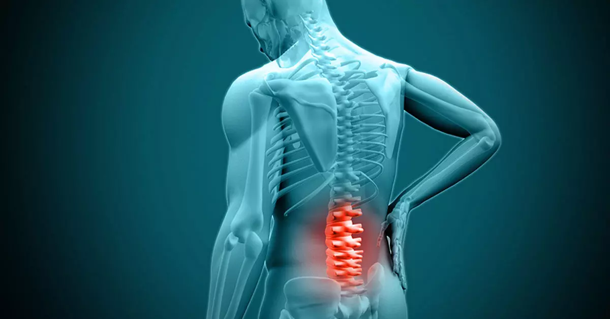
However, fUSI requires a “window” through the bone to image the spinal cord. The researchers found a unique opportunity by working with a group of patients undergoing standard-of-care epidural spinal cord stimulation surgery for chronic low back pain.

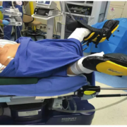
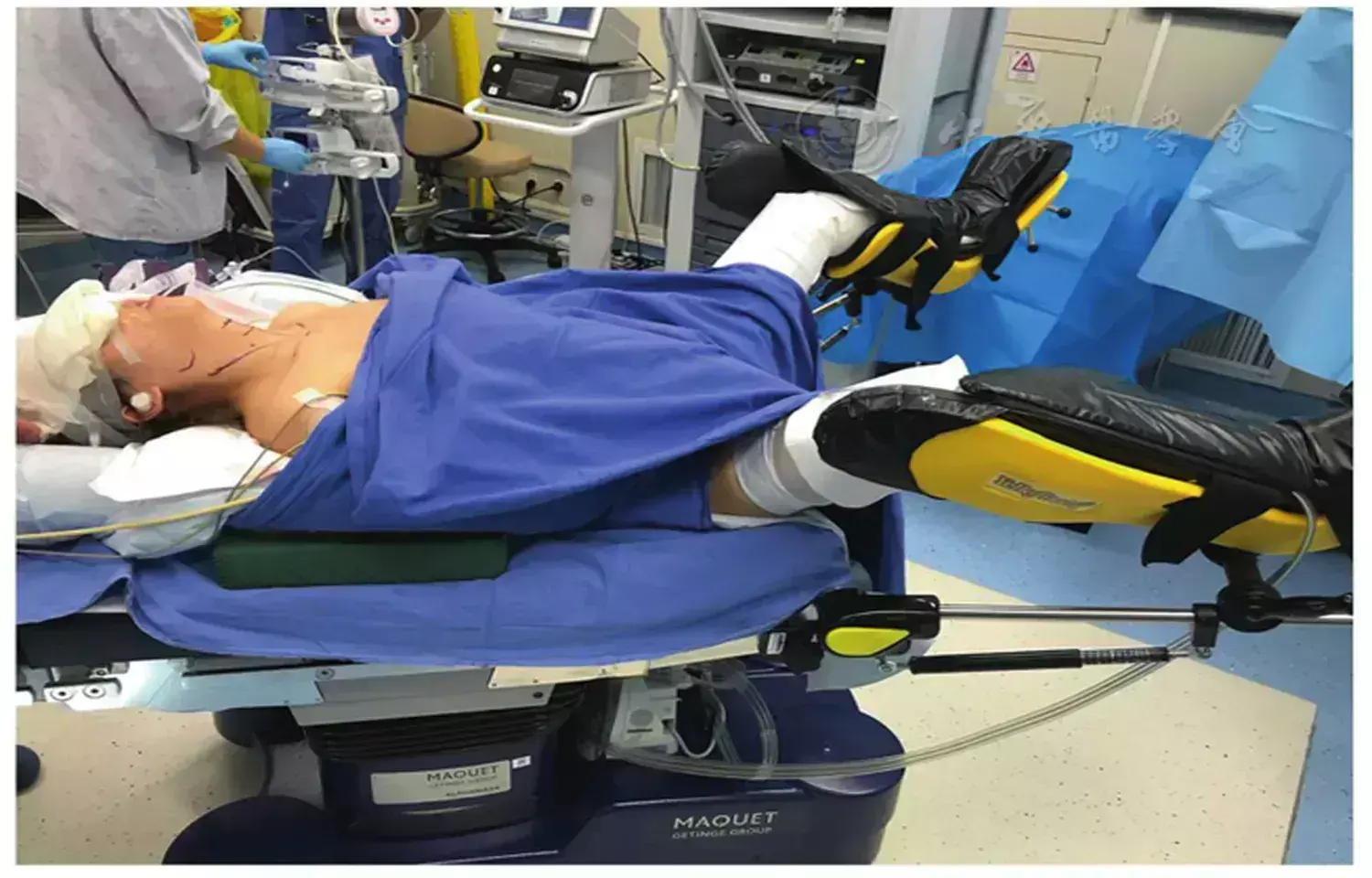
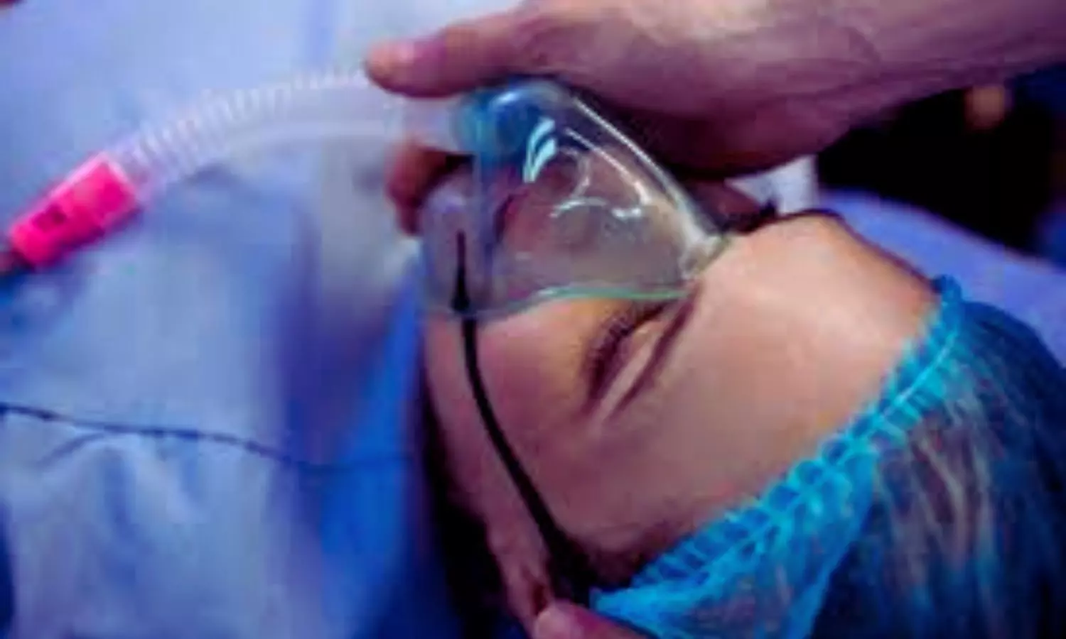
“During the implantation of the spinal cord stimulator, the window we create in the bone through which we insert the leads gives us a perfect and safe opportunity to image the spinal cord using fUSI with no risk or discomfort to the study volunteers,” said Lee, who performed the surgeries. “While the surgical team was preparing the stimulator, we gently filled and emptied the bladder with saline to simulate a full urination cycle under anesthesia while the research team gathered the fUSI data,” added Kreydin, who was already working closely with Liu to study the brain of stroke patients during micturition using fMRI.

“This is the first study where we’ve shown that there are areas in the spinal cord where activity is correlated with the pressure inside the bladder,” Christopoulos said. “Nobody had ever shown a network in the spinal cord correlated with bladder pressure. What this means is I can look at the activity of your spinal cord in these specific areas and tell you your stage of the bladder cycle – how full your bladder is and whether you’re about to urinate”.

Christopoulos said the experiments identified that some spinal cord regions showed positive correlation, meaning their activity increased as bladder pressure rose, while others showed negative (anti-correlation), with activity decreasing as pressure increased. This suggests the involvement of both excitatory and inhibitory spinal cord networks in bladder control. “It was extremely exciting to take data straight from the fUSI scanner in the OR to the lab, where advanced data science techniques quickly revealed results that have never been seen before, even in animal models, let alone in humans,” Agyeman said.

New Hope for Patients

Liu has worked for two decades at the intersection of engineering and medicine to develop transformative strategies to restore function to the nervous system. Christopoulos has spent much of his research career developing neuromodulation techniques to help patients regain motor control. Together, they noted that for patients, retaining control of the autonomic processes that many of us take for granted is more fundamental than even walking.

“If you ask these patients, the most important function they wanted to restore was not their motor or sensory function. It was things like sexual function and bowel and bladder control,” Christopoulos said, noting that urinary dysfunction often leads to poor mental health. “It’s a very dehumanizing problem to deal with.”

Worse still, urinary incontinence leads to more frequent urinary tract infections (UTIs) because patients must often be fitted with a catheter. Due to limited sensory function, they may not be able to feel that they have an infection until it is more severe and has spread to the kidneys, resulting in hospitalization.

This study offers a tangible path toward addressing this critical need for patients suffering from neurogenic lower urinary tract dysfunction. The ability to decode bladder pressure from spinal cord activity provides proof-of-concept for developing personalized spinal cord interfaces that could warn patients about their bladder state, helping them regain control.

Currently, almost all neuromodulation strategies for disorders of micturition are focused on the lower urinary tract, largely because the neural basis of this critical process remains unclear. “One has to understand a process before one can rationally improve it,” Liu said. This latest research marks a significant step forward, opening new avenues for precision medicine interventions that combine invasive and noninvasive neuromodulation with pharmacological therapeutics to make neurorestoration of the genitourinary system a clinical reality for millions worldwide.

Reference:

Agyeman, K.A., Lee, D.J., Abedi, A. et al. Human spinal cord activation during filling and emptying of the bladder. Nat Commun 16, 6506 (2025). https://doi.org/10.1038/s41467-025-61470-1

Powered by WPeMatico