Does a diabetes drug help prevent dementia?

New research in Diabetes, Obesity and Metabolism reveals that metformin, a medication traditionally prescribed to treat diabetes, is linked to lower risks of dementia and early death.

In the study by investigators at Taipei Medical University that included 452,777 adults with varying degrees of overweight and obesity, 35,784 cases of dementia and 76,048 deaths occurred over 10 years. Metformin users exhibited significantly lower risks of both dementia and all-cause death than nonusers.

The benefits of metformin were seen across all categories of overweight, obesity, and severe obesity, with 8–12% lower risks of dementia and 26–28% lower risks of death.

“Although our study results are promising for metformin’s effects on dementia and mortality, further research is required to explore the mechanisms involved,” said co-corresponding author Chiehfeng Chen, MD, PhD, MPH.

Reference:

Yu-Liang Lin, Protective effect of metformin against dementia in patients with obesity: Results from a global federated health network analysis, Diabetes Obesity and Metabolism, DOI: 10.1111/dom.16647.

Powered by WPeMatico

Fiber-based NIRAF enhances parathyroid gland detection during endocrine neck surgery and may help reduce complications: JAMA

A new study published in the Journal of American Medical Association showed that during endocrine neck surgery, fiber-based near-infrared autofluorescence (NIRAF) improves parathyroid gland identification and might potentially lower complications.

Accurately identifying and protecting parathyroid glands (PGs) during parathyroidectomy and thyroidectomy is essential since their removal or injury can result in hypoparathyroidism. Thus, this study determined if fiber-based NIRAF lowers the incidence of hypoparathyroidism and enhances the number of PGs discovered intraoperatively.

From March 2020 to July 2024, this clinical study with a 6-month follow-up was carried out. It comprised 3 junior (less than 5 years of experience) and four senior (more than 10 years of experience) surgeons from 4 medical centers across the US. A total of 398 individuals (2 withdrew) got parathyroidectomy, and 354 patients had total/completion thyroidectomy, out of the 754 patients who were recruited and 752 who were randomized. The use of fiber-based NIRAF during thyroid and parathyroidectomy was one of the primary therapies.

The primary outcome (94.4%) was examined for 712 out of 752 randomly assigned individuals. In all, 159 patients had traditional surgery, and 161 got parathyroidectomy with NIRAF. Furthermore, 178 had conventional surgery, and 176 had thyroidectomies utilizing NIRAF.

Using NIRAF for targeted operations did not substantially increase the mean number of PGs found during parathyroidectomy (mean, NIRAF, 1.6; 95% CI, 1.4-1.8 vs. control, 1.5; 95% CI, 1.4-1.7). The mean number of PGs found by the surgeons utilizing NIRAF during bilateral explorations was higher (mean NIRAF, 3.5; 95% CI, 3.4-3.7 vs. control, 3.2; 95% CI, 3.0-3.4; P <.001).

The mean number of PGs detected during thyroidectomy was higher when NIRAF was used (mean NIRAF, 3.3; 95% CI, 3.2-3.4 versus control, 2.8; 95% CI, 2.7-3.0; P <.001). Hypoparathyroidism following thyroidectomy did not significantly vary at the last follow-up (NIRAF: 3 of 176 patients [1.7%]; control: 6 of 176 patients [3.4%]) or during the transitory period (NIRAF: 48 of 173 patients [27.8%]; control: 44 of 169 patients [26%]).

Overall, this study suggest fiber-based NIRAF, may assist surgeons in confidently identifying more PGs during bilateral exploration, but it offers no further benefit during targeted surgeries. Regardless of the method, the number of PGs found increased during thyroidectomy. Even while NIRAF had no discernible effect on short-term patient outcomes, surgeons may find it useful as an intraoperative tool in situations where alternative techniques for PG tissue confirmation are not accessible.

Source:

Cousart, A. G., Kiernan, C. M., Willmon, P. A., Thomas, G., Wang, T. S., Gauger, P. G., Duh, Q.-Y., Underwood, H. J., Jackson, A., Patel, A., Mahadevan-Jansen, A., & Solórzano, C. C. (2025). Near-infrared autofluorescence for parathyroid detection during endocrine neck surgery: A randomized clinical trial: A randomized clinical trial. JAMA Surgery. https://doi.org/10.1001/jamasurg.2025.2233

Powered by WPeMatico

Ancient practice of blowing through conch shell could help to treat dangerous snoring condition: Study

People who practised blowing through a conch shell regularly for six months experienced a reduction in their symptoms of obstructive sleep apnoea (OSA), according to a small randomised controlled trial published today (Monday) in ERJ Open Research.

OSA is a common sleep disorder where breathing repeatedly stops during the night due to a blocked airway. It leads to loud snoring, restless sleep and daytime sleepiness. It also increases the risk of high blood pressure, heart disease, and stroke.

Blowing the conch shell, or shankh blowing, has been part of Indian culture for thousands of years. The new research showed that people with moderate OSA who practised shankh blowing slept better, felt more alert during the day and had fewer breathing interruptions at night. The researchers say conch blowing is a simple, low-cost intervention that could help reduce symptoms without the need for medication or machines.

The study was led by Dr Krishna K Sharma from the Eternal Heart Care Centre and Research Institute in Jaipur, India. He said: “The standard treatment for OSA is a continuous positive airway pressure machine, or CPAP, which keeps the patient’s airway open by blowing air through a facemask throughout the night. While effective, many patients find it uncomfortable and struggle to use it consistently.

“In my clinical practice, several patients reported feeling more rested and experiencing fewer symptoms after regularly practising shankh blowing – a traditional yogic breathing exercise involving exhaling through a conch shell. These observations led us to design a scientific study to rigorously test whether this simple, ancient practice could serve as a meaningful therapy for people with OSA.”

The study included 30 people with moderate OSA, aged between 19 and 65, who were assessed at the Eternal Heart Care Centre and Research Institute between May 2022 and January 2024. They were tested with polysomnography, meaning they are monitored throughout a night’s sleep, and asked questions about the quality of their sleep and how sleepy they feel during the day.

They were randomly assigned to either be trained to practise blowing through a conch shell (16 patients) or to practise a deep breathing exercise (14 patients). Participants were provided with a traditional shankh used in yogic practices. They were trained in person at the clinic by a study team member before beginning home-based practice. Participants were encouraged to practise at home for a minimum of 15 minutes, five days per week. After six months, the patients were reassessed.

Compared to the people who practised deep breathing, the people who practised shankh blowing were 34% less sleepy during the daytime, they reported sleeping better and polysomnography revealed that they had four to five fewer apnoeas (where breathing stops during sleep) per hour on average. They also had higher levels of oxygen in their blood during the night.

Dr Sharma said: “The way the shankh is blown is quite distinctive. It involves a deep inhalation followed by a forceful, sustained exhalation through tightly pursed lips. This action creates strong vibrations and airflow resistance, which likely strengthens the muscles of the upper airway, including the throat and soft palate – areas that often collapse during sleep in people with OSA. The shankh’s unique spiralling structure may also contribute to specific acoustic and mechanical effects that further stimulate and tone these muscles.

“For people living with OSA, especially those who find CPAP uncomfortable, unaffordable, or inaccessible, our findings offer a promising alternative. Shankh blowing is a simple low-cost, breathing technique that could help improve sleep and reduce symptoms without the need for machines or medication.

“This is a small study, but we are now planning a larger trial involving several hospitals. This next phase will allow us to validate and expand on our findings in a broader, more diverse population and assess how shankh blowing performs over longer periods. We also want to study how this practice affects airway muscle tone, oxygen levels and sleep in greater detail. We’re particularly interested in comparing shankh blowing with standard treatments like CPAP, and in examining its potential help in more severe forms of OSA.”

Professor Sophia Schiza, Head of the ERS group on sleep disordered breathing, based at the University of Crete, Greece, who was not involved in the research said: “Obstructive sleep apnoea is a common disease around the world. We know that OSA patients have poor quality of sleep, and higher risks of high blood pressure, strokes and heart disease. A proportion of patients experience sleepiness during the day. While CPAP and other treatments are available based on careful diagnosis of disease severity, there is still need for new treatments.

“This is an intriguing study that shows the ancient practice of shankh blowing could potentially offer an OSA treatment for selected patients by targeting muscles training. A larger study will help provide more evidence for this intervention which could be of benefit as a treatment option or in combination with other treatments in selected OSA patients.”

Reference:

Krishna K. Sharma,  Rajeev Gupta, Titiksha Choyal, Efficacy of blowing shankh on moderate sleep apnea: a randomised control trial, ERJ Open Research, https://doi.org/10.1183/23120541.00258-2025.

Powered by WPeMatico

Does a parent’s exposure to workplace chemicals affect autism in their children?

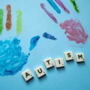
A new study in the International Journal of Hygiene and Environmental Health finds parents’ workplace chemical exposure may be linked to a range of behavioral challenges and developmental delays in their children with autism.

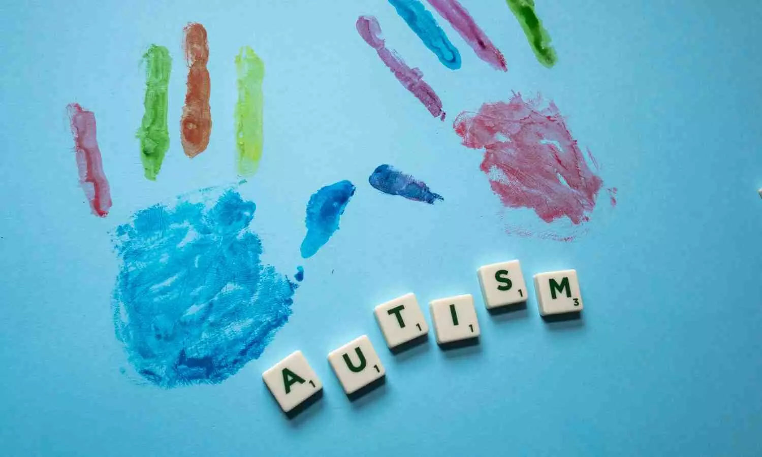
Autism is a neurodevelopmental condition that is marked by challenges with social skills, communication struggles and repetitive behaviors. Autism traits can vary widely in how mild or strong they are.

“Past research explored the impact of environmental factors on the likelihood of a child developing autism,” said Irva Hertz-Picciotto, a co-author and professor in the Department of Public Health Sciences and the UC Davis MIND Institute.

“This study is one of the first to connect parental job exposures to the severity of their child’s autism based on the Autism Diagnostic Observation Schedule, 2nd edition” (ADOS-2),” Hertz-Picciotto said. “The ADOS-2 Calibrated Severity Score is considered the ‘gold standard’ diagnostic assessment of autism,” Hertz-Picciotto said.

Hertz-Picciotto directs the UC Davis Environmental Health Sciences Center and has led a large autism study, CHARGE (ChildHood Autism Risks from Genes and Environment), since 2002. The CHARGE study is funded by the National Institute of Environmental Health Sciences. It includes children with autism or other developmental delays and children with typical development.

Linking parents’ chemical exposures to autism characteristics

In collaboration with the UC Davis CHARGE Study team, researchers from the National Institute for Occupational Safety and Health (NIOSH) studied data from over 500 families in the CHARGE study. They focused entirely on children already diagnosed with autism.

Industrial hygienists assessed both mothers’ and fathers’ job histories from three months before pregnancy to birth. They estimated each parent’s exposure to 16 chemicals or agents. This included plastics, car fluids, disinfectants, medicines and other chemicals.

They then matched the data with the children’s autism severity scores (using the ADOS-2) and their behaviors, cognitive skills and daily living skills.

The researchers found these associations among children with autism:

 Plastics and polymers (like polyethylene, polypropylene and polyvinyl chloride or PVC) are linked to poorer cognitive performance, reduced adaptive skills and increased behavioral issues like hyperactivity and social withdrawal.

• Ethylene oxide, a chemical used for sterilizing, is linked to higher autism severity scores and weaker daily living skills.

• Phenol exposure is tied to increased autism severity and behavioral symptoms like hyperactivity, repeated movements or vocalizations.

“Our findings suggest that parental exposure to certain workplace chemicals during key fetal development periods may influence not just autism likelihood, but also severity and functioning outcomes for children with autism,” said lead author Erin McCanlies, formerly with NIOSH’s Health Effects Laboratory Division.

The authors noted important limitations to the study. The number of families may have been too small to find links for less common exposures. The exposure estimates relied on reported job histories and expert judgment, which might not reflect actual exposures. Lastly, while certain agents showed associations with certain autism traits, the study did not prove that those chemicals caused the traits.

Lessons learned and research needs

The authors say more research is needed to understand how these exposures impact brain development. They also call for including fathers in similar studies on reproductive health and child neurodevelopment, as many associations in this study were linked to paternal exposures.

For instance, the strongest cognitive deficits for children with autism were linked to fathers’ job exposures to plastics and polymers. These exposures correlated with significantly lower skills, including fine motor, visual reception, receptive language, and expressive language.

“This research shows that workplace safety isn’t just about protecting the worker — it’s also about protecting their future children,” said Hertz-Picciotto. “We must consider how workplace chemicals might affect the next generation.”

Reference:

Erin C. McCanlies, Ja Kook Gu, Claudia C. Ma, Wayne T. Sanderson, Yunin J. Ludeña-Rodriguez, Irva Hertz-Picciotto, The effects of parental occupational exposures on autism spectrum disorder severity and skills in cognitive and adaptive domains in children with autism spectrum disorder, International Journal of Hygiene and Environmental Health, https://doi.org/10.1016/j.ijheh.2025.114613.

Powered by WPeMatico

Half of Abdominal TB Patients on ATT Develop Liver Abnormalities, Many Recover via Hepatic Adaptation: AIIMS Patna Study

India: A new prospective cohort study published in the Journal of Clinical and Experimental Hepatology has shed light on the patterns of liver injury and recovery in patients with abdominal tuberculosis (ATB) undergoing anti-tuberculosis treatment (ATT).   

Conducted by Abhishek Kumar and colleagues from the Department of Gastroenterology, All India Institute of Medical Sciences (AIIMS), Patna, the research highlights how early monitoring and nutritional assessment can play a critical role in preventing severe drug-induced liver injury (DILI).

Patients with extra-pulmonary tuberculosis, including ATB, are known to have a higher risk of developing DILI during ATT. However, in some cases, liver function abnormalities resolve spontaneously through a process known as hepatic adaptation (HA). While previous studies have looked at DILI risk in pulmonary tuberculosis, prospective data specifically focusing on ATB patients have been scarce.

The study followed 140 patients with confirmed ATB and normal baseline liver function tests (LFTs). All participants received the standard four-drug ATT regimen, and their LFTs were monitored at regular intervals. Potential predictors of liver injury progression were assessed using multivariable logistic regression.

The study revealed the following notable findings:

  • LFT abnormalities occurred in 71 patients (50.7%) during treatment.
  • Twenty patients (14.2%) met the criteria for DILI at the first detection of abnormality.
  • Of the remaining 51 patients, 18 (35.3%) progressed to DILI, while 33 (64.7%) showed spontaneous normalization of liver function, indicating hepatic adaptation.
  • Overall, 27.1% of the study population developed DILI.
  • Nearly half (46.4%) of those with LFT abnormalities recovered without requiring treatment interruption.
  • Most DILI cases (89%) occurred within the first eight weeks of initiating ATT.
  • The median time for hepatic adaptation was 21 days.

The analysis revealed that low serum albumin levels and vitamin D deficiency were independent predictors of progression from mild LFT abnormalities to DILI. These findings suggest that nutritional status may influence the liver’s ability to adapt during ATT.

When ATT was temporarily halted due to liver injury, complete reintroduction was successful in 65.8% of patients. Pyrazinamide was the drug most frequently linked to reintroduction failure. Importantly, none of the patients who developed DILI progressed to acute liver failure.

The authors note that while liver test abnormalities are common during ATT in ATB patients, nearly half can recover naturally due to hepatic adaptation. Identifying those at higher risk—particularly patients with hypoalbuminemia or vitamin D deficiency—could help clinicians prevent severe outcomes through closer monitoring and early intervention.

The study highlights the importance of regular LFT monitoring during the initial weeks of ATT, alongside nutritional assessment, to improve treatment safety and outcomes in abdominal TB patients.

Reference:

Kumar, A., Kumar, R., Kumar, V., Kumar, S., Marrapu, S., Anand, U., & Priyadarshi, R. N. (2025). Patterns of Liver Injury and Adaptation in Patients with Abdominal Tuberculosis on Antituberculosis Treatment: A Prospective Cohort Study. Journal of Clinical and Experimental Hepatology, 103124. https://doi.org/10.1016/j.jceh.2025.103124

Powered by WPeMatico

Stopping HRT leads to a period of higher risk of bone fracture for most women: Study

A new study has found that the bone fracture protection women get from menopausal hormone therapy (MHT, also known as HRT) disappears within a year of stopping treatment.

In the new study, published in Lancet Healthy Longevity, experts from the School of Medicine at the University of Nottingham, also found that in most cases, stopping treatment is then followed by some years of elevated fracture risk compared to women who have never used MHT. Fracture risks then falls to be similar to, and then lower than women who have never used MHT.

The study was funded by the National Institute for Health and Care Research (NIHR) SPCR.

During menopause, all women experience a drop in hormone levels, particularly of oestrogen. This can cause a range of distressing mental and physical side effects, requiring use of MHT. However, oestrogen deficiency in women also leads to increased age-related bone weakening. Previous studies have confirmed a protective role of the oestrogen component in MHT treatments, and MHT is known to decrease fracture risk when it is being used.

However, MHT is also associated with increased risk of breast cancer and blood clots, so long-term MHT use is not recommended. For women using MHT to counteract increasing bone fragility, it is, therefore, important to know the strength and persistence of any protective effect after stopping treatment. Detailed information on this aspect from past studies has been unclear – covering only the first couple of years, and also being somewhat conflicting.

In this new study, experts used data for 6,000,000 women from around 2,000 GP surgeries in the UK, which allowed them to follow-up of fracture risk levels for up to 25 years. The researchers identified all women with records of first fracture (cases) and matched each to a number of women of the same age and from the same practice, but without record of fracture (controls). They then compared the MHT use in cases before their fracture with the MHT use among their matched controls.

Dr Yana Vinogradova, from the Centre for Academic Primary Care in the School of Medicine, and lead author of the study said: “The findings of our study confirmed that women on MHT show a progressively reducing fracture risk compared with women not using MHT. More importantly, we also observed a clear pattern of risk change after therapy was discontinued.

“For most women, the bone protective effect of MHT use disappears completely within about one year of treatment being stopped, then their fracture risk rises compared to never users, peaking after about three years, before declining to become again equivalent to never users – about 10 years after discontinuation – and then again continuing to decline relative to never users. So, even after stopping MHT, women should benefit from notably reduced fracture risk in their later decades.”

This observed risk pattern was the same for all menopausal hormonal treatments, but the level of excess risk depended on the treatment type and the length of past MHT use.

“Our comparative illustration of observed patterns of fracture risk for short and long use can help doctors and patients when discussing MHT treatment options, and to consider how fracture risk may change after stopping MHT use. Anticipating periods of increased risk might prompt doctors to check patients’ bone health at discontinuation, particularly for patients most at risk with other fracture risk factors such as smoking or inactivity.

“These novel findings may also usefully stimulate further clinical and biological research into these treatments,” adds Dr Vinogradova.

Reference:

Vinogradova, Yana et al. Discontinuation of menopausal hormone therapy and risk of fracture: nested case–control studies using routinely collected primary care data, The Lancet Healthy Longevity, DOI: 10.1016/j.lanhl.2025.100729.

Powered by WPeMatico

Study Uncovers role of vitamin C in skin regeneration

The skin acts as the body’s first line of defense against external threats. However, as we age, the epidermis—the outermost layer of skin—gradually becomes thinner and loses its protective strength. About 90% of the cells in this layer are keratinocytes, which originate from deeper layers of the epidermis and migrate upward, ultimately forming the skin’s protective barrier. To combat aging’s impact on skin, numerous studies have emphasized the benefits of vitamin C (VC), a vitamin well known for its role in skin health and antioxidant properties.

Now, researchers in Japan have discovered that VC helps thicken the skin by directly activating genes that control skin cell growth and development. Their findings, published online in the Journal of Investigative Dermatology on April 20, 2025, suggest that VC may restore skin function by reactivating genes essential for epidermal renewal.

This study was led by Dr. Akihito Ishigami, Vice President of the Division of Biology and Medical Sciences at Tokyo Metropolitan Institute for Geriatrics and Gerontology (TMIG), Japan, in collaboration with Hokuriku University, and ROHTO Pharmaceutical Co., Ltd. Associate Professor Ayami Sato from TMIG (currently at the Toyo University); Associate Professor Yasunori Sato, Professor Toshiyuki Kimura, and Mr. Hideki Tanaka (currently at the University of Fukui Hospital) from Hokuriku University; and Ms. Florence, Ms. Akari Kuwano, Mr. Yasunari Sato, and Mr. Tsuyoshi Ishii from ROHTO Pharmaceutical Co., Ltd also co-authored the study.

“VC seems to influence the structure and function of epidermis, especially by controlling the growth of epidermal cells. In this study, we investigated whether it promotes cell proliferation and differentiation via epigenetic changes,” explains Dr. Ishigami, while talking about this study.

To investigate how VC affects skin regeneration, the team used human epidermal equivalents, which are laboratory-grown models that closely mimic real human skin. In this model, skin cells are exposed to air on the surface while being nourished from underneath by a liquid nutrient medium, replicating the way human skin receives nutrients from underlying blood vessels while remaining exposed to the external environment.

The researchers used this model and applied VC at 1.0 and 0.1 mM—concentrations comparable to those typically transported from the bloodstream into the epidermis. On assessing its effect, they found that VC-treated skin showed a thicker epidermal cell layer without significantly affecting the stratum corneum (the outer layer composed of dead cells) on day seven. By day 14, the inner layer was even thicker, and the outer layer was found to be thinner, suggesting that VC promotes the formation and division of keratinocytes. Samples treated with VC showed increased cell proliferation, demonstrated by a higher number of Ki-67-positive cells—a protein marker present in the nucleus of actively dividing cells.

Importantly, the study revealed that VC helps skin cells grow by reactivating genes associated with cell proliferation. It does so by promoting the removal of methyl groups from DNA, in a process known as DNA demethylation. When DNA is methylated, methyl groups attach to cytosine bases, which can prevent the DNA from being transcribed or read, thereby suppressing gene activity. Conversely, by promoting DNA demethylation, VC promotes gene expression and helps cells to grow, multiply, and differentiate.

The study suggests that VC supports active DNA demethylation by sustaining the function of TET enzymes (ten-eleven translocation enzymes), which regulate gene activity. These enzymes convert 5-methylcytosine (5-mC) into 5-hydroxymethylcytosine (5-hmC), a process in which Fe2+ is oxidized to Fe3+. VC helps maintain TET enzyme activity by donating electrons to regenerate Fe2+ from Fe3+, enabling continued DNA demethylation.

The researchers further identified over 10,138 hypomethylated differentially methylated regions in VC-treated skin and observed a 1.6- to 75.2-fold increase in the expression of 12 key proliferation-related genes. When a TET enzyme inhibitor was applied, these effects were reversed, confirming that VC functions through TET-mediated DNA demethylation.

These findings reveal how VC promotes skin renewal by triggering genetic pathways involved in growth and repair. This suggests that VC may be particularly helpful for older adults or those with damaged or thinning skin, boosting the skin’s natural capacity to regenerate and strengthen itself.

“We found that VC helps thicken the skin by encouraging keratinocyte proliferation through DNA demethylation, making it a promising treatment for thinning skin, especially in older adults,” concludes Dr. Ishigami.

Reference:

Sato, Yasunori, Vitamin C Promotes Epidermal Proliferation by Promoting DNA Demethylation of Proliferation-Related Genes in Human Epidermal Equivalents, Journal of Investigative Dermatology, DOI: 10.1016/j.jid.2025.03.040 

Powered by WPeMatico

Predicting the Unpredictable: Study finds Insights into Intraoperative Hypotension through HPI Evaluation

In a recent study comparing the Hypotension Prediction Index (HPI) values obtained from Acumen™ arterial pressure transducer and ClearSight™ continuous non-invasive blood pressure monitor, the primary objective was to assess the potential impact on intraoperative hypotension incidence and perioperative complications. Intraoperative hypotension is a significant concern due to its association with increased post-operative morbidities and mortalities, with 40% to over 90% of patients experiencing at least one hypotensive episode during general anesthesia. The HPI provides early warning of critical hypotension, aiding in earlier interventions to reduce its incidence. The HPI algorithm analyzes arterial pressure waveform features to predict hypotension, demonstrating over 80% sensitivity and specificity in previous studies.

Comparison of Monitoring Systems

The study compared HPI values obtained from invasive and non-invasive pressure inputs in adult patients undergoing general anesthesia. The data analysis included Bland-Altman analysis, correlation calculations, and concordance assessments to evaluate the agreement between the two monitoring systems. The results showed a bias of -8.4 between invasive and non-invasive HPI values, with a high correlation of 0.76. The concordance rates remained consistent across different exclusion zones, indicating a reliable agreement between the two systems.

Clinical Performance Evaluation

The clinical performance evaluation included Receiver Operator Characteristic (ROC) analyses and HPI alerts agreement analysis. Both invasive and non-invasive systems showed comparable performance in predicting hypotensive events, with similar Area Under the Curve (AUC) measurements, sensitivity, specificity, positive predictive values (PPV), and negative predictive values (NPV) at different time intervals before hypotensive events.

Conclusion

The study concluded that the HPI derived from the non-invasive ClearSight monitor exhibited reliable predictive performance comparable to the gold standard invasive arterial catheter. This suggests that the non-invasive HPI can be used effectively for continuous blood pressure monitoring in various surgical scenarios, potentially improving patient outcomes by predicting and preventing hypotensive episodes. However, the study highlighted the need for caution in generalizing the results to other patient populations and emphasized the importance of further validation in larger cohorts. Overall, utilizing the HPI parameter from non-invasive monitoring systems could enhance the management of intraoperative hypotension and reduce perioperative complications in a wider range of surgical procedures.

Key Points

– The primary objective of the study was to evaluate the impact of using the Hypotension Prediction Index (HPI) from Acumen™ arterial pressure transducer and ClearSight™ continuous non-invasive blood pressure monitor on intraoperative hypotension incidence and perioperative complications.

– Intraoperative hypotension is a major concern during general anesthesia as it is linked to increased post-operative morbidities and mortalities, affecting a significant percentage of patients. The HPI algorithm, with over 80% sensitivity and specificity, can predict critical hypotension early, allowing for timely interventions.

– The study compared HPI values from invasive and non-invasive pressure inputs in adult patients under general anesthesia, revealing a bias of -8.4 between the two systems with a high correlation of 0.76, demonstrating reliable agreement.

– Clinical performance evaluation using Receiver Operator Characteristic (ROC) analyses and HPI alerts agreement analysis showed that both invasive and non-invasive systems had similar predictive performance in detecting hypotensive events, with comparable Area Under the Curve (AUC) measurements, sensitivity, specificity, positive predictive values (PPV), and negative predictive values (NPV).

– The study concluded that the non-invasive HPI derived from the ClearSight monitor exhibited reliable predictive performance similar to invasive arterial catheters, suggesting its effectiveness for continuous blood pressure monitoring in various surgical settings. However, caution is advised in generalizing the results to other patient populations, emphasizing the importance of further validation in larger cohorts.

– Utilizing the HPI parameter from non-invasive monitoring systems could enhance the management of intraoperative hypotension and decrease perioperative complications across a broader spectrum of surgical procedures, potentially leading to improved patient outcomes.

Reference –

Kamron Sarhadi et al. (2025). Hypotension Prediction Index: Comparison Between Invasive And Non-Invasive Pressure Inputs. *BMC Anesthesiology*, 25. https://doi.org/10.1186/s12871-025-03086-y.

Powered by WPeMatico

Recent advancements in treatment of age-related macular degeneration

Age-related macular degeneration (AMD) remains a leading cause of vision loss globally, with significant advancements in treatment options for both dry and wet AMD. As highlighted by Huang and colleagues, “Current therapies for dry AMD have limited effectiveness in halting the progression of geographic atrophy (GA), underscoring the need for innovative approaches”.

One of the most notable breakthroughs is the FDA approval of pegcetacoplan and avacincaptad pegol, targeting the complement system to slow GA progression. Pegcetacoplan, a C3 inhibitor, reduced GA lesion growth by 19–22% in clinical trials, while avacincaptad pegol, a C5 inhibitor, showed a 35% reduction. These therapies address the inflammatory component of AMD, offering hope for a condition previously deemed untreatable.

For wet AMD, anti-VEGF therapies continue to dominate, but newer options like faricimab—a bispecific antibody targeting VEGF and angiopoietin-2—stand out. Faricimab allows extended dosing intervals (up to 16 weeks), reducing the burden of frequent injections. As Gao et al. note, “Combining complement inhibition with anti-VEGF therapy holds significant potential,” exemplified by IBI302, a dual-targeting drug currently in Phase III trials.

Surprisingly, photobiomodulation (PBM) emerged as a non-invasive option for dry AMD, improving visual acuity by 2.4 letters in the LIGHTSITE III trial. Meanwhile, gene therapy (e.g., RGX-314) and stem cell-based treatments show promise but remain experimental.

These advancements not only refine existing treatments but also challenge traditional paradigms. For instance, the shift from monthly injections to longer-acting therapies could revolutionize patient care. However, challenges like retinal vasculitis with pegcetacoplan remind us of the need for cautious optimism.

In summary, the AMD treatment landscape is evolving rapidly, with novel mechanisms and improved delivery systems offering brighter prospects for patients. As research continues, the focus on combination therapy and personalized precision medicine may further transform outcomes.

Reference:

Shan Liu, Danyang Lv, Junran Sun, Huixun Jia, Fang Zheng, Felipe Pereira, Siddharth Narendran, Zhenyu Dong, Haiyun Liu, Shunxiang Gao, Peirong Huang, Recent Advancements in the Treatment of Age-Related Macular Degeneration, https://doi.org/10.1002/mdr2.70009

Powered by WPeMatico

Rotary Instrumentation May Improve Pediatric Root Canal Outcomes, but Standardized Research Needed: Study

A new study suggests that using rotary instrumentation in root canal treatments for primary teeth could improve obturation quality and postoperative comfort in children. The findings indicate potential benefits over traditional hand instrumentation, including more consistent filling of root canals and reduced discomfort after the procedure.

However, researchers noted that the current evidence base is limited. Few high-confidence systematic reviews exist, and studies often use varied outcome measures, making it difficult to draw definitive conclusions. This lack of standardization in pediatric endodontic research hinders direct comparison between trials and weakens the ability to create evidence-based guidelines.

The authors recommend developing and adopting standardized protocols for outcome reporting in pediatric root canal studies. Consistent methodology, they argue, would help clarify whether rotary systems should be adopted as a standard approach in managing primary teeth requiring endodontic care.

For now, while rotary instrumentation shows promise, clinicians are advised to weigh individual patient needs, operator skill, and equipment availability until more rigorous, uniform research is available.

Reference:
Swaminathan K, Shan S, Kirubakaran E, et al. (July 06, 2025) Cleaning Effectiveness and Postoperative Pain Associated With Rotary Instrumentation in Primary Teeth: An Umbrella Review of Systematic Evidence. Cureus 17(7): e87369. doi:10.7759/cureus.87369

Keywords: Rotary instrumentation, pediatric dentistry, primary teeth, root canal, obturation quality, postoperative comfort, endodontics, standardized research

Powered by WPeMatico