20-minute mindful breathing could effectively alleviate variety of symptoms in kidney transplant recipients: Study

A new study published in the journal of BMC Nephrology showed that 20 minutes of focused breathing may quickly alleviate a variety of symptoms in kidney transplant recipients.

Even after receiving a kidney transplant, many recipients still experience a variety of symptoms, despite the fact that this is the preferred therapy for people with end-stage renal failure. Dialysis patients had an average of nine symptoms, while kidney transplant patients had an average of seven symptoms.

Many symptoms persisted after transplantation, including weakness (55%), sleep disturbances (45%), dyspnea (42%), drowsiness (35%), anxiety (36%), weight gain (33%), body image dissatisfaction (35%), and depression (32%), even though some symptoms, like pruritus, fatigue, xeroderma, cramps, and anorexia, undoubtedly improved after the procedure.

Since a high symptom load may impact the patient’s and the transplant’s prognosis, it is critical to identify and treat symptoms in transplant recipients. It could exacerbate melancholy and anxiety. The cultivation of mindfulness involves paying attention in the present moment in a certain way, as open-heartedly, non-judgmentally, and non-reactively as possible. Pain acceptance, exhaustion, sleeplessness, stress, anxiety, and depression have all been demonstrated to improve with mindfulness-based therapies, such as mindfulness-based stress reduction (MBSR) and mindfulness-based cognitive therapy (MBCT).

The effectiveness of short mindfulness exercises in the transplant context is yet unknown, despite the fact that a traditional 8-week mindfulness intervention has been shown to be successful in lowering anxiety, depression, and sleeplessness symptoms in solid organ transplant recipients. Therefore, this replication research is to ascertain if a single 20-minute mindful breathing exercise may effectively reduce a variety of symptoms in kidney transplant recipients.

From November 2020 to May 2021, adult kidney transplant recipients who had at least one symptom with an Edmonton Symptom Assessment Scale (ESAS) score of ≥ 4/10 were sought for. Participants were randomized to either the control group or the 20-minute mindful breathing session.

After recruiting 60 individuals, they were randomized to either the control group (N = 30) or the intervention group (N = 30). When compared to the control group, the intervention group’s overall ESAS score decreased by a statistically significant amount.

Overall, kidney transplant recipients can experience rapid alleviation from a variety of symptoms after only one 20-minute mindful breathing practice. In addition to participating in a typical 8-week mindfulness program to get long-term symptom reduction, 20-minute mindful breathing provides kidney transplant recipients with an additional low-cost alternative for quick symptom alleviation.

Source:

Lim, K. W., Ng, K. P., Ooi, S. H., Siow, W. S., & Tan, S. B. (2025). Symptom reduction in kidney transplant patients from 20-minute mindful breathing: a randomized controlled trial. BMC Nephrology, 26(1), 194. https://doi.org/10.1186/s12882-024-03820-7

Powered by WPeMatico

Home based pulmonary Rehab Plus NPPV Improves QoL in COPD Patients, confirms study

Researchers have identified in a new study that the integration of home-based pulmonary rehabilitation with non-invasive positive pressure ventilation (NPPV) improves the quality of life, lung function, and exercise capacity in patients with moderate to severe chronic obstructive pulmonary disease (COPD) to a great extent. This is following a trial comparing the effectiveness of this combined therapy (CPRNG group) with standard treatment alone (CTG group). The study was conducted by Shu Xie and colleagues published in BMC Pulmonary Medicine.

Chronic obstructive pulmonary disease (COPD) continues to be a global cause of morbidity and mortality, with the symptoms of breathlessness, tiredness, and decreased physical activity profoundly affecting day-to-day functioning. NPPV facilitates breathing by assisting airflow on inhalation and is typically applied in more severe instances to enhance oxygenation and alleviate carbon dioxide retention. Simultaneously, pulmonary rehabilitation enhances the strength of respiratory muscles, improves tolerance to exercise, and enhances the well-being of patients. This research aimed to assess whether a home-based rehabilitation program combined with NPPV would offer better outcomes in the management of COPD symptoms than conventional treatments alone.

269 patients with COPD of moderate to severe degree were included and divided into two groups: 132 patients received combined pulmonary rehabilitation and NPPV (CPRNG group), and 137 patients received conventional therapy (CTG group). Both groups were assessed with a range of standardized tools, including the COPD Assessment Test (CAT), modified Medical Research Council scale (mMRC), forced expiratory volume in 1 second (FEV₁%), 6-minute walk test, and arterial oxygen pressure (PaO₂). Patient-reported quality-of-life scores were also directed toward three domains: impact, symptoms, and activity.

Results

Early comparisons revealed no substantial differences between the groups regarding age, gender, duration of illness, or baseline symptoms, making a fair comparison possible. Following the intervention, the patients in the CPRNG group had statistically significant improvements in all primary outcomes than those in the CTG group:

  • CAT score: Improved substantially in CPRNG group (p = 0.028)

  • mMRC score: Improved symptom control (p = 0.015)

  • FEV₁%: Increased improvement in lung function (p = 0.008)

  • 6-min walk distance: Enhanced exercise tolerance (p = 0.001)

  • PaO₂ levels: Increased oxygenation (p < 0.001)

Home pulmonary rehabilitation combined with non-invasive positive pressure ventilation greatly enhances lung function, physical performance, symptom control, and quality of life in patients with moderate to severe COPD. This combination is a useful strategy in the management of COPD, particularly in improving patient-centered outcomes like day-to-day activity and comfort.

Reference:

Xie, S., Li, X., Liu, Y. et al. Effect of home noninvasive positive pressure ventilation combined with pulmonary rehabilitation on dyspnea severity and quality of life in patients with severe stable chronic obstructive pulmonary disease combined with chronic type II respiratory failure: a randomized controlled trial. BMC Pulm Med 25, 185 (2025). https://doi.org/10.1186/s12890-025-03656-3

Powered by WPeMatico

Neutrophil Profiling May Aid Surgical and Palliative Care Decisions in Elderly Hip Fracture Patients: Study Shows

Netherlands: A recent study published in the European Journal of Trauma and Emergency Surgery highlights the potential of neutrophil profiling as a valuable tool to guide clinical decisions in elderly patients with hip fractures. The research conducted on a large cohort found a strong link between systemic inflammation levels and patient outcomes.

Those in higher inflammation categories had elevated leukocyte counts, increased C-reactive protein levels, more infections, and an 83% 30-day palliative care mortality rate, compared to 50% in the low-inflammation group. These findings suggest that assessing systemic inflammation may help tailor treatment strategies and improve care for this high-risk population.

Geriatric patients with hip fractures often face a high risk of complications following surgery, making timely and accurate clinical decision-making essential. A previous pilot study demonstrated that analyzing neutrophils—key immune cells—can help assess systemic inflammation in these patients, offering a potential tool to predict outcomes more effectively.

Building on this, E. J. de Fraiture, Department of Trauma Surgery, UMC Utrecht, Utrecht, Netherlands, and colleagues aimed to explore the relationship between neutrophil inflammation categories and clinical outcomes in a larger group of elderly hip fracture patients.

For this purpose, the researchers conducted a prospective cohort study that collected blood samples from elderly patients soon after they experienced a hip fracture. They also included healthy older adults as a control group. The neutrophils in the blood were analyzed and categorized on a scale from 0 to 6, representing levels of inflammation ranging from none to severe. These inflammation categories were then compared with the patient’s clinical outcomes to understand how systemic inflammation might impact recovery and prognosis.

The study led to the following findings:

  • The study included 289 geriatric patients with hip fractures (median age 82) and 45 age-matched healthy individuals.
  • Severe infections were reported in 8% of the patients.
  • The 30-day mortality rate among patients was 9%.
  • Patients showed a full range of neutrophil inflammation categories (0–6), whereas healthy controls showed only categories 0, 1, and 3.
  • A newly identified neutrophil category was associated with higher leukocyte counts, and elevated C-reactive protein (CRP) levels, and a trend toward more infections and higher mortality.
  • Among patients receiving palliative care, 30-day mortality was 50% in the low-inflammation group (categories 0–1) and 83% in those with higher inflammation levels.

In conclusion, the researchers highlight that “neutrophil profiling offers a practical and effective method to assess systemic inflammation in elderly patients with hip fractures.” According to their findings, “the identification of neutrophil categories may help guide shared decision-making, especially in determining the suitability of patients for surgery or palliative care.” They further noted that “patients falling into categories 0–1 appear more likely to be fit for surgery when no other major risk factors are present.”

However, the researchers emphasize that “further studies are needed to assess the long-term quality of life in patients who survive beyond 30 days” and suggest that “ongoing immune monitoring throughout hospitalization and treatment could provide deeper insights.” They believe that “a better understanding of these immune patterns is key to improving care and optimizing outcomes for geriatric trauma patients.”

Reference:

de Fraiture, E.J., Nijdam, T.M.P., van Eerten, F.J.C. et al. Exploring the role of systemic inflammation in guiding clinical decision making for geriatric patients with a hip fracture. Eur J Trauma Emerg Surg 51, 192 (2025). https://doi.org/10.1007/s00068-025-02875-x

Powered by WPeMatico

FDA Approves At-Home Cervical Cancer Screening Device

The US Food and Drug Administration (FDA) has Approved first and only self-collection At-Home Cervical Cancer Screening Device. Patients can now collect cervical cancer screening samples themselves at home using a newly approved device. According to manufacturers, the device is as accurate as traditional screening methods.

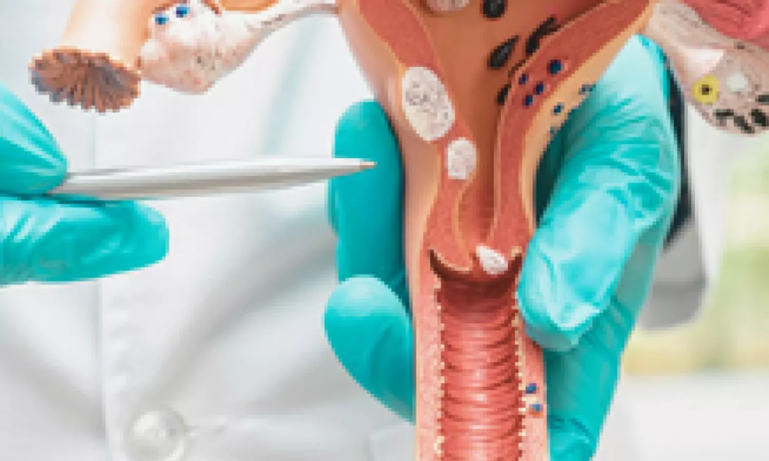
Teal Health®, a women’s health company on a mission to eliminate cervical cancer, today announced the Food and Drug Administration’s (FDA) approval of the Teal Wand™, the first and only at-home vaginal sample self-collection device for cervical cancer screening in the United States. Cervical cancer screenings, commonly referred to as the Pap smear, are critical to a woman’s health, but they are inconvenient and uncomfortable for most.

Now women have a new way to screen that is as accurate as going to the doctor’s office, comfortable, and done from home. The Teal Wand is a prescription device that will soon be available at getteal.com for individuals aged 25–65 at average risk. The at-home screening includes both the Teal Wand collection kit and an end-to-end telehealth service providing virtual access to Teal medical providers who prescribe the kit, review the results from the lab, and support women throughout their at-home screening experience.

Cervical cancer is one of the only cancers that is almost entirely preventable with regular screening, yet more than 1 in 4 women in the U.S. are behind. Whether it’s because a woman can’t get time off work, is unable to find an available appointment, or avoids the discomfort of an in-clinic exam, Teal can provide a comfortable and convenient option. The Teal Wand is a preferred alternative, one that has been built with empathy, is driven by science, and designed to make screening easy, so that more women can take control of their health on their own terms.

“As a mom and a woman, I get how easy it is to put your own health last,” said Kara Egan, CEO and Co-Founder of Teal Health. “That’s why this FDA approval means so much; it’s not just about an innovative new product, it’s about finally giving women an option that makes sense for their lives – something that can be done quickly and comfortably at home. Because when we make care easier to get, we help women stay healthy, for themselves and for the people who rely on them every day.”

With the Teal Wand, women are testing a sample for HPV (human papillomavirus), the virus that causes nearly all cervical cancers, using the same highly accurate HPV test that medical guidelines recommend and providers use in the clinic – cobas® HPV from Roche. The Teal Wand simply provides a different method of sample collection. Cervical cancer screening has evolved from the Pap smear to HPV primary screening. HPV primary screening demonstrates higher sensitivity compared to the Pap test to identify women who may be at risk of cervical cancer. With this FDA approval, women can use the same test as the doctor’s office, with the same accuracy, but collect their own sample from the privacy of their home, the sample is then conveniently shipped to a certified lab for processing.

Teal Health’s FDA approval was supported by their SELF-CERV study, the largest U.S.-based comparative study of its kind. The study confirmed that self-collected samples using the Teal Wand have the same performance as clinician-collected samples, proven to detect cervical precancer 96% of the time, and that the Teal Wand is a much preferred experience. Study participants reflected the racial, ethnic, and socioeconomic diversity of the U.S. population, underscoring Teal Health’s commitment to clinical excellence, equity, and inclusivity in women’s health research.

Notably, 86% of participants said they’d be more likely to stay up to date with cervical cancer screening if they could do it at home, and 94% said they would prefer to self-collect at home with the Teal Wand if they knew it was accurate. The clinical performance and preference for the Teal Wand, alongside Teal’s comprehensive telehealth service, highlight the potential of at-home self-collection to expand access to high-quality cervical cancer screening and improve outcomes.

“As a Principal Investigator in the SELF-CERV trial, I saw firsthand how receptive and excited women were to use the Teal Wand. Cervical cancer is largely preventable, yet screening rates in the U.S. continue to lag, and the FDA approval of this at-home Teal Wand self-collection device is a critical step forward. It offers an evidence-based way to expand access without compromising accuracy.” Said Dr. Christine Conageski, Associate Professor, OB-GYN and Director of the Complex Dysplasia Clinic at the University of Colorado, “But access is only part of the solution. Comprehensive screening must go hand in hand with structured, reliable follow-up. That’s why Teal Health’s approach to not only advancing screening technology but also providing education and support to women through every step of their care and follow-up journey is crucial. That’s how we ensure this breakthrough truly closes the gap.”

With FDA approval in hand, Teal Health is moving quickly to get the Teal Wand to as many women as possible. Kits become available in June, starting in California and expanding nationwide as soon as possible thereafter. Teal is working with major insurance providers and plans to have flexible payment options, helping to remove financial concerns and ensuring more women have access to this preferred at-home screening if they want it.

“The FDA prioritized the review of at-home self-collection, recognizing its potential to increase cervical cancer screening adherence, as emphasized in the recent USPSTF (United States Preventive Services Task Force) draft guidelines,” said Trena Depel, Teal Health’s VP of Clinical, Regulatory, and Quality. “After awarding the Teal Wand Breakthrough Device Designation, the FDA stayed committed to a timely review, leading to approval of a technology that delivers meaningful performance, benefits, and choice. This isn’t just a win for Teal—it’s a win for every woman who deserves a rigorously tested and FDA reviewed at-home cervical cancer screening option.”

The approval of the Teal Wand and the shift toward at-home cervical cancer screening signals a new era in healthcare, one designed around women’s comfort, control, and real-life preferences. Join the waitlist at getteal.com to be the first to know when Teal Health expands to your state. Waitlisters get early access to updates and availability.

Powered by WPeMatico

Maternal Depression Linked to 40 Percent Higher Risk of Early Childhood Tooth Decay, Meta-Analysis Finds

China: A new systematic review and meta-analysis has highlighted a significant connection between maternal depression and the increased risk of early childhood caries (ECC), shedding light on the critical influence of parental mental health on children’s oral wellbeing. The findings were published in the Journal of Clinical Pediatric Dentistry and represent one of the first quantitative evaluations of this association.    

Wentao Bian, Shaanxi Provincial People’s Hospital, 710000 Xi’an, Shaanxi, China, and colleagues conducted an extensive review of existing literature to assess whether depressive symptoms in mothers could influence their children’s risk of developing dental caries in early childhood. Drawing from multiple scientific databases including PubMed, Embase, Web of Science, and the Cochrane Library, the team screened studies dating up to June 1, 2024.

Key findings were as follows:

  • Seven studies, with 22,764 participants, were included in the final analysis.
  • Data were combined using either fixed or random effects models based on study heterogeneity.
  • The quality of evidence was assessed using the GRADE framework.
  • Maternal depression was linked to a 40% higher risk of early childhood caries.
  • The calculated odds ratio was 1.40, with a 95% confidence interval of 1.09 to 1.80.
  • The analysis showed substantial variability between studies, with an I² value of 71.9%, indicating potential inconsistencies in the data.

Although the number of studies included was relatively small, the results indicate a potential link between maternal mental health and children’s dental outcomes. Researchers noted that while this study offers important initial evidence, more robust and detailed studies are needed to confirm the relationship and identify underlying mechanisms.

One possible explanation is that maternal depression might impact parenting behaviors such as maintaining children’s oral hygiene routines, attending dental visits, or ensuring a balanced diet. Additionally, depression could reduce a mother’s ability to detect early signs of dental problems or prioritize preventive care.

The authors emphasized the importance of integrating mental health support into maternal and child health programs. “Improving awareness and education around maternal mental health could play a key role in preventing early dental problems among children,” they noted.

The study brings attention to the often-overlooked role of maternal well-being in shaping child health outcomes. It suggests that oral health interventions should consider the broader psychosocial environment of young children.

“Maternal depression significantly raises the risk of early dental caries in children, highlighting the need for greater emphasis on educating parents about maternal mental health,” the authors concluded.

Reference:

Wentao Bian, TingTing Gao, Wenkai Bian, Qiang Zhang. Maternal depression increases the risk of early childhood caries (ECC): a systematic review and meta-analysis. Journal of Clinical Pediatric Dentistry. 2025; 49(3): 30-38. doi: 10.22514/jocpd.2025.047.

Powered by WPeMatico

Higher BMI in early pregnancy Linked to Child Obesity Risk, finds Australian study

New research being presented at this year’s European Congress on Obesity (ECO 2025) highlights the need to support women and their families with overweight or obesity to optimise their health and weight before they become pregnant.

The Australian study found the greater a woman’s BMI in pregnancy, the greater her child’s weight from birth to the age of ten. This was the case regardless of whether the woman took part in a dietary and lifestyle intervention (LI) while pregnant or received standard antenatal care (SC). They also reported that the BMI of the father significantly influences the child’s weight at age 10.

The results are the latest from the LIMIT trial which involved 2121 pregnant women with overweight or obesity (average age 29.4 years, median BMI 31.1 in early pregnancy).

Half of the women took part in the LI, which consisted of advice and support for eating a healthy diet (for example, eating more fruit and fibre and reducing intake of refined carbohydrates and saturated fat) and increasing physical activity. The other participants received standard antenatal care (standard care group, SC).

“Women with overweight or obesity are at increased risk of pregnancy complications, like gestational diabetes, high blood pressure, caesarean birth and high infant birth weight, and for their children to develop obesity,” says researcher Professor Jodie Dodd, of the University of Adelaide, South Australia, Australia.

“With approximately 50% of women entering pregnancy with overweight or obesity, the trial was carried out to see if changes in diet and physical activity during pregnancy could reduce these risks.”

The initial results1, which were published in 2014, showed the infants whose mothers took part in the LI group were 18% less likely to have birth weight above 4kg (8lb 13oz), a known risk for childhood obesity. However, there were no other differences in maternal or birth outcomes, including pregnancy complications, between the two groups.

The children were also followed-up throughout childhood. There was no evidence of a difference in health or growth of the children from women in either the LI and SC group.

For the latest study, Professor Dodd and colleagues examined the effect of maternal BMI in early pregnancy on childhood weight and other measures of growth taken at birth and aged 6 months, 18 months, 3-5 years old and 8-10 years old.

They found that a child’s growth was linked to its mother’s BMI in early pregnancy – and that every 5kg/m2 increase in a woman’s BMI was associated with an increase in their child’s BMI by 0.11kg/m2 at birth, to 0.74kg/m2 at 8-10 years of age.

The effect of maternal BMI became more prominent at age 3-5 years and was particularly noticeable at 8-10 years. This was the case across the different measures of child growth studied – BMI, weight and their standardised measures. Additionally, paternal BMI impacted child weight and BMI, particularly at ages 8-10 years.

Professor Dodd says: “We know that women living with overweight or obesity are at increased risk of complications like high blood pressure in pregnancy. We also know that lifestyle interventions during pregnancy are ineffective for improving health outcomes for women and their baby/child.(1, 2, 3)

“What we found here was that a woman’s BMI in early pregnancy impacts how her child grows from birth to 8-10 years of age –so, if a woman’s BMI is higher going into pregnancy her child’s risk of overweight and obesity increases too. Father’s also have a role to play, as paternal BMI also contributes to child obesity.

“It’s vital that healthcare providers focus on supporting women and their families to optimise their health and weight before pregnancy, if we are to intervene and potentially reduce the intergenerational cycle of obesity.”

The greater a woman’s BMI in early pregnancy, the more likely her child is to develop overweight or obesity, Australian study finds European Congress on Obesity (ECO2025)

Powered by WPeMatico

Preconception Sexual Dysfunction may delay Conception, suggests study

Researchers have found in a new study that Preconception sexual dysfunction, particularly distress and frequent painful intercourse, was linked to delayed conception. Therefore, assessing sexual function before conception, including addressing specific domains, may help identify and address modifiable factors affecting fertility.

Fertility success among mixed-sex couples often depends on frequency and timing of sexual intercourse, yet little research has evaluated the association between preconception sexual function and time-to-pregnancy. A study was done to evaluate the effects of female sexual dysfunction, distress related to sexual functioning, and painful intercourse on time-to-pregnancy. They followed 2500 participants from Pregnancy Study Online, a prospective cohort study of self-identified females attempting pregnancy without the use of fertility treatments. Participants enrolled between 2021 and 2024. Thirty days after enrollment, participants completed a supplemental questionnaire that contained questions about sexual health, including a modified version of the 6-item Female Sexual Function Index (score range 2–30, score ≤19 defined as sexual dysfunction) and the Female Sexual Distress Scale (score range 0–48, score ≥20 defined as clinically relevant distress), which assess experiences in the previous 4 weeks. Participants completed the supplemental questionnaire no later than 6 months after initiating conception attempts. We estimated time-to-pregnancy based on self-reported pregnancy status on follow-up questionnaires completed every 8 weeks for up to 12 months. We used proportional probabilities regression to calculate fecundability ratios and 95% confidence intervals relating exposure measures with time-to-pregnancy, adjusting for a range of prespecified confounders. As an exploratory analysis, we evaluated individual domains of sexual function (ie, interest, arousal, orgasm, lubrication, and satisfaction) in relation to time-to-pregnancy. Results: The study population was primarily non-Hispanic White, high income, with college or graduate education. Exposure prevalence was 20.1% for female sexual dysfunction, 8.8% for distress, and 29.6% for any pain with intercourse. We observed no association between female sexual dysfunction and time-to-pregnancy (adjusted fecundability ratio 1.00, 95% confidence interval 0.890, 1.13) when female sexual dysfunction was defined using a clinically validated cut point, but observed that those in the first, second, and third quartile of scores had delayed conception compared to those in the fourth (highest function) (adjusted fecundability ratios 0.90, 95% confidence interval 0.76, 1.06; 0.88, 95% confidence interval 0.75, 1.04; and 0.90, 95% confidence interval 0.77, 1.04, respectively). We found 18% reduced fecundability among those with sexual distress as defined by a clinically validated cut point compared to those without (adjusted fecundability ratio 0.82, 95% confidence interval 0.69, 0.98). Participants reporting painful intercourse most or all the time had a longer time-to-pregnancy than those reporting no pain (adjusted fecundability ratio 0.81, 95% confidence interval 0.62, 1.06). In exploratory analyses, lower function in orgasm and lubrication domains, but not interest, desire, and arousal, were associated with longer time-to-pregnancy.Preconception sexual dysfunction, specifically distress and frequent painful intercourse, was associated with delayed conception. Preconception clinical assessment of sexual function, including discussion of individual domains of sexual function, may elucidate important modifiable issues.

Reference:

Julia C. Bond, Brenda Heaton, Katharine O. White, Jasmine A. Abrams, Wendy Kuohung, Rebecca R. Fisher, Amelia K. Wesselink, Matthew P. Fox, Lauren A. Wise, Female sexual function and distress and time-to-pregnancy in a prospective preconception cohort. American Journal of Obstetrics and Gynecology, Volume 232, Issue 4, 2025, Pages 375.e1-375.e24, ISSN 0002-9378, https://doi.org/10.1016/j.ajog.2024.09.117.(https://www.sciencedirect.com/science/article/pii/S0002937824010482)

Keywords:

Preconception, Sexual, Dysfunction, delay, Conception, suggests, study , American Journal of Obstetrics and Gynecology, Julia C. Bond, Brenda Heaton, Katharine O. White, Jasmine A. Abrams, Wendy Kuohung, Rebecca R. Fisher, Amelia K. Wesselink, Matthew P. Fox, Lauren A. Wise

Powered by WPeMatico

Neutrophils Could Improve Survival in Ruptured AAA Cases: Study Suggests

China: A recent case-control study published in the Journal of Inflammation Research has shed light on a potential breakthrough in the early diagnosis of ruptured abdominal aortic aneurysms (rAAAs), highlighting the role of venous blood neutrophil count as a promising clinical indicator.   

“Elevated neutrophil levels effectively distinguished ruptured abdominal aortic aneurysms (rAAAs) from unruptured cases,” the researchers reported. “Neutrophil count (OR: 1.36) and D-dimer levels were significantly higher in patients with rAAAs, while abdominal pain (OR: 32.61) and back pain (OR: 91.95) showed strong associations with rupture. Neutrophils also demonstrated high diagnostic accuracy (AUC: 0.847), supporting their potential role in early detection.”

Abdominal aortic aneurysms (AAAs) can become life-threatening when ruptured, but timely differentiation between ruptured (rAAAs) and unruptured cases remains a significant clinical challenge, as there are currently no effective, convenient, or widely accepted biomarkers to aid in early diagnosis.

To bridge this gap, Jing Zhang, Department of General Surgery, Department of Vascular Surgery, The First Hospital of Hebei Medical University, Shijiazhuang, People’s Republic of China, and colleagues aimed to identify potential markers that can aid in the early clinical distinction between ruptured and unruptured AAAs.

For this purpose, the researchers conducted a case–control study involving 276 patients over the past five years—220 with unruptured AAAs and 56 with rAAAs. After subgroup analysis, 229 cases (186 unruptured, 43 ruptured) were included. Potential diagnostic indicators were identified using univariate and logistic regression analyses, while their diagnostic performance and clinical utility were evaluated through ROC curves, decision curve analysis (DCA), and clinical impact curves (CIC).

The study led to the following findings:

  • Elevated venous blood neutrophil counts were a significant risk factor for ruptured AAAs (OR = 1.316) in the initial analysis.
  • Subgroup analysis confirmed that neutrophil levels (OR = 1.394) and D-dimer levels (OR = 1.023) were significantly higher in patients with rAAAs.
  • Clinical symptoms such as abdominal pain (OR = 32.613) and back pain (OR = 91.946) showed a strong association with aneurysm rupture.
  • ROC analysis indicated strong diagnostic performance for neutrophils (AUC: 0.847) and the neutrophil-to-lymphocyte ratio (NLR) (AUC: 0.795).
  • Decision curve analysis (DCA) revealed that neutrophils offered the highest net clinical benefit among all indicators.
  • Clinical impact curve (CIC) analysis further supported the model’s practical utility in a clinical setting.

The study highlights the potential of neutrophil counts as a valuable indicator for distinguishing ruptured from unruptured abdominal aortic aneurysms. The association between elevated neutrophil levels and rAAAs suggests their usefulness in enhancing early diagnostic accuracy and guiding timely clinical decisions.

“These findings offer promising insight into improving diagnostic strategies and warrant further exploration for broader clinical application,” the researchers concluded.

Reference:

Zhang J, He Z, Zheng L, He X, Li J, Zhang L. Factors Influencing Early Diagnosis of Ruptured Abdominal Aortic Aneurysms: The Role of Neutrophils. J Inflamm Res. 2025;18:5777-5790 https://doi.org/10.2147/JIR.S512895

Powered by WPeMatico

Prenatal Syphilis Exposure Linked to Higher Hospitalization Risk in Children: JAMA

A new study published in the Journal of American Medical Association found that children exposed to syphilis during pregnancy, even without congenital infection at birth, faced increased risk of hospital admissions and longer hospital stays. The study highlighted the need for close follow-up and prevention efforts in women of childbearing age.

The long-term consequences and effects on children exposed to syphilis during pregnancy but without a congenital infection identified at birth are still unknown, despite the fact that the short-term effects of congenital syphilis are well established. Thus, to examine the rates of hospitalization for all causes in children under five years old between those who were exposed to syphilis during pregnancy (with or without congenital syphilis) and those who were not, Enny Paixão and her colleagues carried out this study.

This population-based study included singleton live births from January 1, 2011, to December 31, 2015, after utilizing linked data from the Center of Data and Knowledge Integration for Health Birth Cohort. Until they turned five, passed away, or died on December 31, 2018, the children were monitored. Between March and September 2024, data that had been made public in 2020 were examined. Maternal syphilis, congenital syphilis, and no exposure, for individuals lacking syphilis records, were the three categories into which syphilis during pregnancy was divided in this study.

Of the 82,86,867 singleton newborns in the research, 30,039 had maternal syphilis exposure, and 36,443 had congenital syphilis. Black, unmarried, and less educated women were more likely to give birth in an exposed manner. those with congenital syphilis and those exposed to maternal syphilis were more likely to be hospitalized for the first time than those without exposure.

Congenital syphilis patients had the highest likelihood of their initial hospitalization within the first month of life. Up until the age of 36 months, children exposed to syphilis had greater hospitalization rates than the non-exposed group, despite the fact that the risk reduced with age.

Additionally, hospital stays for live-born infants exposed to syphilis during pregnancy were longer and more frequent. When compared to the non-exposed group and those with maternal syphilis, those with congenital syphilis had a decreased rate of respiratory and digestive diagnoses.

Overall, the live-born children exposed to syphilis during pregnancy, including those who were exposed but did not have congenital syphilis identified at birth, had a consistently higher risk of hospitalization and longer hospital stays during their first five years of life. This study emphasizes how important it is to closely monitor and follow up with children who have been exposed.

Source:

Paixão, E. S., Carroll, O., Rodrigues, L. C., de Oliveira, G. L., Cardoso, A. M., de Cássia Ribeiro-Silva, R., Barreto, M. L., & Ichihara, M. Y. (2025). Syphilis exposure during pregnancy and childhood hospital admissions in Brazil. JAMA Network Open, 8(4), e257471. https://doi.org/10.1001/jamanetworkopen.2025.7471

Powered by WPeMatico

TN Health invites Applications from In-Service Candidates for Super Speciality Courses, Check all admission details here

Tamil Nadu- The Tamil Nadu (TN Health) is inviting online applications from eligible candidates for admission into the Super Speciality courses in TN Government Medical Colleges only for in-service candidates for the 2024 session. In this regard, TN Health has released a prospectus detailing the important details such as eligibility criteria, procedure for filling & submission of application, counselling procedure, tuition fee, method of selection, admission, etc.

As per the prospectus, the online application has already been made available and the window is open till 13 May 2025 Up to 12:00 Noon. However, the Tentative date of declaration of Rank, Tentative dates for counselling, Commencement of courses and Closure of admission will be notified later.

PROCEDURE FOR FILLING & SUBMISSION OF APPLICATION

1 The online submission of the Application form for admission to Super Speciality Courses 2024 session in Tamil Nadu Government Medical Colleges can be accessed from the TN Health official website.

2 Candidates should submit their filled-in online application form by uploading the details in the required fields.

3 After submission, requests for change in any particulars in the online Application shall not be entertained under any circumstances. It is mandatory to upload all relevant Documents with the Online application.

4 Incomplete applications will be rejected.

APPLICATION FEE

The fee for application of Rs 5000/- (Non-refundable) must be paid through Debit Card, Credit Card and Net Banking. Necessary facilities have been provided for making payments through nationalised banks via the bank payment portal. Candidates must note that mere deduction of the fee from the Bank account is not proof of fee payment.

DOCUMENTS

Candidates must upload the following certificates/documents visibly-

1 NEET-SS 2024 Admit Card.

2 NEET-SS 2024-Score Card.

3 MBBS Degree Certificate and Post Graduate Degree Certificate or Provisional Pass Certificate.

4 Permanent Medical Registration Certificate issued by the Medical Council of India / State Medical Council or a provisional Registration Certificate issued by the Medical Council of India / State Medical Council.

5 TNPSC / MRB (through competitive written examination) Selection & Posting Order.

6 Eligibility Certificate obtained from TN Dr. MGR Medical University for candidates who did Broad Speciality PG in other Universities.

7 Same photo uploaded in NEET-SS 2024 must be uploaded in the filled–in online application.

8 Postcard-size colour photographs (4”x6”) (size: 50kb to 300kb) to be uploaded.

9 Aadhaar Card / Driving License / Voter ID.

10 Service Particular Proforma issued by the competent authority.

ELIGIBILITY CRITERIA

1 Candidates should be Citizens of India.

2 Candidates must be in-service candidates of Tamil Nadu and their service should have been regularised.

3 Candidates seeking admission to Super Speciality Courses, will have to qualify for the National Eligibility cum Entrance Test-NEET-SS 2024 conducted by the National Board of Examination. The eligibility criteria are as prescribed by the Director General of Health Services, Government of India, New Delhi, in accordance with Post Graduate Medical Education Regulations notified by Board of Governors, the National Medical Commission with prior approval of Ministry of Health and Family Welfare, Government of India, New Delhi shall be taken into account for admission to Super Speciality Course in Tamil Nadu for the NEET-SS 2024 session.

4 The candidates should have completed MD, MS and DNB courses on or before 30.04.2025.

NON-ELIGIBILITY

1 Candidates who are undergoing a Super Speciality Course in Tamil Nadu are not eligible to apply for any Super Speciality Courses in Tamil Nadu.

2 Candidates who have already completed a Super Speciality Course / Equivalent Courses are not eligible to apply for admission to any other Super Speciality Course in Tamil Nadu.

3 Non-Service Candidates are not eligible to apply for admission to Super Speciality Course in Tamil Nadu for State Quota.

4 The service candidates who applied under the service quota should have a minimum of five years left in their service after completion of the study period of the Super Speciality courses.

TUITION FEE

Fees Payable in Government Institutions-

1 FEE PER ANNUM

i Tuition Fee Rs 50,000/- (Non-refundable).

ii Special Fee- Rs 10,000/-(Non-refundable).

2 Discontinuation Fees: Rs 20,00,000/-.

3 In case a candidate discontinues, he/she shall remit a discontinuation fee of Rs 20,00,000/-. In addition to the discontinuation fee, he/she shall also be debarred from applying for the next session of the Super Speciality Courses.

To view the prospectus, click the link below

https://medicaldialogues.in/pdf_upload/tn-health-opens-applications-for-2024-super-speciality-medical-courses-for-in-service-candidates-details-286403.pdf

Powered by WPeMatico