NAFLD tied to brain damage in patients with type 2 diabetes without cognitive impairment: Study

China: A recent study has suggested that controlling lipid levels, blood glucose levels, and abdominal obesity may reduce brain damage in patients with type 2 diabetes and non-alcoholic fatty liver disease (NAFLD).

The findings, published in Diabetes, Obesity and Metabolism, provide novel insights into neuroimaging correlates for underlying pathophysiological processes inducing brain damage in patients with type 2 diabetes (T2D) with NAFLD.

T2D and NAFLD are the two most prevalent metabolic dysfunction types and have overlapping pathophysiological aspects. There is an increasing incidence of both conditions, posing severe health threats. About 55.5% of type 2 diabetes patients have NAFLD.

Cognitive impairment is a recognizable and clinically significant complication of T2D; however, recently, the neurocognitive implications of NAFLD have garnered significant attention. T2D patients are at 1.5 to 2.5 times higher risk of developing dementia than those without diabetes. NAFLD has been associated with cerebral perfusion, cognitive dysfunction, and brain ageing. However, there is no clarity on whether NAFLD worsens brain damage in T2D patients.

Bing Zhang, Nanjing University, Nanjing, China, and colleagues aimed to investigate the neural static and dynamic intrinsic activity of intra-/inter-network topology among patients with type 2 diabetes with NAFLD and those without NAFLD (T2NAFLD group and T2noNAFLD group, respectively) and to evaluate the relationship with metabolism.

The study included fifty-six patients with T2NAFLD, 78 with T2noNAFLD, and 55 healthy controls (HCs). Participants had normal cognition and underwent clinical measurements, functional magnetic resonance imaging (MRI) scans, and local cognition evaluation. Static functional network connectivity, frequency spectrum parameters, and temporal properties of dynamic functional network connectivity were identified using independent component analysis.

The study led to the following findings:

  • T2NAFLD patients had more disordered glucose and lipid metabolism, had more severe insulin resistance, and were more obese than T2noNAFLD patients
  • Type 2 diabetes patients exhibited disrupted brain function, as evidenced by alterations in intra-/inter-network topology, even without clinically measurable cognitive impairment.
  • T2NAFLD patients had more significant reductions in the frequency spectrum parameters of cognitive executive and visual networks than those with T2noNAFLD.
  • Altered brain function in T2D patients was correlated with postprandial glucose, high-density lipoprotein cholesterol, and waist-hip ratio.

“Our study indicates the presence of brain function disruption before the clinically measurable cognitive in T2D patients, and T2NAFLD worsens intra-network rather than inter-network disruption,” the researchers wrote. “Therefore, the impact of T2NAFLD is less extensive for cognitively normal T2D patients.

“Brain alterations were related to dysregulated lipid metabolism, hyperglycaemia, and waist-hip ratio (WHR); therefore, controlling lipid and blood glucose levels and abdominal obesity may potentially reduce the brain damage in these patients,” they concluded.

Reference:

Li, Xin, et al. “Non-alcoholic Fatty Liver Disease Is Associated With Brain Function Disruption in Type 2 Diabetes Patients Without Cognitive Impairment.” Diabetes, Obesity & Metabolism, 2023.

Powered by WPeMatico

FDA approves Fabhalta as oral treatment for paroxysmal nocturnal hemoglobinuria

The US Food and Drug Administration has approved Fabhalta (iptacopan) as first oral monotherapy for adults suffering from paroxysmal nocturnal hemoglobinuria (PNH). PNH is a rare and chronic blood disorder where red blood cells are prone to premature destruction by the complement system. Despite prior anti-C5 treatments, a good number of patients with PNH still suffer with anemia, requiring blood transfusions.

The approval was based on the APPLY-PNH trial that revealed patients switching to Fabhalta experienced remarkable increases in hemoglobin levels, surpassing those continuing on anti-C5 treatment. Fabhalta demonstrated superior increases of hemoglobin levels ≥ 2 g/dL (82.3% vs. 0%) and hemoglobin level ≥ 12 g/dL (67.7% vs. 0%), both achieved without the need for red blood cell transfusions. This underscores the efficacy of Fabhalta in addressing the persistent unmet need for improved hemoglobin levels in PNH patients.

Fabhalta is a Factor B inhibitor targeting the complement pathway of immune system, crucial in complement-mediated hemolysis in PNH. The importance of this approval not only lies in its efficacy but also in being the first oral monotherapy for PNH, potentially offering a paradigm shift in the management of this rare blood disorder.

The approval comes after rigorous evaluation in the Phase III APPLY-PNH trial, where Fabhalta showcased its superiority in hemoglobin improvement in the absence of transfusions compared to anti-C5 treatments. Notably, patients avoiding transfusion reached an impressive rate of 95.2% with Fabhalta compared to 45.7% with anti-C5.

The most commonly reported adverse reactions with Fabhalta were headache, nasopharyngitis, diarrhea, abdominal pain, bacterial infection, nausea, and viral infection. Serious adverse reactions were reported in a small percentage of patients, including instances of pyelonephritis, urinary tract infection, COVID-19, and bacterial pneumonia.

Fabhalta is expected to be available in the US market soon, offering hope to those living with PNH and setting the stage for further exploration of its potential in other complement-mediated diseases. 

Source:

Novartis receives FDA approval for Fabhalta® (iptacopan), offering superior hemoglobin improvement in the absence of transfusions as the first oral monotherapy for adults with PNH. (2023, December 6). Novartis. 

Powered by WPeMatico

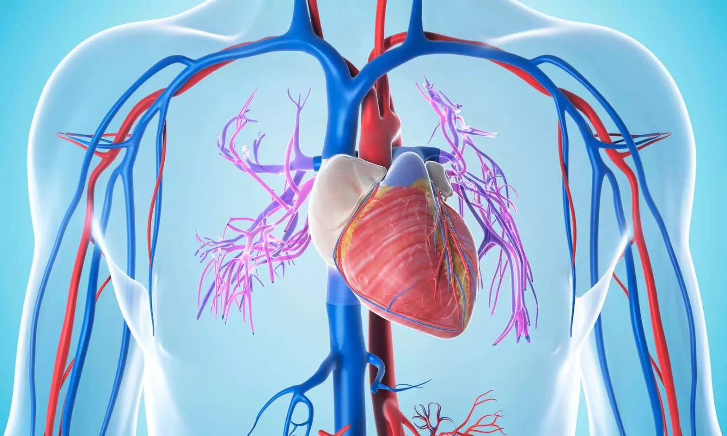
Low PUFA intake may magnify risk of developing CVD among people with family history of heart disease

A recent study delving into the interplay between dietary intake of polyunsaturated fatty acids (PUFA) and family history in relation to cardiovascular disease (CVD) risk has uncovered a significant association. Researchers have found in a new study that Low PUFA intake may magnify risk of developing CVD among people with family history of heart disease. 

This study was published in the Circulation journal by Laguzzi A. and colleagues.

Researchers examined data from over 40,000 adults, exploring the impact of low PUFA intake, particularly eicosapentaenoic/docosahexaenoic acids (EPA/DHA), in individuals with and without a family history of CVD. The study, conducted across a large consortium, assessed blood and tissue PUFA levels, categorizing PUFA levels ≤25th percentile as indicative of low intake of specific fatty acids. A family history of CVD was defined as having at least one first-degree relative who had experienced a CVD event.

The most striking finding revealed a significant interaction between low EPA/DHA intake and a family history of CVD. After meticulous adjustments for various influencing factors, the combined exposure to low EPA/DHA and a family history of CVD was associated with a substantial increase in relative risk for CVD (pooled relative risk 1.41; 95% CI, 1.30–1.54). In comparison, the relative risk was lower for those with a family history alone (pooled relative risk 1.25; 95% CI, 1.16–1.33) and even lower for individuals with low EPA/DHA intake alone (pooled relative risk 1.06; 95% CI, 0.98–1.14).

The study highlighted that low intake of EPA/DHA in conjunction with a family history of CVD significantly amplified the risk of developing CVD, emphasizing the potential impact of this dietary factor on cardiovascular health.

Although the relative excess risk due to interaction did not show statistical significance, the findings underscore the need for individuals with a family history of CVD to pay particular attention to their dietary intake of EPA/DHA, primarily found in oily fish. This novel insight could inform healthcare strategies, advocating for the consumption of oily fish as a preventive measure against CVD for those with a family history.

These findings pave the way for further investigation into personalized dietary interventions targeting high-risk populations. Emphasizing the inclusion of EPA/DHA-rich foods in the diet, especially for individuals with a genetic predisposition to CVD, may prove pivotal in reducing cardiovascular risk and promoting long-term heart health.

Reference:

Laguzzi, F., Åkesson, A., Marklund, M., Qian, F., Gigante, B., Bartz, T. M., Bassett, J. K., Birukov, A., Campos, H., Hirakawa, Y., Imamura, F., Jäger, S., Lankinen, M., Murphy, R. A., Senn, M., Tanaka, T., Tintle, N., Virtanen, J. K., Yamagishi, K., … Fatty Acids and Outcomes Research Consortium (FORCE). Role of polyunsaturated fat in modifying cardiovascular risk associated with family history of cardiovascular disease: Pooled DE Novo results from 15 observational studies. Circulation,2023. https://doi.org/10.1161/circulationaha.123.065530 

Powered by WPeMatico

Placement of 6-mm implants as good as 11-mm implants for combined maxillary sinus floor augmentation

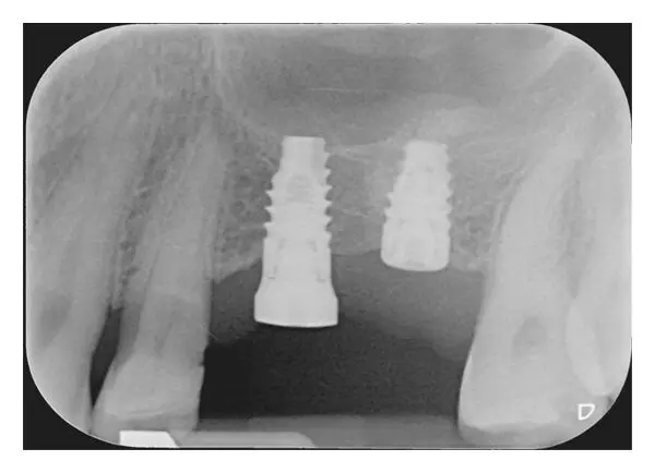
Placement of 6-mm implants as good as 11-mm implants for combined maxillary sinus floor augmentation suggests a new study published in the Clinical Oral Implants Research.

A study was done to compare the clinical performance of single crowns in the posterior maxilla supported by either 11-mm long implants combined with maxillary sinus floor augmentation (MSFA) surgery or by 6-mm long implants during a 10-year follow-up period.

Subjects were randomly allocated to receive one 11-mm long implant in combination MFSA or to receive one 6-mm long implant without any grafting. Twenty-one implants in 20 patients were placed in the 6-mm group and 20 implants in 18 patients were placed in the 11-mm group. Both groups were followed by clinical and radiographic examinations up to 10 years. Patients’ satisfaction was also scored before treatment.

Results

Two patients died and eight patients moved during the follow-up. Two patients lost an implant in the 6-mm group and one implant was lost in the 11-mm group (implant survival 89.5% and 90.9%, respectively). From loading to 10 years’ follow-up, mean ± SE marginal bone loss in the 6-mm group and 11-mm group was 0.18 ± 0.10 mm and 0.26 ± 0.12 mm, respectively, without a significant difference between the groups at 10 years (p = .650). In both groups, indices scores for plaque, calculus, gingiva and bleeding were low as well as mean pocket probing depth. Patients’ satisfaction at 10 years was high in both groups; mean overall satisfaction in the 6-mm group and 11-mm group was 9.6 ± 0.6 and 9.2 ± 0.8, respectively (p = .168).

Placement of 6-mm implants or 11-mm implants combined MFSA are equally successful during a 10-year follow-up period when applied for supporting a single restoration.

Reference:

Guljé, F. L., Raghoebar, G. M., Gareb, B., Vissink, A., & Meijer, H. J. A. (2023). Single crowns in the posterior maxilla supported by either 11-mm long implants with sinus floor augmentation or by 6-mm long implants: A 10-year randomized controlled trial. Clinical Oral Implants Research, 00, 1–12. https://doi.org/10.1111/clr.14200

Keywords:

Placement, 6-mm, implants, as, good, 11-mm, implants, combined, maxillary, sinus, floor, augmentation, Guljé, F. L., Raghoebar, G. M., Gareb, B., Vissink, A., & Meijer, H. J. A.

Powered by WPeMatico

Prenatal maternal inflammation tied to ADHD diagnosis by age 10

The available preclinical and registered data indicate that maternal inflammation during pregnancy may impact early neurodevelopment in offspring. However, there is limited clinical evidence of the risk of abnormal neurodevelopment in children later in life. In the population-based COPSAC2010 mother-child cohort, a team of researchers examined the relationship between maternal inflammation levels during pregnancy and the likelihood of a child being diagnosed with ADHD, as well as the severity of ADHD symptoms in children at age 10.

A recent study published in Brain, Behavior, and Immunity has concluded that Prenatal maternal inflammation, assessed by hs-CRP, is linked to ADHD by age 10. Maternal inflammation associated with ADHD symptom load in the full cohort. This study identified inflammation as a marker crucial for future prevention during pregnancy to improve neurodevelopmental outcomes in children.

This study followed 700 mothers and their children since pregnancy week 24. The mother’s hs-CRP level at week 24 was investigated about the child’s neurodevelopment at age 10. The children underwent a neurodevelopment exam using K-SADS-PL and ADHD-RS. Logistic and linear regression models were used with confounder adjustment, including socioeconomic status and maternal polygenic risk of ADHD.

Key findings from the study are:

  • Total 604/ 700 children in the COPSAC2010 cohort participated in the COPSYCH visit at age 10.
  • Sixty-five (16 girls and 49 boys), constituting 10.8 %, fulfilled a research diagnosis of ADHD.
  • Higher maternal hs-CRP level in pregnancy at week 24 was associated with increased risk for ADHD diagnosis with adjusted OR 1.40.
  • Higher maternal hs-CRP was associated with increased ADHD symptom load in the entire cohort, reflected by ADHD-RS raw scores.

Discussing further, they said the clinical data demonstrates a robust association of prenatal maternal inflammation assessed by hs-CRP with ADHD diagnosis by age 10.

Attention-Deficit/Hyperactivity Disorder is the most frequent neurodevelopmental disorder in childhood and adolescence, with an estimated worldwide prevalence of around 5–7 %. To our knowledge, this is the first study combining deep phenotyping with lege artis clinical evaluations to confirm associations between exposure to prenatal inflammation and subsequent risk of ADHD in the offspring in mid-childhood. Furthermore, we report positive associations between maternal inflammation and a load of ADHD symptoms in the entire cohort, they said.

Reference:

Rosenberg, J. B. et al. Maternal inflammation during pregnancy is associated with the risk of ADHD in children at age 10. Brain Behavior and Immunity, 115, 450–457. https://doi.org/10.1016/j.bbi.2023.10.023

Powered by WPeMatico

Body fat and nutrition strongly associated with diabetic kidney disease in type 2 diabetes patients: Study

China: A recent study published in Diabetes Therapy showed increased odds of diabetic kidney disease (DKD) in subjects with diabetes having poor nutritional parameters and high body fat.

“Nutritional parameters and body fat were strongly associated with and considerably contributed to DKD presence, indicating that nutrition and body fat might be promising markers representing a metabolic state in the DKD pathogenesis and clinical utility of bioelectrical impedance analysis (BIA) might provide valuable recommendations to patients with type 2 diabetes mellitus (T2DM),” the researchers wrote.

Diabetic kidney disease develops in about 40% of patients with diabetes and has become a leading cause of chronic kidney disease and end-stage renal failure in most developed and developing countries. Therefore, there arises an urgent need for strategies aimed at identifying potentially modifiable risk factors for DKD.

To address the same, Junwei Yang, The Second Affiliated Hospital of Nanjing Medical University, Nanjing, Jiangsu, China, and colleagues investigated the association between clusters of body fat and nutritional parameters with DKD in adults with type 2 diabetes in a cross-sectional study.

The study included 184 participants with T2DM. Biochemical parameters including haemoglobin A1c, fasting blood glucose, haemoglobin, creatinine, albumin, and urinary albumin-to-creatinine ratio (UACR) were measured. BIA was used to obtain data for visceral fat area (VFA), percentage of body fat mass (PBF), body cell mass (BCM), and phase angle at 50 kHz (PA50).

DKD diagnosis was made by UACR and estimated glomerular filtration rate (eGFR). Factor analysis was used for dimensionality reduction clustering among variables. Binary logistic regression analysis was used to assess the association of clusters with the presence of DKD.

Based on the study, the researchers reported the following findings:

· Factor analysis identified two clusters which were interpreted as a body fat cluster with positive loadings of VFA, waist circumstance, body mass index, and PBF and a nutritional parameters cluster with positive loadings of PA50, haemoglobin, albumin, and BCM.

· Participants were divided into four groups based on the sex-specific cutoff value (median) of each cluster score calculated using the cluster weights and the original variable values.

· Only participants with high body fat and poor nutritional parameters (OR 3.43) were associated with increased odds of having DKD.

“Although the BIA method and consequent findings require further studies for confirmation, our findings indicate that nutritional parameters and body fat could be strong risk markers of DKD and are promising for clinical diagnosis and interventions,” the researchers concluded.

Reference:

He, A., Shi, C., Wu, X. et al. Clusters of Body Fat and Nutritional Parameters are Strongly Associated with Diabetic Kidney Disease in Adults with Type 2 Diabetes. Diabetes Ther (2023). https://doi.org/10.1007/s13300-023-01502-5

Powered by WPeMatico

Cancer antigen-125 useful biomarker for preeclampsia severity?

India: A recent case-control study published in The Journal of Obstetrics and Gynecology of India explored the potential of cancer Antigen-125 (CA-125) as a biomarker for predicting and correlating with the severity of preeclampsia. 

The researchers found higher levels of serum CA-125 in the severe preeclampsia group versus the normotensive and non-severe preeclampsia group, but the difference was not statistically significant.

Preeclampsia is a potentially dangerous pregnancy complication characterised by high BP and is often associated with unfavourable feto-maternal outcomes. There is a lack in its pathophysiology, emphasizing the need to research for tests that can correlate with or predict the severity of preeclampsia.

Cancer antigen-125 is a readily available, simple biomarker with evidence of its secretion at the choriodecidual unit and may have a possible role. Pooja Bhatia, Department of Obstetrics and Gynaecology, Government Medical College and Hospital, Chandigarh, India, and colleagues compared serum CA-125 levels between normal pregnant women and women with preeclampsia. They determined its clinical usefulness in correlating with preeclampsia severity.

For this purpose, the researchers conducted a case-control study involving 58 women with preeclampsia. they were further divided into severe and non-severe groups and 62 gestational age-matched healthy, pregnant controls. A comparison was drawn of the serum CA-125 levels between the two groups.

The study revealed the following findings:

· The mean serum CA-125 in the controls was 16.44 ± 8.28 IU/ml, 13.82 ± 9.18 IU/ml in the non-severe and 23.55 ± 30.55 IU/ml in the severe pre-eclampsia group.

· Serum CA-125 had a significant association with systolic blood pressure (SBP), foetal growth restriction, diastolic blood pressure (DBP), pre-term birth and a highly significant association with 24-h urinary protein, liver enzymes, placental abruption, need for maternal intensive care as well as with poor neonatal outcome including stillbirth and neonatal mortality.

“More studies on a larger scale are needed to prove the usefulness of this marker concerning perinatal and maternal outcome and its association with pre-eclampsia and its severity,” the researchers concluded.

Reference:

Bhatia, P., Goel, P., Mehra, R. et al. Correlation of Serum Cancer Antigen-125 (CA-125) Levels with Severity of Pre-eclampsia. J Obstet Gynecol India (2023). https://doi.org/10.1007/s13224-023-01869-2

Powered by WPeMatico

Treating Pituitary Apoplexy: Medical Management Versus Surgery

The first prospective study comparing outcomes in patients with pituitary apoplexy-sudden bleeding or death of a pituitary tumor-found that individuals managed medically fared as well as those treated surgically in the majority of cases. The multicenter international study, led by Cedars-Sinai investigators, was published in The Journal of Clinical Endocrinology & Metabolism.

“This is the best data to date on the question of surgery versus medical management in patients with this rare but serious condition,” said Adam Mamelak, MD,co-director of the Pituitary Center, director of the Functional Neurosurgery Program at Cedars-Sinai and lead author of the study. “It demonstrates in a prospective, controlled way what previous studies had begun to suggest, namely that pituitary apoplexy is very rarely a surgical emergency.”

The pituitary gland, located at the base of the brain, controls the function of several hormone-producing glands. Pituitary apoplexy occurs when a benign tumor in the pituitary gland area begins to bleed or dies, causing the tumor to grow and press on the surrounding brain tissues. This, in turn, causes symptoms such as severe headache, fatigue, confusion and vision problems that may include vision loss. Most commonly, these tumors are undetected prior to the apoplexy event.

To relieve the pressure on brain tissues and mitigate the symptoms of apoplexy, patients can either have the tumor surgically removed or be treated with medications to relieve pain and other symptoms while they wait for it to naturally shrink over time. In both cases, patients generally also need hormone-replacement therapy, Mamelak said.

Pituitary apoplexy has long been considered an emergency requiring rapid surgical treatment to achieve best results. Small retrospective studies, where investigators look back at previously collected data, have suggested that pituitary apoplexy patients have equally good outcomes with medical management, but these observations have failed to significantly change clinical practice.

“While we’ve seen a growing trend toward clinicians being a little more comfortable with medical management of pituitary apoplexy, in general, most patients still end up getting surgery,” Mamelak said. “Those small retrospective studies really didn’t move the needle that much.”

To provide a more solid basis for clinical decision-making, Mamelak and fellow investigators from the Department of Neurosurgery and the Pituitary Center at Cedars-Sinai-as well as colleagues from 11 other medical centers in North America, Japan, South Korea and Europe-developed the Pituitary Apoplexy Surgical Timing and Outcomes Registry (PASTOR).

The registry enrolled 97 patients with pituitary apoplexy at the time of diagnosis and compared outcomes for the 67 patients who had surgery with those for the 30 patients whose symptoms were medically managed. This type of prospective registry allows investigators to draw stronger conclusions because data collection is the same for all participants.

Investigators found that:

  • Length of hospital stay was the same for patients who had surgery as for those who did not.
  • Patients who had surgery experienced the same outcomes whether they had surgery right away or days after the onset of symptoms.
  • There were no statistically significant differences in hormone function, vision or quality of life between the two groups of patients three and six months after treatment.

Mamelak noted that patients experiencing more severe vision symptoms were more likely to have surgery than those experiencing milder symptoms but that surgery didn’t necessarily lead to better outcomes.

“Cedars-Sinai and the other sites taking part in this study all have expert pituitary neurosurgery and endocrinology teams, which could help account for the positive outcomes experienced by surgical patients,” said , chair of the DepKeith L. Black, MD artment of Neurosurgery and the Ruth and Lawrence Harvey Chair in Neuroscience at Cedars-Sinai. “A wealth of solid research tells us that outcomes from pituitary surgery are directly tied to surgical experience.”

Mamelak said that further research specifically looking at outcomes for people with visual field deficits would be needed to determine whether surgery is best in those cases. Meanwhile, the current findings could especially benefit medical centers that do not have the expertise to surgically treat pituitary tumors.

“These findings take some of the urgency out of managing these cases,” Mamelak said. “Clinicians at these centers now know that they can safely manage these patients medically and transfer them when it’s reasonable and available, or allow patients to forgo surgery entirely if they are comfortable with that.”

Reference:

Adam N Mamelak, Andrew S Little, Paul A Gardner, João Paulo Almeida, Pablo Recinos, Pranay Soni, Varun R Kshettry, John A Jane, Garni Barkhoudarian, Daniel F Kelly, Robert Dodd, Debraj Mukherjee, Zachary C Gersey, Noriaki Fukuhara, Hiroshi Nishioka, Eui-Hyun Kim, Claude-Fabien Litré, Elliott Sina, Mia W Mazer, Yujie Cui, Vivien Bonert, A Prospective, Multicenter, Observational Study of Surgical vs Nonsurgical Management for Pituitary Apoplexy, The Journal of Clinical Endocrinology & Metabolism, 2023;, dgad541, https://doi.org/10.1210/clinem/dgad541

Powered by WPeMatico

Refuses stay on neet pg counseling

kayakalp award for hospital in bhopal