Is there a relation between stillbirth Risk in Grandchild and Grandmother’s Obesity?

Women’s obesity and overweight problems have reached epidemic levels, making them the most significant preventable risk factors for stillbirth.

Eduardo Villamor, in a recent investigation published in the American Journal of Epidemiology, said grandmaternal overweight and obesity are associated with grand offspring stillbirth.

In this Swedish population-based study, researchers investigated the association between maternal grandmaternal early pregnancy BMI and grand offspring stillbirth risk. This three-generation cohort included nearly 176,908 grandmothers, 197,579 mothers, and more than 316,459 grand offspring born 1997-2016, using Swedish Medical Birth Register.

Key findings are:

  • There were 998 stillbirths reported in grand offspring with risk, 3.2 per 1000 births.
  • Compared to the grandmaternal normal BMI of 18.5-24.9 mg/kg2, stillbirth risk increased by 41 % (Relative risk 1.41).
  • With increasing BMI ≥30, stillbirth risk increased by 62 % (relative risk 1.62).
  • Maternal overweight and obesity in early pregnancy increased stillbirth risk in offspring by 32% and 77%, respectively.
  • As indicated by causal mediation analysis, Maternal BMI mediated only 19% of this relation.

They said, In 101,368 pregnancies, we studied the relationship between maternal full sisters’ BMI and stillbirth risk to determine if shared familial factors explain the association. The stillbirth risk for full sisters with a BMI of 25.0-29.9 and ≥30 compared to 18.5-24.9 was 0.76 and 0.88, respectively. Our conclusion is that shared familial factors do not fully explain the association.

This study revealed an association between grandmaternal body mass index (BMI) and grand offspring stillbirth.

Observational design and selection bias due to missing data were the limitations of this study.

National Institutes of Health funded the study.

Reference:

Eduardo Villamor, Sven Cnattingius, Grandmaternal body mass index in early pregnancy and risk of grand offspring stillbirth: A nationwide, three-generation cohort study, American Journal of Epidemiology, 2023; kwad235, https://doi.org/10.1093/aje/kwad235

Powered by WPeMatico

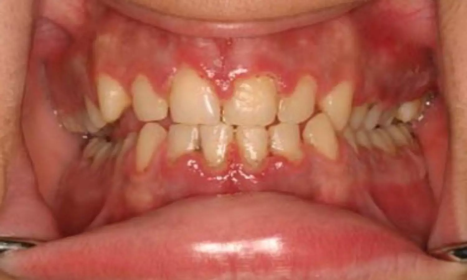
Diffuse gingival enlargement can be manifestation of hematological malignancies- Case report

Greece: A recent case study published in Cureus describes an unusual case of acute myeloid leukaemia (AML) in a 26-year-old male patient who presented to the clinic with a complaint of persistent bilateral gingival bleeding in the posterior areas of the maxilla.

“Hematological malignancies can manifest in the oral cavity and that manifestation could be the only diagnostic sign as observed in this case,” Vasileios Zisis, Oral Medicine/Pathology, Aristotle University of Thessaloniki, Thessaloniki, GRC, and colleagues wrote.

They added, “It is, therefore, vital for the clinician to not rule out malignancy when such a clinical situation presents and to make an appropriate early referral.”

Acute myeloid leukaemia is a malignancy characterized by its extreme aggressiveness. It accounts for about 25% of leukaemia cases in the pediatric population. Therefore, in the case described, the age of the patient may be considered atypical, according to the authors.

Gingival hyperplasia may occur due to microbial-related local irritation, drug administration, mouth breathing, leukaemia, genetic disorders, Crohn’s disease, Wegener granulomatosis, and sarcoidosis. The background may be fibrotic, inflammatory, or combined. In the study, Dr. Zisis and colleagues aimed to present the diagnostic procedure for a case of gingival enlargement, which was the only sign of severe systemic disease in a young male adult.

The 26 years old male patient was referred by his dentist to the Department of Oral Medicine and Pathology, School of Dentistry, Aristotle University of Thessaloniki, Greece, complaining of persistent gingival bleeding in the posterior area of the maxilla, bilaterally, and mild pain due to food impaction in the interdental area. The patient provided written informed consent before the examination. Subsequently, the patient was examined thoroughly. The physical examination revealed diffuse reddish swelling, palpable gingival masses, bleeding on probing, and the presence of pseudopockets of 4-6mm. Clinically, a diffuse gingival enlargement was seen without lymphadenopathy.

The histopathological examination demonstrated abundant neoplastic cells of hemopoietic origin with strong and diffuse positivity for CD45 and CD68. In addition, scattered neoplastic cells exhibited mild to moderate positivity for c-kit (CD117), indicating the diagnosis of acute myeloid leukaemia, which diffusely infiltrated the lamina propria of the gingiva.

“The several conditions leading to gingival enlargement other than periodontitis or gingivitis are diagnostic challenges in clinical practices,” the team wrote. “The dentist plays an important role in the timely identification of hematologic disorders.” They note the significant role that dentists play in commencing leukaemia diagnosis in cases of oral manifestations.

“Therefore, these manifestations must be easily identifiable, prompting a comprehensive inquiry involving supplementary tests or a referral to a qualified practitioner to arrive at a definitive diagnosis,” they concluded.

Reference:

Zisis V, Zisis S, Anagnostou E, et al. (October 30, 2023) Gingival Enlargement Can Constitute the Only Diagnostic Sign of Leukemia: Report of an Unusual Case. Cureus 15(10): e47959. doi:10.7759/cureus.47959

Powered by WPeMatico

Maternal influenza vaccination during pregnancy may protect infant in first few months of life: JAMA

A recent study exploring the impact of maternal influenza vaccination during pregnancy has shown promising outcomes in reducing influenza-related hospitalizations and emergency department (ED) visits in infants under 6 months of age. The study, conducted using data from the New Vaccine Surveillance Network across multiple influenza seasons, sheds light on the protective benefits of maternal vaccination against influenza-associated illnesses in young infants.

The study was published in the journal of JAMA Pediatrics by Leila Sahni and colleagues. The study included 3764 infants, among whom 53% were born to mothers who received influenza vaccination during pregnancy. The overall effectiveness of maternal vaccination in infants against influenza-related hospitalizations and ED visits was estimated at 34%. Specifically, the vaccine demonstrated a 39% effectiveness in preventing hospitalizations and a 19% effectiveness in mitigating ED visits associated with influenza.

Among infants younger than 3 months, the effectiveness of maternal vaccination soared to 53%, indicating a stronger protective effect in this vulnerable age group. Notably, infants born to mothers vaccinated during the third trimester exhibited a higher effectiveness rate of 52%, whereas those whose mothers were vaccinated during the first or second trimesters showed a lower effectiveness rate of 17%.

The study’s findings underscore the significance of maternal influenza vaccination during pregnancy in reducing the risk of influenza-associated hospitalizations and ED visits in infants below 6 months of age. The observed effectiveness was particularly notable in younger infants and those born to mothers vaccinated during the later stages of pregnancy.

These results emphasize the importance of promoting maternal influenza vaccination as a crucial preventive measure to safeguard both maternal and infant health. Encouraging vaccination, especially during the third trimester, could offer substantial protection against influenza-related complications in vulnerable newborns.

Reference:

Sahni, L. C., Olson, S. M., Halasa, N. B., Stewart, L. S., Michaels, M. G., Williams, J. V., Englund, J. A., Klein, E. J., Staat, M. A., Schlaudecker, E. P., Selvarangan, R., Schuster, J. E., Weinberg, G. A., Szilagyi, P. G., Boom, J. A., Patel, M. M., Muñoz, F. M., McNeal, M., Rohlfs, C., … New Vaccine Surveillance Network Collaborators. Maternal vaccine effectiveness against influenza-associated hospitalizations and emergency department visits in infants. JAMA Pediatrics,2023. https://doi.org/10.1001/jamapediatrics.2023.5639

Powered by WPeMatico

IPC issues Safety warning against use of common painkiller Meftal

India: The Indian Pharmacopoeia Commission (IPC) has recently issued a drug safety alert on the common painkiller Meftal. The IPC is an autonomous institution of the Ministry of Health that sets standards for all drugs that are manufactured, sold, and consumed in India.

The IPC has advised consumers and healthcare professionals (HCPs) to exercise caution concerning the potential adverse drug reaction (ADR) linked to the use of Mefenamic acid, the constituent of Meftal. 

A preliminary analysis of ADRs from the database of the Pharmacovigilance Programme of India has revealed cases of Drug Rash with Eosinophilia and Systemic Symptoms (DRESS) syndrome, which affects internal organs. These symptoms may appear 2-8 weeks after taking the drug.

“The IPC has advised healthcare professionals, patients/consumers to closely monitor the possibility of the above adverse drug reaction associated with the use of the suspected drug,” stated the alert, issued on 30 November.

The alert further stated, “If such a reaction is encountered, people should report the matter to the national coordination centre of the PvPI under the commission by filling a form on the website — www.ipc.gov.in — or through the Android mobile app ADR PvPI, as well as PvPI helpline number 1800-180-3024.”

DRESS syndrome, also known as Drug-Induced Hypersensitivity Syndrome (DIHS), is a severe allergic reaction affecting around 10% of individuals, potentially deadly and caused by certain medications. It is characterized by high fever, skin rash, swollen lymph nodes and complications in internal organs.

The key features of DRESS syndrome are:

· Skin Rash: Patients with DRESS often develop a widespread rash that can involve various body parts. The rash may be maculopapular (flat and raised lesions), erythematous (red), and may resemble other skin conditions.

· Internal Organ Involvement: DRESS can affect multiple organs, including the liver, kidneys, lungs, and heart. This can lead to hepatitis, nephritis, pneumonitis, and myocarditis.

· Fever: Individuals with DRESS typically experience a high fever.

· Lymphadenopathy: Swollen lymph nodes (lymphadenopathy) are common in DRESS.

· Haematologic Abnormalities: Blood abnormalities, such as atypical lymphocytes, eosinophilia (elevated eosinophil count), and other hematologic changes, may be present.

Use and Associated Side Effects of Meftal

Meftal is used commonly as a non-steroidal anti-inflammatory drug (NSAID). It is indicated for the treatment of osteoarthritis, rheumatoid arthritis, dysmenorrhoea, mild to moderate pain, fever, inflammation, and dental pain.

Although the drug is not available over the counter (OTC) and requires a prescription for purchase, it is extensively used in India for various purposes such as relieving headaches, menstrual pain, and muscle and joint pain, and is even prevalent among children for high fever.

Prolonged use of Meftal is reported to increase the risk of stomach ulcers, bleeding, and related complications. It has been associated with potential adverse effects on the cardiovascular system. Some experts have flagged renal complications as a potential side effect of Meftal.

If someone suspects DRESS or experiences symptoms after taking a medication, it is important to seek prompt medical attention. DRESS is a medical emergency, and early recognition and intervention are required for a better prognosis. Consultation with healthcare professionals is recommended for accurate diagnosis and appropriate management.

Powered by WPeMatico

Body dissatisfaction in late childhood tied to risk of depression by age 14: Lancet

UK: Findings from a recent longitudinal study published in The Lancet Psychiatry suggest that greater body dissatisfaction in late childhood is an important risk factor for depression during adolescence, irrespective of the child’s body mass index (BMI). 

“Body dissatisfaction at age 11 is linked to increased risk of depression by age 14,” UCL researchers report. The findings show that body image concerns explain a large proportion of an association between BMI and depression in children, particularly in girls.

The study, supported by Wellcome, involved 13,135 participants of the Millennium Cohort Study, a UCL-led nationally representative birth cohort study of people born between 2000 and 2002.

The researchers found that high BMI at age seven was linked with increased depressive symptoms (which can include low mood, loss of pleasure and poor concentration) by age 14, as well as with greater body dissatisfaction at age 11.

They found that body dissatisfaction was a major contributor to the link between BMI at age seven and subsequent depressive symptoms, explaining 43% of the association.

All three of these associations were twice as large in girls compared to boys.

Lead author Dr Francesca Solmi (UCL Psychiatry) said: “Depression has become more common among young people, as have having an overweight BMI, and body dissatisfaction.

“Here we have found strong longitudinal evidence that a high BMI in childhood is linked with an increased risk of depressive symptoms multiple years later.

“But we were particularly interested in how much body dissatisfaction might be the driver of this link. We found strong evidence that being unhappy with one’s appearance is linked with increased depressive symptoms years later. Our findings suggest that any efforts to reduce weight in childhood need to consider their potential mental health impacts, so that we can avoid stigmatising weight and instead support children’s mental health and wellbeing.”

The research did not cover what other factors, besides body dissatisfaction, could explain why children with high BMI are more likely to develop depressive symptoms, but they say that other biological (for instance inflammation) or environmental (for instance bullying) pathways might explain part of the association.

First author Emma Blundell, trainee clinical psychologist at UCL Psychology & Language Sciences, said: “Many public health strategies seek to reduce weight in childhood. Primary school children are being taught about the importance of calories and exercise, and all young people in England are being weighed at school to determine whether weight loss efforts are needed. Whilst promoting healthy diet and exercise is important, it may be that some public health messaging could be fostering feelings of guilt or shame.

“It is important to ensure that any interventions to reduce BMI in childhood do not inadvertently increase body dissatisfaction and harm children’s mental health.”

The researchers say that some strategies to target body image concerns in early adolescence have been developed, such as with psychological interventions or media literacy training that could address self-esteem, social comparisons, and social media influences, but more research is needed to more effectively tackle body image concerns in young people.

Dr Solmi added: “Reducing body dissatisfaction in young people could be an important way of preventing depression, particularly in girls, at ages when social environments and peer relations become increasingly impactful.”

Reference:

Emma Blundell, Prof Bianca L De Stavola, Madelaine Davies Kellock, Gemma Lewis, Prof Anne McMunn, Longitudinal pathways between childhood BMI, body dissatisfaction, and adolescent depression: an observational study using the UK Millenium Cohort Study, DOI:https://doi.org/10.1016/S2215-0366(23)00365-6.

Powered by WPeMatico

Early Pharmacological Treatment of Type II SMA Patients Slows Scoliosis Progression

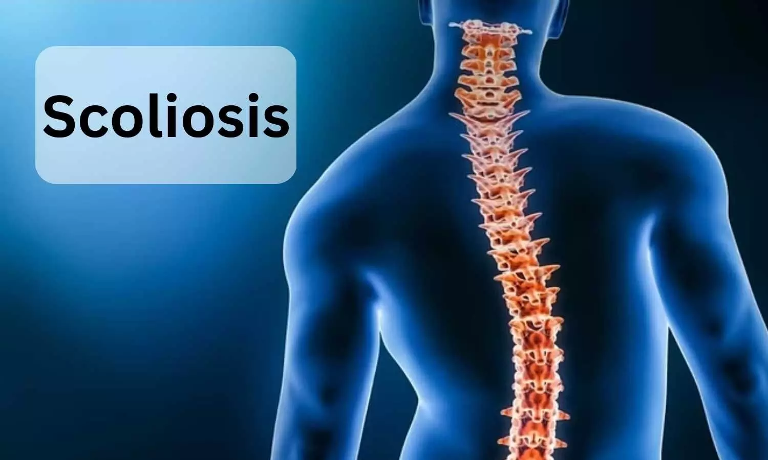
A recent study has shed new light on the potential impact of early pharmacological interventions on scoliosis progression in individuals with Type II spinal muscular atrophy (SMA). Scoliosis, affecting up to 90% of Type II SMA cases, poses a significant challenge, and while treatments have shown improvements in motor function, their effect on scoliosis remains uncertain. This study was published in the Journal Of Neurology Neurosurgery and Psychiatry by Giorgia Coratti and colleagues.

The study aimed to investigate differences in scoliosis progression between treated and untreated Type II SMA patients. Researchers analyzed the effect of treatment on Cobb’s angle annual changes and the likelihood of reaching a critical 50° Cobb angle in patients with a minimum 1.5-year follow-up. The study utilized a sliding cut-off approach to identify the optimal treatment subpopulation based on age, initial Cobb angle, and Hammersmith Functional Motor Scale Expanded at the initial visit. The Mann-Whitney U-test assessed statistical significance.

Comparing a cohort of 46 untreated individuals with 39 treated patients, the study initially found no significant differences in baseline characteristics. However, when analyzing the impact of pharmacological treatment on scoliosis progression, intriguing patterns emerged. The mean Cobb angle variation did not significantly differ between the treated and untreated groups initially. Yet, upon exploring optimal cut-off values for a better outcome, the treated group, especially those with a Cobb angle <26° or an age <4.5 years, demonstrated a significantly lower mean Cobb angle variation. The treated group showed a lower mean Cobb variation compared to the untreated group (5.61 (SD 4.72) degrees/year vs 10.05 (SD 6.38) degrees/year; p=0.01). Furthermore, Cox-regression analysis indicated a protective effect of treatment in preventing patients from reaching a 50° Cobb angle, particularly pronounced in patients younger than 4.5 years old (p=0.016).

The study’s results underscore a potential correlation between early pharmacological treatment initiation and slowed scoliosis progression in Type II SMA patients. These findings emphasize the importance of early intervention, particularly in individuals under 4.5 years old or those with a lower initial Cobb angle, suggesting a more favorable response to treatment. However, researchers caution that larger-scale studies are imperative to delve deeper into the efficacy of individual pharmacological treatments on scoliosis progression in this patient population.

Reference:

Coratti, G., Lenkowicz, J., Pera, M. C., D’Amico, A., Bruno, C., Gullì, C., Brolatti, N., Pedemonte, M., Antonaci, L., Ricci, M., Capasso, A., Cicala, G., Cutrona, C., de Sanctis, R., Carnicella, S., Forcina, N., Cateruccia, M., Damasio, M. B., Labianca, L., … Mercuri, E. Early treatment of type II SMA slows rate of progression of scoliosis. Journal of Neurology, Neurosurgery, and Psychiatry,2023, jnnp-2023-332084. https://doi.org/10.1136/jnnp-2023-332084

Powered by WPeMatico

Dapagliflozin may improve symptoms in patients with HFrEF with limited impact on exercise capacity

Dapagliflozin may improve symptoms in patients with HFrEF with limited impact on exercise capacity suggests a new study published in the Circulation.

Sodium-glucose cotransporter 2 inhibitors reduce the risk of worsening heart failure (HF) and cardiovascular death in patients with HF irrespective of left ventricular ejection fraction. It is important to determine whether therapies for HF improve symptoms and functional capacity.

The DETERMINE (Dapagliflozin Effect on Exercise Capacity Using a 6-Minute Walk Test in Patients With Heart Failure) double-blind, placebo-controlled, multicenter trials assessed the efficacy of the sodium-glucose cotransporter 2 inhibitor dapagliflozin on the Total Symptom Score (TSS) and Physical Limitation Scale (PLS) of the Kansas City Cardiomyopathy Questionnaire (KCCQ) and 6-minute walk distance (6MWD) in 313 patients with HF with reduced ejection fraction (DETERMINE-Reduced) and in 504 patients with HF with preserved ejection fraction (DETERMINE-Preserved) with New York Heart Association class II or III symptoms and elevated natriuretic peptide levels. The primary outcomes were changes in the KCCQ-TSS, KCCQ-PLS, and 6MWD after 16 weeks of treatment.

RESULTS:

Among the 313 randomized patients with HF with reduced ejection fraction, the median placebo-corrected difference in KCCQ-TSS from baseline at 16 weeks was 4.23 (95% CI, 0.96, 8.22; P=0.022) in favor of dapagliflozin. The median placebo-corrected difference in KCCQ-PLS was 4.2 (95% CI, 0.00, 8.3; P=0.058). The median placebo-corrected difference in 6MWD from baseline at 16 weeks was 3.2 meters (95% CI, −6.5, 13.0; P=0.69). In the 504 patients with HF with preserved ejection fraction, the median placebo-corrected 16-week difference in KCCQ-TSS and KCCQ-PLS was 3.2 (95% CI, 0.4, 6.0; P=0.079) and 3.1 (−0.1, 5.4; P=0.23), respectively. The median 16-week difference in 6MWD was 1.6 meters (95% CI, −5.9, 9.0; P=0.67). In an exploratory post hoc analysis of both trials combined (DETERMINE-Pooled), the median placebo-corrected difference from baseline at 16 weeks was 3.7 (1.5, 5.9; P=0.005) for KCCQ-TSS, 4.0 (0.3, 4.9; P=0.036) for KCCQ-PLS, and 2.5 meters (−3.5, 8.4; P=0.50) for 6MWD.

Dapagliflozin improved the KCCQ-TSS in patients with HF with reduced ejection fraction but did not improve KCCQ-PLS or 6MWD. Dapagliflozin did not improve these outcomes in patients with HF with preserved ejection fraction. In a post hoc analysis including all patients across the full spectrum of ejection fraction, there was a beneficial effect of dapagliflozin on KCCQ-TSS and KCCQ-PLS but not 6MWD.

Reference:

Effect of Dapagliflozin Versus Placebo on Symptoms and 6-Minute Walk Distance in Patients With Heart Failure: The DETERMINE Randomized Clinical Trials

John J.V. McMurray, Kieran F. Docherty, Rudolf A. de Boer, Ann Hammarstedt, Dalane W. Kitzman, Mikhail N. Kosiborod, Anna Maria Langkilde, Barry Reicher, Michele Senni, Sanjiv J. Shah, Ulrica Wilderäng, Subodh Verma and Scott D. Solomon

Originally published 7 Dec 2023https://doi.org/10.1161/CIRCULATIONAHA.123.065061Circulation. 2023;0

Keywords:

Dapagliflozin, improve, symptoms, patients, HFrEF, limited, impact, exercise, capacity,John J.V. McMurray, Kieran F. Docherty, Rudolf A. de Boer, Ann Hammarstedt, Dalane W. Kitzman, Mikhail N. Kosiborod, Anna Maria Langkilde, Barry Reicher, Michele Senni, Sanjiv J. Shah, Ulrica Wilderäng, Subodh Verma and Scott D. Solomon

Powered by WPeMatico

New AI tool predicts post-surgery mortality

USA: The researchers at the Smidt Heart Institute at Cedars-Sinai and colleagues at two other institutions have developed an artificial intelligence (AI) tool that accurately predicts how patients would fare after surgeries and procedures. The results, published in The Lancet Digital Health, include data from patients from three healthcare systems: Cedars-Sinai, Stanford University and Columbia University.

All patients in the study underwent a surgical procedure, including open heart surgery, other major surgeries, and minimally invasive procedures involving a catheter or endoscope.

Investigators trained an AI model on pre-operative electrocardiograms, discovering a new use for the 130-year-old test. Invented in the late-1800s, an electrocardiogram is a commonly deployed test that involves placing electrodes on the skin to measure the heart’s electrical activity and assess how well the heart is functioning.

“This is the first electrocardiogram-based AI algorithm that predicts post-operative mortality,” said David Ouyang, MD, a cardiologist in the Department of Cardiology in the Smidt Heart Institute at Cedars-Sinai, and a corresponding author of the study. “Previously, algorithms have been used to assess long-term mortality as well as individual disease states, but determining post-surgical outcomes helps inform the actual decision to do surgery.”

Investigators paired patients’ electrocardiograms from before the surgery or procedure, with their outcomes after the surgery or procedure, and asked the algorithm to find associations or patterns in the electrocardiogram waveforms.

While classifying most patients as low risk, those individuals the algorithm identified as high risk had nearly a 9-fold increased probability of post-operative mortality.

“As it now stands, clinicians only have a modest ability to predict how a patient is going to do after surgery,” said Ouyang, also a faculty member in the Division of Artificial Intelligence in Medicine at Cedars-Sinai. “Current clinical risk prediction tools are insufficient. This AI model could potentially be used to determine exactly which patients should undergo an intervention and which patients might be too sick.”

Every medical intervention comes with risk, and doctors currently rely on guidelines from medical societies to determine a patient’s individual risk.

“In cardiology, we’re fortunate to have many life-saving procedures, everything from catheter-based procedures to open-heart surgery, so we’re often trying to think about who are the right patients for the right procedure,” said Christine M. Albert, MD, MPH, chair of the Department of Cardiology in the Smidt Heart Institute and the other corresponding author of the study. “A better understanding of risk, particularly by using a commonly obtained diagnostic test, can inform important medical decisions.”

Investigators are currently studying how the algorithm could be translated into a web application that would be widely available for physicians and patients.

Reference:

David Ouyang, John Theurer, Nathan R Stein, J Weston Hughes, Pierre Elias, Bryan He, Electrocardiographic deep learning for predicting post-procedural mortality: a model development and validation study, DOI:https://doi.org/10.1016/S2589-7500(23)00220-0.

Powered by WPeMatico

Lupin gets CDSCO Panel nod for Rifapentine Dispersible Tablet 150 mg

New Delhi: Granting the local clinical trial waiver, the Subject Expert Committee (SEC) functional under the Central Drug Standard Control Organisation (CDSCO) has approved drug major Lupin’s Rifapentine dispersible Tablets 150mg for already approved indication with the condition of conducting a Phase IV Clinical study.

This came after the firm presented their proposal for a grant of permission to manufacture and marketing of Rifapentine Dispersible Tablets 150mg (Additional Dosage Form) for an already approved indication along with the bioequivalence study report before the committee.

Rifapentine is an antitubercular agent and a leprostatic drug. Rifapentine is an antibacterial prescription medicine approved by the U.S. Food and Drug Administration (FDA) for the treatment of active tuberculosis (TB) of the lungs. (Active TB is also called TB disease.) Rifapentine is also FDA-approved for the treatment of latent TB infection to prevent the infection from advancing to active TB disease.

It inhibits DNA-dependent RNA polymerase activity in susceptible cells. Specifically, it interacts with bacterial RNA polymerase but does not inhibit the mammalian enzyme.

Rifapentine is used with other medications to treat active tuberculosis (TB; a serious infection that affects the lungs and sometimes other parts of the body) in adults and children 12 years of age and older.

At the recent SEC meeting for Antimicrobial and Antiviral held on 29th November 2023, the expert panel reviewed the proposal for grant of permission to manufacture and marketing of Rifapentine Dispersible Tablets 150mg (Additional Dosage Form) for an already approved indication along with the Bioequivalence study report presented by drug major Lupin.

After detailed deliberation, the committee recommended for approval of Rifapentine Dispersible Tablets 150mg for an already approved indication with a local clinical trial waiver with a condition to conduct a Phase IV Clinical study.

Accordingly, the expert panel suggested that the firm should submit a Phase IV protocol to CDSCO within 3 months of approval for further evaluation by the committee.

In addition, the committee stated that the firm should fulfill the requirements of CMC (chemistry, manufacturing, and controls) data before approval of the product.

Also Read: AstraZeneca gets CDSCO panel Nod to study anti-cancer drug Volrustomig in women with cervical cancer

Powered by WPeMatico

Consumption of Fresh vegetables has potential benefit of improving eGFR among elderly

Diet rich in Fresh vegetables has potential benefit of improving eGFR among elderly suggests a new study published in the BMC Nephrology.

This study aimed to investigate the relationship between the consumption of fresh and salt-preserved vegetables and the estimated glomerular filtration rate (eGFR), which requires further research.

The prevalence of chronic kidney disease (CKD) generally increases with age owing to nephron loss and a decline in the glomerular filtration rate (GFR). The estimated GFR (eGFR) assumes a widely recognized role as a marker that plays a pivotal role in evaluating overall kidney health and diagnosing various renal disorders.

For this purpose, the data of those subjects who participated in the 2011–2012 and 2014 surveys of the Chinese Longitudinal Healthy Longevity Survey (CLHLS) and had biomarker data were selected. Fresh and salt-preserved vegetable consumptions were assessed at each wave. eGFR was assessed using the Chronic Kidney Disease Epidemiology Collaboration (CKD-EPI) equation based on plasma creatinine. Furthermore, a linear mixed model was used to evaluate associations between fresh/salt-preserved vegetables and eGFR.

Results

The results indicated that the median baseline and follow-up eGFRs were 72.47 mL/min/1.73 m² and 70.26 mL/min/1.73 m², respectively. After applying adjusted linear mixed model analysis to the data, the results revealed that compared to almost daily intake, occasional consumption of fresh vegetables was associated with a lower eGFR (β=-2.23, 95% CI: -4.23, -0.23). Moreover, rare or no consumption of salt-preserved vegetables was associated with a higher eGFR (β = 1.87, 95% CI: 0.12, 3.63) compared to individuals who consumed salt-preserved vegetables daily.

Fresh vegetable consumption was direct, whereas intake of salt-preserved vegetables was inversely associated with eGFR among the oldest subjects, supporting the potential benefits of diet-rich fresh vegetables for improving eGFR.

Reference:

Zheng, H., Li, H., Pan, L. et al. Association of fresh vegetable and salt-preserved vegetable consumptions with estimated glomerular filtration rate. BMC Nephrol 24, 369 (2023). https://doi.org/10.1186/s12882-023-03353-5

Keywords:

Diet, rich, Fresh, vegetable, potential, benefit, of, improving, eGFR, among, elderly, BMC Neph8rology, Zheng, H., Li, H., Pan, L

Powered by WPeMatico