Children with severe Cerebral Palsy have poor bone mineralization

Researchers have found in a new study that Children with severe Cerebral Palsy were more likely to have poor bone mineralization.  The new study has been published in the Frontiers in Pediatrics.

A study was conducted by researchers to describe bone health and associated factors in children with severe cerebral palsy.

In a retrospective, single-centre study, the  researchers performed a comprehensive bone evaluation (including clinical, densitometric and bone biomarker assessments) of children with severe cerebral palsy.

Results:

In the  research, None of the 19 included children had a normal BMCTBLH Z score, and only one had a BMDTBLH Z score greater than −2. Six children had a BMDLS Z score greater than −2. The bone biomarker data were suggestive of excessive bone remodelling. Levels of bone remodelling markers factors and densitometric variables were not significantly related. Further Age, weight and pubertal stage were significantly related to bone mass.

The results highlights the insufficient increase in bone mass with age (probably due to excessive bone remodelling) and confirms the high prevalence of low bone mineral density in children with severe cerebral palsy. Possible preventive measures might include calcium + vitamin D supplementation and the systematic management of underweight and delayed puberty. Bone remodelling markers might be of value for follow-up.

An insufficient increase in bone mass was seen with age and high prevalence of low bone mineral densityBone biomarker data indicated excessive bone remodelling; weight and pubertal stage were also related to bone mass

Reference:

Barbier V, Goeb V, Gouron R, Fritot S, Mentaverri R, Klein C. Bone health in children with severe cerebral palsy. Front Pediatr. 2023 Nov 29;11:1264111. doi: 10.3389/fped.2023.1264111. PMID: 38094189; PMCID: PMC10716435.

Keywords:

Children, severe, Cerebral Palsy, more, likely, have, poor, bone, mineralization, Barbier V, Goeb V, Gouron R, Fritot S, Mentaverri R, Klein C, Frontiers in Pediatrics

Powered by WPeMatico

Inflammatory tongue conditions increase the risk of oral cancer

The prevalence of oral tongue cancers has risen among both male and female individuals in the United States since the 1980s, despite the reasons for this trend remaining unclear.

In a study published in the Journal of Clinical Oncology, researchers found that Inflammatory tongue conditions raised oral tongue cancer risks and preceded diagnosis by years, emphasizing the importance of increased clinical surveillance for patients with apparently benign conditions.

A team of researchers conducted a case-control study using the SEER-Medicare dataset (1992-2013) comprising 2,534 oral tongue cancers, 6,832 other oral cavity cancers, 9,373 oropharyngeal cancers, and 200,000 controls.

Medicare data was used to identify patients with clinically diagnosed inflammatory tongue conditions (glossitis, benign migratory glossitis, median rhomboid glossitis, atrophic glossitis, glossodynia, other specified conditions [e.g., atrophy and hypertrophy], and other unspecified conditions) and oral precancer (leukoplakia/erythroplakia). Only conditions preceding cancer/control selection by more than 12 months were included.

Key findings from the study are:

  • Compared to controls, the prevalence of inflammatory tongue conditions was higher in tongue cancer patients (6.0% vs. 0.6%).
  • The odds ratio was 5.8.
  • The overall association primarily arose from glossitis (5.6), other specified conditions (9.1), and unspecified conditions (13.7).
  • These associations were elevated for >5 years preceding tongue cancer, diagnosed by specialists, and among patients who received an oral biopsy, arguing against reverse causation, misclassification, and missed cancer, respectively.

 In 2013, 1 in 11 oral tongue cancer patients had a prior inflammatory tongue condition diagnosis. The association between inflammatory tongue conditions and other oral cavity cancers and oropharyngeal cancer were weak and observed only near cancer diagnosis.

Concluding further, they said that inflammatory tongue conditions, including glossitis, atrophy, hypertrophy, and other unspecified conditions, were found to increase the risk of oral tongue cancer among US individuals aged 65 or older. The risk associations ranged from 6-fold to 15-fold, and inflammatory tongue conditions often preceded tongue cancer diagnosis by at least five years. Approximately one in 11 tongue cancers had a preceding diagnosis of inflammatory tongue condition. As such, clinicians should consider referring patients with glossitis to specialists (like oral medicine, oral surgeons) for oral cancer surveillance.

Reference:

Tota et al. Inflammatory tongue conditions and risk of oral tongue cancer among the US elderly individuals. Journal of Clinical Oncology.

Powered by WPeMatico

Last date admission for NEET SS candidates will be 31st December 2023, says NMC

Through a recent notice, The National Medical Commission (NMC) has informed the candidates about the starting of the academic session 2023-24 for the Super Speciality Courses. 

As per the notice, for the current academic session, i.e. for 2023-24, the Post Graduate Super Special Courses have already commenced from 25.11.2023, and the last date of admission will be 31st December 2023.

The notice informed that while the academic sessions could not have commenced as per PGMER 2000 for the past three years due to COVID-19 situation, the Post Graduate Super Special Courses have already commenced from 25.11.2023 and last date admission will be 31st December 2023.

For more details, check out the link given below:

Last Date Admission For NEET SS Candidates Will Be 31st December 2023, Says NMC

Powered by WPeMatico

Promise of MBBS Seat: Doctor booked for defrauding parents of two medical aspirants of Rs 1.2 crore

A shocking case of MBBS admission scam has come to light from the city where a doctor and two others have been accused of defrauding two businessmen of Rs 1.2 crore. They promised to secure their children a seat at Parel’s KEM hospital under management quota.

Following their alleged crime, the accused persons – Dr Hiten Kenia, Heena and Jeevan were booked on Monday under sections 406 (criminal breach of trust), 420 (cheating) and 34 (common intention) of the Indian Penal Code. They are yet to face action as the police are investigating for more evidence in connection with the case.

For more details, check out the link given below:

MBBS Seat Scam: Doctor Booked For Defrauding Parents Of Two Medical Aspirants Of Rs 1.2 Crore

Powered by WPeMatico

Mediterranean diet effective nutritional option for improving chance of success in IVF

Adjuvant therapies to help infertile women conceive by IVF – especially those whose treatments have been unsuccessful in the past – are now a common feature both before and during the treatment cycle. Now, a new analysis of the evidence for many nutritional supplements and diets thought to improve outcome in IVF has concluded that adopting a Mediterranean diet during treatment would offer a single ‘straightforward approach’ with good evidence of benefit in contrast to that of a Western diet.

Evidence from studies of nine commonly used nutritional supplements was found to be inconsistent and not always of good quality. The analysis, by Professor Roger Hart, University of Western Australia and City Fertility, Perth, Australia, is published today in the peer-reviewed journal Reproductive Biomedicine Online.

He explained that the extent to which nutritional supplements are used in IVF is largely unknown. ‘Nutritional supplements are usually not prescribed,’ explained Professor Hart, ‘but bought online or over-the-counter. They’re self-medicated and solid data on usage is impossible to determine. Our information is largely anecdotal but it’s quite clear from online IVF discussion forums that they are widely used and of great public interest.’

The nutritional supplements analysed in the study were dehydroepiandrosterone (DHEA), melatonin, co-enzyme Q10 (CoQ1O), carnitine, selenium, Vitamin D, myo-inositol, Omega-3, Chinese herbs and several diets (as well as weight loss). Many were reviewed as adjuvants for poor response to previous IVF treatment, of which DHEA and COQ10 appeared in studies to have more benefit than control therapies. Similarly, there was some evidence of benefit from melatonin, but it remains unclear which patient groups might benefit, nor at which dose.

However, the evidence in favour of a Mediterranean diet is much stronger and includes several well designed randomised clinical trials, showing benefits in both embryo development and pregnancy outcome (even from a six-week intervention programme). Professor Hart explained that the common features of these favourable diets were those with high intakes of fruits and vegetables, whole grains, legumes, nuts, fish, and monounsaturated or polyunsaturated oils, but with a limited intake of highly processed foods. ‘These diets are high in B-vitamins, antioxidants, omega-3 poly-unsaturated fatty acids and fibre and are low in saturated fat, sugar and sodium,’ he said. Omega-3 fatty acids, often taken as combined preparations, are the most studied dietary fatty acids in the IVF literature, largely, said Professor Hart, because of their perceived benefits in general health and reproduction. The evidence suggests that omega-3 fatty acids ‘may be beneficial’ in improving IVF clinical outcomes and embryo quality.

Similarly, antioxidants are usually taken as combinations of supplements but evidence from one major review (of 63 antioxidant studies in reproduction) described them as of low quality and was unable to demonstrate any benefit in terms of live birth rate.

Thus, from this mass of evidence Professor Hart advised that a simple nutritional approach to assist conception via IVF would be the adoption of a Mediterranean diet. The use of COQ-10 and DHEA before starting IVF may be a useful adjunct for women who had a previously poor response to ovarian stimulation, while supplementation with omega-3 free-fatty acids may indeed improve some clinical and embryological outcomes.

Professor Hart also advised that all women attempting to conceive should be taking adequate folate supplementation and should seek the advice of their general practitioner or specialist to ensure they are in their best general health for conception.

Reference:

R.J. Hart, Nutritional supplements and IVF: an evidence based approach, Reproductive BioMedicine Online, DOI: https://doi.org/10.1016/j.rbmo.2023.103770

Powered by WPeMatico

Sleep Deprivation can affect our emotional health, finds study

“Slept like a baby” is most heard among people whose has a sound sleep,  however this sleep is lot among todays people where changing lifestyles and daily habit.

Sleep loss does more than just make us tired. It can undermine our emotional functioning, decrease positive moods and put us at higher risk for anxiety symptoms, according to a study published by the American Psychological Association that synthesized more than 50 years of research on sleep deprivation and mood.

“In our largely sleep-deprived society, quantifying the effects of sleep loss on emotion is critical for promoting psychological health,” said study lead author Cara Palmer, PhD, of Montana State University.

“This study represents the most comprehensive synthesis of experimental sleep and emotion research to date, and provides strong evidence that periods of extended wakefulness, shortened sleep duration, and nighttime awakenings adversely influence human emotional functioning.”

The study was published in the journal Psychological Bulletin.

Palmer and her colleagues, including co-lead author Joanne Bower, PhD, of East Anglia University, analyzed data from 154 studies spanning five decades, with 5,715 total participants. In all those studies, researchers disrupted participants’ sleep for one or more nights.

In some experiments, participants were kept awake for an extended period. In others, they were allowed a shorter-than-typical amount of sleep, and in others they were periodically awakened throughout the night.

Each study also measured at least one emotion-related variable after the sleep manipulation, such as participants’ self-reported mood, their response to emotional stimuli, and measures of depression and anxiety symptoms.

Overall, the researchers found that all three types of sleep loss resulted in fewer positive emotions such as joy, happiness and contentment among participants, as well as increased anxiety symptoms such as a rapid heart rate and increased worrying.

“This occurred even after short periods of sleep loss, like staying up an hour or two later than usual or after losing just a few of hours of sleep,” Palmer said. “We also found that sleep loss increased anxiety symptoms and blunted arousal in response to emotional stimuli.”

Findings for symptoms of depression were smaller and less consistent, as were those for negative emotions such as sadness, worry and stress.

One limitation to the study is that the majority of participants were young adults – the average age was 23. Future research should include a more diverse age sample to better understand how sleep deprivation affects people at different ages, according to the researchers.

Other directions for future research could include examining the effects of multiple nights of sleep loss, looking at individual differences to find out why some people may be more vulnerable than others to the effects of sleep loss, and examining the effects of sleep loss across different cultures, as most of the research in the current study was conducted in the United States and Europe, according to the researchers.

“Research has found that more than 30 percent of adults and up to 90 percent of teens don’t get enough sleep,” Palmer said.

“The implications of this research for individual and public health are considerable in a largely sleep-deprived society. Industries and sectors prone to sleep loss, such as first responders, pilots and truck drivers, should develop and adopt policies that prioritize sleep to mitigate against the risks to daytime function and well-being.”

Powered by WPeMatico

Colchicine fails to prevent atrial arrhythmia recurrence after ablation for AF, finds trial

Canada: Colchicine administration for ten days following catheter ablation reduced post-ablation chest pain and increased diarrhoea, however, it failed to reduce atrial arrhythmia recurrence or atrial fibrillation (AF)-associated clinical events, a small pilot trial showed.

“Through 14-day Holter monitoring, colchicine failed to prevent the recurrence of atrial arrhythmia after ablation (31% versus 32% with placebo; HR 0.98) nor at three months (14% versus 15%; HR 0.95),” Alexander P. Benz, Population Health Research Institute at McMaster University in Hamilton, Ontario, and colleagues reported in the study published in Circulation: Arrhythmia and Electrophysiology.

During a median follow-up of 1.3 years, the anti-inflammatory drug also did not lower the composite of cardiovascular hospitalizations, emergency department visits, cardioversions, or repeat ablations (29 vs 25 per 100 patient-years; HR 1.18).

Inflammation may promote the recurrence of atrial fibrillation after catheter ablation. Colchicine is a widely prescribed anti-inflammatory agent that has cardiovascular prevention benefits. Therefore, Dr. Benz and colleagues aimed to evaluate a short-term anti-inflammatory treatment with colchicine following AF ablation.

For this purpose, the patients scheduled for ablation were randomized to receive twice daily colchicine 0.6 mg or placebo for 10 days. The study drug’s first dose was administered within 4 hours before ablation.

Atrial arrhythmia recurrence was defined as atrial fibrillation, atrial flutter, or atrial tachycardia >30 s on two 14-day Holters performed immediately and at 3 months following ablation.

Based on the study, the researchers reported the following findings:

· The modified intention-to-treat population included 199 patients (median age, 61 years; 22% female; 70% first procedure) who underwent radiofrequency (79%) or cryoballoon ablation (21%) of AF.

· Antiarrhythmic drugs were prescribed at discharge in 75% of patients.

· Colchicine did not prevent atrial arrhythmia recurrence at 2 weeks (31% versus 32%; hazard ratio [HR]) or 3 months following ablation (14% versus 15%; HR, 0.95).

· Postablation chest pain consistent with pericarditis was reduced with colchicine (4% versus 15%; HR, 0.26) and colchicine increased diarrhoea (26% versus 7%; HR, 4.74).

· During a median follow-up of 1.3 years, colchicine did not reduce a composite of emergency department visits, cardioversion, cardiovascular hospitalization, or repeat ablation (29 versus 25 per 100 patient-years; HR, 1.18).

Limitations include small samples, recurrent arrhythmias were not looked at using implantable loop recorders that would likely result in an undercounting in both treatment groups.

“For patients undergoing catheter ablation for AF there was no signal that a brief course of colchicine reduced atrial arrhythmia recurrence or improved clinical outcomes when taken for 10 days starting right before the procedure,” the researchers concluded.

Reference:

Benz AP, et al “Colchicine to prevent atrial fibrillation recurrence after catheter ablation: a randomized, placebo-controlled trial” Circ Arrhythm Electrophysiol 2023; DOI: 10.1161/CIRCEP.123.012387.

Powered by WPeMatico

Cardiosphere-derived stem cells effective for treatment of pulmonary arterial hypertension

USA: Intravenous cardio-sphere-derived cells (CDCs) are safe in both the short and long term in subjects with pulmonary arterial hypertension (PAH), according to findings from Phase I clinical trial published in eBioMedicine, a Lancet journal.

CDCs are heart-derived progenitor cells exhibiting immunomodulatory and anti-inflammatory effects, are anti-oxidative, anti-fibrotic, and anti-apoptotic to potentially impact several aspects of PAH pathobiology

“Infusions of potentially therapeutic cells derived from the heart are safe for people with pulmonary arterial hypertension, a form of high blood pressure that occurs in the blood vessels of the lungs and typically affects middle-aged women,” Cedars-Sinai investigators report. 

“Although several drugs are approved for pulmonary arterial hypertension, mortality remains high,” said Eduardo Marbán, MD, PhD, executive director of the Smidt Heart Institute at Cedars-Sinai, the Mark S. Siegel Family Foundation Distinguished Professor and senior author of the study. “We tried a fundamentally different approach-cell therapy delivered into the pulmonary artery-and found encouraging results, in patients already on combination conventional therapy.”

Pulmonary arterial hypertension is a rare disease, affecting fewer than 100 people per million. There currently is no cure and the average median life expectancy on treatment for most patients is roughly 6.2 years after diagnosis.

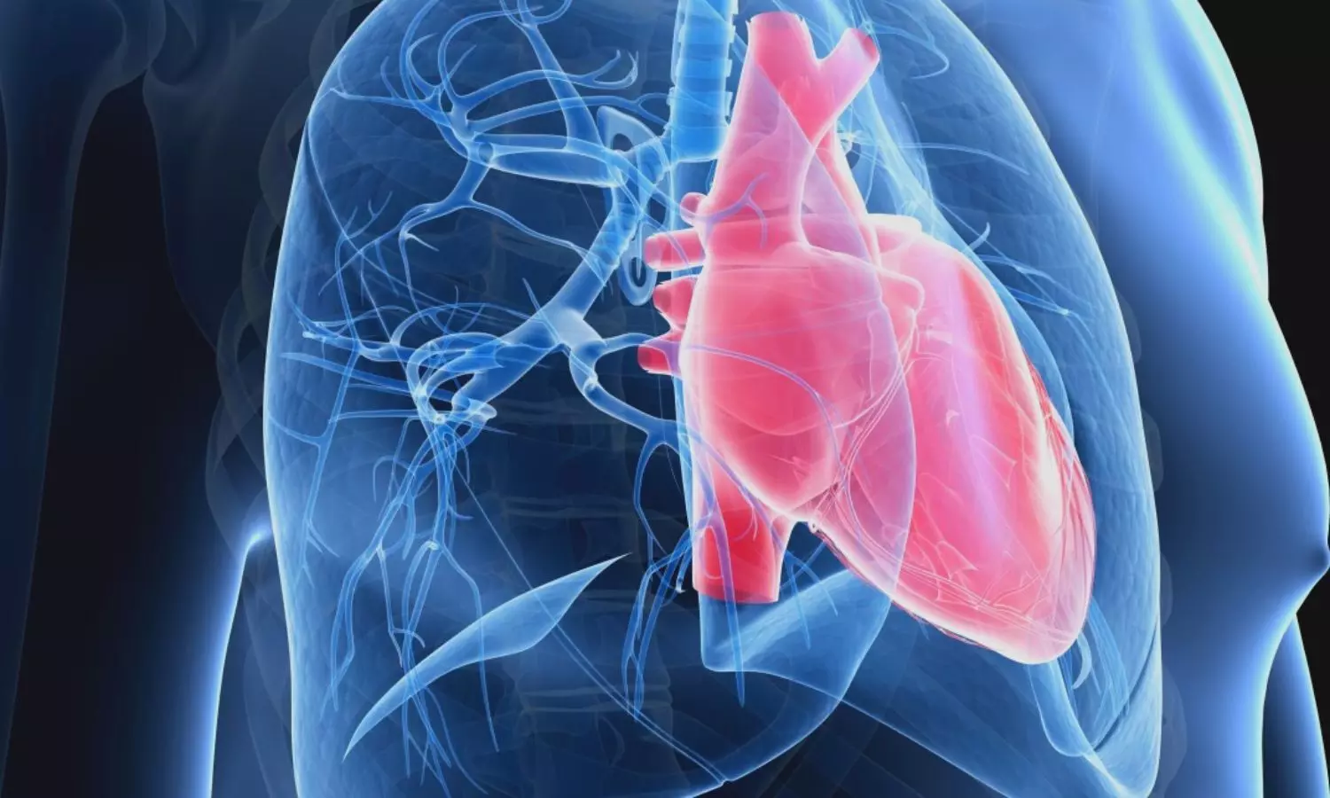
Currently approved medications for the condition aim to open up blood vessels in the lungs, allowing for better blood flow; however, studies on lungs in patients on treatment still show severe occlusive vessel changes. Further, these medications don’t address many of the complex underlying mechanisms that cause the high pulmonary pressures. Even on medication, people with pulmonary arterial hypertension can develop severe dysfunction in the right ventricle of the heart, the part that pumps blood to the lungs and whose function correlates best with survival.

Cedars-Sinai investigators are experimenting with using cardiosphere-derived cells (CDCs) to address some of the biological processes involved in pulmonary arterial hypertension. CDCs were first developed and characterized by Marbán. They have been used in multiple clinical trials for a variety of diseases, most recently, Duchenne muscular dystrophy. These are cells derived from human heart tissue that Marbán and colleagues have discovered reduce inflammation in the body and exert beneficial effects on the immune system.

Called the ALPHA study, this clinical trial was conducted in two phases. In the first, six people with pulmonary arterial hypertension received an infusion of CDCs into their lungs. Three patients received an infusion of 50 million CDCs and the other three received an infusion of 100 million CDCs.

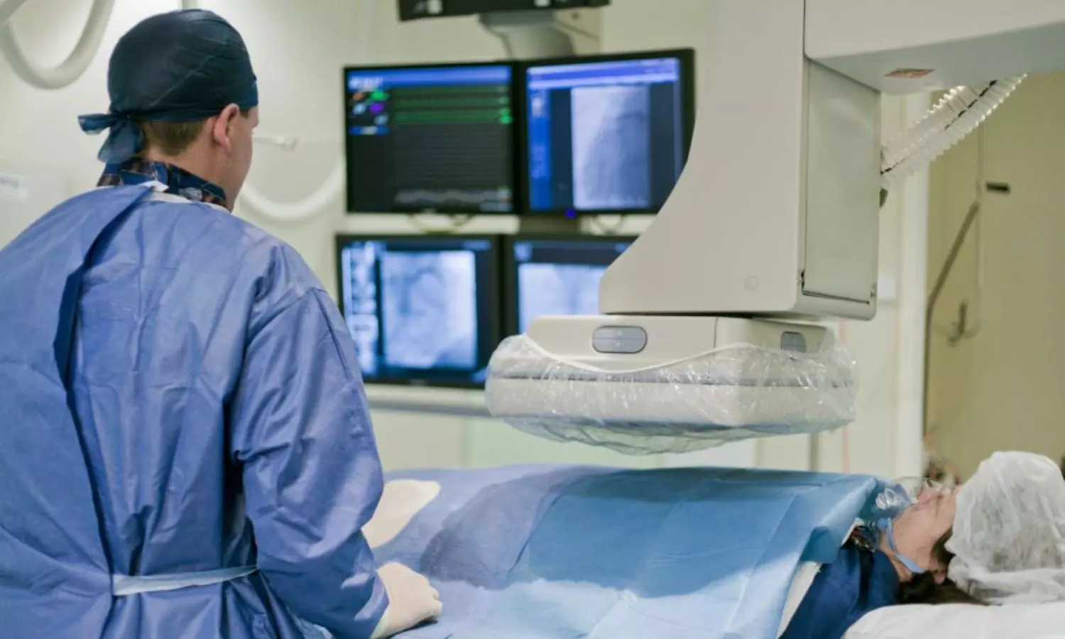
In the second phase, 10 people with pulmonary arterial hypertension were randomized to receive an infusion of 100 million CDCs and 10 people were randomized to receive infusions containing a placebo. Investigators performed right heart catheterization and cardiac MR imaging on each study participant before the infusions and four months after the infusions.

All the participants were on combination pulmonary arterial hypertension-specific medications throughout the course of the study.

The investigators tracked the health of participants for 12 months after the infusions. No adverse effects related to the infusions occurred during this time. Although this study was only designed to assess the safety of the CDC infusions, the investigators observed encouraging changes that might indicate the 16 people who had received the CDC infusions had improved cardiopulmonary health. People who received the infusions, for example, showed improved functioning in the heart’s right ventricle and, at two months post-infusion, were able to walk a greater distance during a six-minute test than people who received placebo.

“The most important takeaway is that this approach is safe and feasible to do in people with pulmonary arterial hypertension,” said Michael I. Lewis, MD, director of Respiratory Care Services at Cedars-Sinai, and first and corresponding author of the study. “These are encouraging exploratory findings that motivate moving on to more advanced studies.”

The investigators plan additional trials to study the effects of repeated infusions of CDCs given to people with pulmonary arterial hypertension.

Reference:

Michael I. Lewis, Shelley Shapiro, Ronald J. Oudiz, Mamoo Nakamura, Dael Geft, Yuri Matusov,The ALPHA phase 1 study: pulmonary ArteriaL hypertension treated with CardiosPHere-Derived allogeneic stem cells, DOI: https://doi.org/10.1016/j.ebiom.2023.104900

Powered by WPeMatico

Smartphone-based technology helps in prevention of suicidal behavior

The use of smartphone technologies could be beneficial in preventing suicide among at-risk adults, says a study conducted by investigators from Italy and Spain, published online in the Journal of Psychiatric Research.

Suicide stands among the primary causes of unnatural deaths globally. According to the WHO mortality database, suicides in the United States saw a 53% increase from 2000 to 2020 (WHO, 2022). Essential for preventing suicide is the implementation of secondary prevention strategies. Digital medicine holds the potential to address numerous mental disorders, making it a promising complement to conventional mental health care. Hence, Alejandro Porras-Segovia and the research team conducted this study to investigate the viability and acceptability of a safety plan integrated into a smartphone application.

Scientists carried out a retrospective pilot analysis to assess the practicality and approval of a safety plan incorporated into a smartphone application (MEmind) among adult patients aged 18 years and older who had recently exhibited suicidal behavior.

The key findings of this study were:

Out of the 128 participants, 82% had the safety plan integrated into their smartphones, with approximately half completing end-of-study surveys.

Over an average follow-up duration of 285 days, participants activated their safety plan an average of 9.1 times, with some opting to memorize it.

Ratings on a scale of 1 to 10 indicated a perceived usefulness of 7.4, usability of 8.9, likelihood to recommend it to others at 8.6, and satisfaction with the overall project at 9.6. Internal coping strategies and personal contact functions were identified as the most beneficial features.

Higher usefulness scores correlated with a greater likelihood of recommending the app to family or friends. Feeling sad or lonely was the most commonly reported warning sign, while walking or exercise was the frequently used coping strategy, with visits to public spaces also being popular.

Overall, participants expressed a desire for direct contact with professionals through the app. The smartphone-based safety plan, as demonstrated in this study, appears to be a practical intervention. The data gathered from this pilot study indicated elevated participation rates and strong acceptance among patients. 

Reference:

Porras-Segovia, A., De Granda-Beltrán, A. M., Gallardo, C., Abascal-Peiró, S., Barrigón, M. L., Artés-Rodríguez, A., López-Castroman, J., Courtet, P., & Baca-García, E. (2023). Smartphone-based safety plan for suicidal crisis: The SmartCrisis 2.0 pilot study. In Journal of Psychiatric Research. Elsevier BV. https://doi.org/10.1016/j.jpsychires.2023.11.039

Powered by WPeMatico

Ocular imaging may provide early information on microvascular damage in PAD patients

Peripheral artery disease (PAD) is a prevalent vascular disorder associated with atherosclerosis and reduced blood flow to the extremities. Affecting 8-10 million Americans and over 200 million people globally, it is a complex condition influenced by various risk factors like diabetes, smoking, hypertension, high cholesterol, and coronary artery disease. PAD leads to structural and functional changes in the limbs, resulting in disability, decreased quality of life, increased mortality, etc.

A recent study published in Vascular Medicine concluded that a more specific biomarker for PAD is necessary due to the poor predictive value of clinical assessment. A noninvasive ophthalmic imaging technique can significantly improve the diagnosis rate without putting the patient at risk for complications. Early detection and treatment of PAD can enhance quality of life and improve clinical outcomes.
This study reviewed the current literature on noninvasive ocular imaging for the PAD diagnosis. The search included Medline, Scopus, Embase, Cochrane, and other databases, with five articles selected. Two studies used retinal colour fundus photography, one used optical coherence tomography (OCT), and two used optical coherence tomography angiography (OCTA) to assess ocular changes in PAD.
Key findings from the study are:
· PAD patients showed both structural and functional changes in the retina.
· Structural alterations were found around PAD patients’ optic disc and temporal retinal vascular arcades.
· Retinal hemorrhages, exudates, and microaneurysms, detected through color fundus photography, were associated with an increased future risk of PAD.
· The RNFL (retinal nerve fibre layer) was thinner in the nasal quadrant of PAD patients compared to age-matched healthy individuals in OCT.
· choroidal thickness in the subfoveal region was thinner in PAD patients controls.
· OCTA revealed a significant reduction in the retinal and choroidal circulation in PAD patients compared to healthy controls.
As PAD causes thinning and ischemic changes in retinal vessels, retinal imaging techniques can provide valuable information about early microvascular damage in PAD. Ocular imaging may serve as a potential biomarker for PAD.
Study limitations include a smaller number of studies, bias, only two studies using OCTA, and none of the studies investigating the association between retinal changes and disease severity.
Reference:
Prem Senthil et al. Role of noninvasive ocular imaging as a biomarker in peripheral artery disease: A systematic review. Vascular Medicine. 2023;0(0).

Powered by WPeMatico