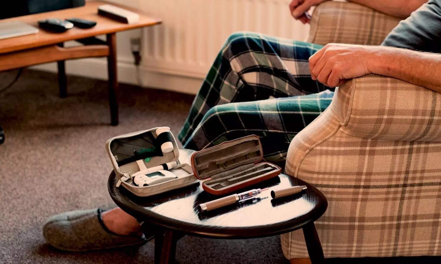
Intensive Food-as-Medicine Program fails to improve Glycemic control in diabetes patients: JAMA

The growing prevalence of food-as-medicine programs necessitates robust evidence to assess their impact on health.

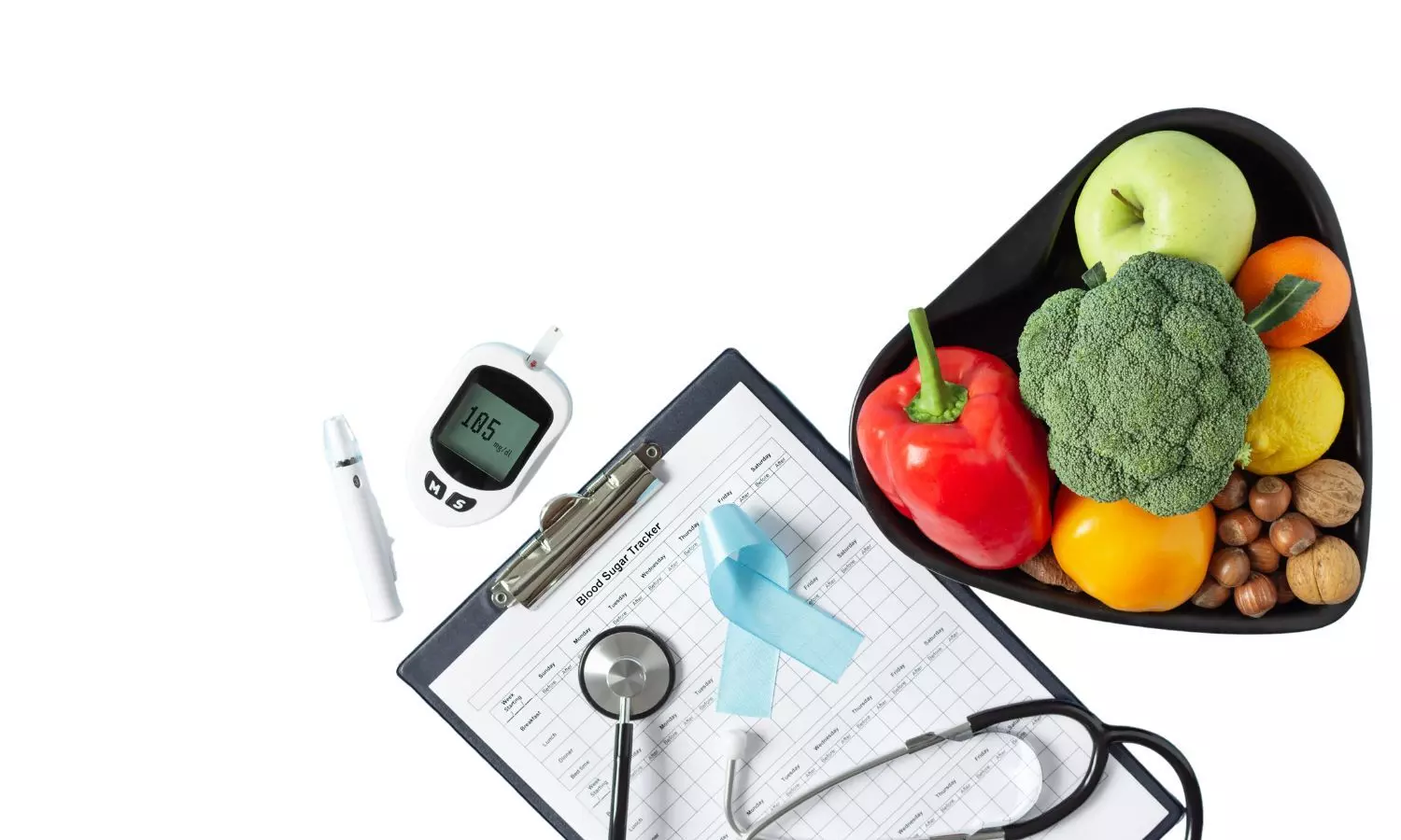
A recent JAMA Internal Medicine concluded that an intensive food-as-medicine program boosts engagement in preventive health care. Still, it didn’t enhance glycemic control compared to standard care among adult participants. Programs for people with high biomarkers need a control group to show effectiveness. More research is required to develop food-as-medicine programs promoting better health outcomes.

This study determined if an intensive food-as-medicine program for diabetes patients and food insecurity can improve glycemic control and affect healthcare use. The study was conducted as a stratified randomized clinical trial with a waitlist design from April 19, 2019, to September 16, 2022. Participants were followed for one year and were randomly assigned to either the treatment group, which participated in the program immediately, or the control group, which waited six months. The trial took place at two sites, one rural and one urban. To be eligible for the study, participants required a T2D diagnosis, HbA1c level of 8% or higher, food insecurity, and residence within the service area of the participating clinics.

The program offered healthy groceries for ten meals/week, plus consultations, evaluations, coaching, and diabetes education for a year. The primary outcome was HbA1c at six months, with secondary outcomes including biometric measures, healthcare use, and self-reported diet/healthy behaviours at 6 and 12 months

The summary of this study includes the following points:

· Of 3712 patients assessed, 3168 were contacted, 1064 were deemed eligible, 500 consented and were randomized, and 465 patients completed the study.

· Of these, 349 patients had laboratory test results at six months. Both treatment and control groups experienced a decline in HbA1c levels, resulting in a non-significant adjusted mean difference of -0.10.

· Access to the program increased preventive health care, including more dietitian visits, active prescription drug orders for metformin and glucagon-like peptide one medication, and participants reporting improved diets.

This clinical trial showed that an intensive food-as-medicine program improved engagement with preventive health care but didn’t affect glycemic control compared to usual care. Control groups are necessary to demonstrate effectiveness in programs targeting individuals with elevated biomarkers. More research is needed to design food-as-medicine programs that improve health.

Reference:

Doyle J et al.Effect of an Intensive Food-as-Medicine Program on Health and Health Care Use: A Randomized Clinical Trial. JAMA Intern Med.

Powered by WPeMatico

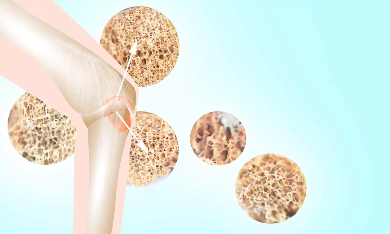
Preoperative carbohydrate intake linked to Success of surgery of Osteoporotic fractures in Elderly

In a groundbreaking study conducted at a prominent Chinese institution, researchers have unveiled a promising connection between preoperative oral carbohydrate (CHO) intake and improved outcomes for elderly patients undergoing surgical treatment for osteoporotic fractures (OPFs). The study addressed the notable gap in the utilization of this practice and concluded that preoperative oral CHO intake may correlate with reduced mortality risk and improved outcomes in elderly patients undergoing surgical treatment for OPFs.

The study results were published in the Journal of Orthopedic Surgery and Research.

Despite the well-established benefits of preoperative CHO intake, its underutilization, especially among older patients facing orthopedic surgery for OPFs, has raised eyebrows. This prompted a comprehensive retrospective cohort study, enrolling 879 elderly patients, with a median age of 71 years, to investigate the potential correlation between preoperative oral CHO consumption and surgical outcomes.

The study, conducted at a single Chinese institution, implemented stringent exclusion criteria and utilized propensity score matching to ensure a balanced analysis. The  exclusion criteria were: 

  1. The necessity for urgent surgical intervention
  2. The existence of hypoglycemia, hyperglycemia, or diabetes mellitus with blood glucose levels lower than 2.8 mmol/L
  3. A medical history of gastrointestinal motility disorders or delayed gastric emptying
  4. The utilization of local anesthesia
  5. A Charlson comorbidity index (CCI) score over 2
  6. An American Society of Anesthesiologists (ASA) score exceeding 3.

 After matching, 264 patients in each cohort were subjected to rigorous evaluation, focusing on primary outcomes such as the 60-day all-cause mortality rate, and secondary outcomes, including the length of hospital stay (LOS), hospitalization costs, blood transfusions, and postoperative complications. 

Key Findings:

  • Post-propensity score matching, the study yielded compelling results.
  • Patients who received preoperative oral CHO intake exhibited a significant negative association with 60-day mortality in the fully adjusted model.
  • Moreover, these individuals experienced a shorter LOS and incurred lower hospitalization costs compared to their counterparts.
  • Notably, no significant associations were found between CHO intake and postoperative nausea and vomiting (PONV) or blood transfusion risks.
  • Equally noteworthy was the absence of reported cases of aspiration in either cohort.

The study’s findings suggest that incorporating preoperative oral CHO intake could be a game-changer in enhancing outcomes for elderly patients undergoing surgical treatment for OPFs. By demonstrating a potential reduction in mortality risk and improved postoperative experiences, the study paves the way for a shift in preoperative care practices.

Limitations:

While the results are promising, the study acknowledges its limitations, including its retrospective nature and the confined scope of participation to a single institution. To solidify and expand upon these findings, the researchers call for future endeavors with larger sample sizes and extended follow-up periods. This would enable a more comprehensive exploration of the long-term impact of preoperative CHO intake and a deeper understanding of the underlying mechanisms at play. 

Further reading: Zhu et al. The association between oral carbohydrate intake before orthopedic surgery for osteoporotic fractures and outcomes in elderly patients. Journal of Orthopaedic Surgery and Research (2023) 18:966. https://doi.org/10.1186/s13018-023-04458-1

Powered by WPeMatico

Rare case of Severe Hypoglycemia in diabetes patient Following Anesthesia Induction: A report

Acute severe hypoglycemia immediately following anesthesia induction is a rare but life-threatening complication that is frequently underdiagnosed due to insufficient awareness

A recent case analysis found acute severe hypoglycemia immediately following anesthesia induction in the perioperative setting. The key observations by Qin Tian and team were published in the Journal of Medicine.

The case report found that after examining the case of an 89 year old man with type 2 diabetes mellitus, he experienced a dramatic drop in blood glucose levels from a baseline of 4.0 mmol/L to a perilous 0.96 mmol/L immediately after the induction of anesthesia. More importantly, the patient displayed normal insulin, C-peptide, and cortisol levels, thereby eliminating other potential causes that lead to hypoglycemia. The temporal association and rapid decline implicated opioids as the likely impact.

After the diagnosis, the patient received a prompt intervention of 40 ml of 50% dextrose intravenously which was followed by a 5% dextrose infusion. Fortunately, recovery from anesthesia was uneventful with no further hypoglycemic episodes during the period of hospitalization.

This case report highlights the often-neglected connection between opioids and severe hypoglycemia during anesthesia. Thereby, the outcomes observed emphasize the necessity for vigilant glucose monitoring in the perioperative period. The implications of this research extend beyond just this one case which points to a broader need for awareness among clinical anesthesiologists. This case report altogether reassures that timely diagnosis can be pivotal in averting life-threatening hypoglycemic events with safer outcomes in the field of anesthesia. Prompted by the life-threatening complications of this rare but underdiagnosed occurrence, this case report underscores the critical need for increased awareness and vigilant glucose monitoring during procedures involving anesthesia.

Source:

Tian, Q., Yi, M. L., Wan, J. L., & Yin, H. (2023). Acute severe hypoglycemia immediately after induction of anesthesia in an elderly patient with type 2 diabetes mellitus: A case report. In Medicine (Vol. 102, Issue 51, p. e36683). Ovid Technologies (Wolters Kluwer Health). https://doi.org/10.1097/md.0000000000036683

Powered by WPeMatico

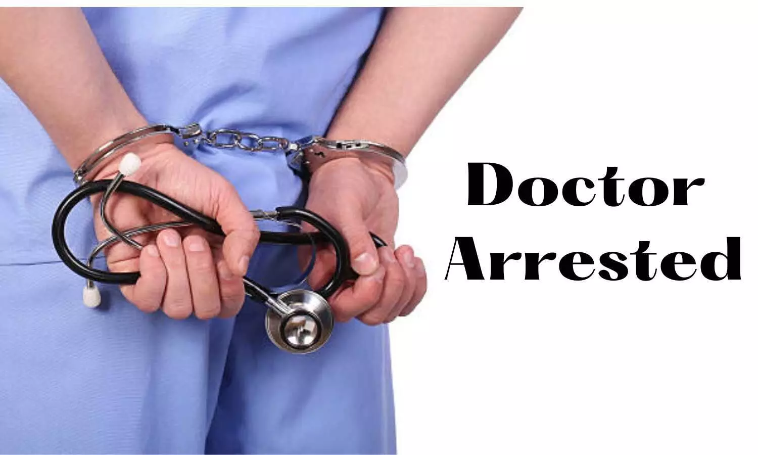
Kolkata doctor arrested for allegedly raping, holding18-year-old nurse captive

Kolkata: In a shocking incident, a doctor has been arrested for allegedly raping, holding a 18-year-old nurse captive for several days at his private nursing home in New Town area in Kolkata.

Subsequently, the nurse filed a complaint at the local police station on December 23, leading to the registration of an FIR under various relevant sections. The doctor was arrested on Sunday after substantial evidence was gathered during the preliminary investigation.

According to the complainant, she had been employed at the nursing home for the past two months. On December 12, the doctor allegedly summoned her to his chamber around noon and subjected her to sexual assault. Disturbingly, she asserted that he forcibly confined her within the nursing home, issuing threats of harm if she revealed the incident. 

Also read- Doctor Arrested For Allegedly Sexually Assaulting Nurse At Delhi Clinic

As per a TOI news report, the nurse alleged that she was raped and held captive for around five days in the doctor’s chamber in the nursing home. In search of an opportunity, the nurse managed to escape on December 17. 

After the alleged assault, she made her way home. It was only later that she disclosed the traumatic experience to her family. Summoning courage, she officially reported the incident on December 23, prompting the authorities to take swift action.

An officer involved in the case remarked, “The nurse alleged that she had been raped by the doctor who owned the nursing home. Following her complaint, we nabbed the accused on Sunday and produced him in Barasat court. The nurse underwent a medical test.”

An investigation into the case is underway. 

Medical dialogues team had earlier reported that a doctor was booked for allegedly sexually assaulting a nurse on the pretext of marriage in Bhopal’s Sukhi Sewania. The nurse was a 40-year-old woman who worked at the doctor’s private hospital when she was subject to sexual assault.

Also read- Bhopal Nurse Files Complaint Alleging Rape By Doctor On Pretext Of Marriage

Powered by WPeMatico

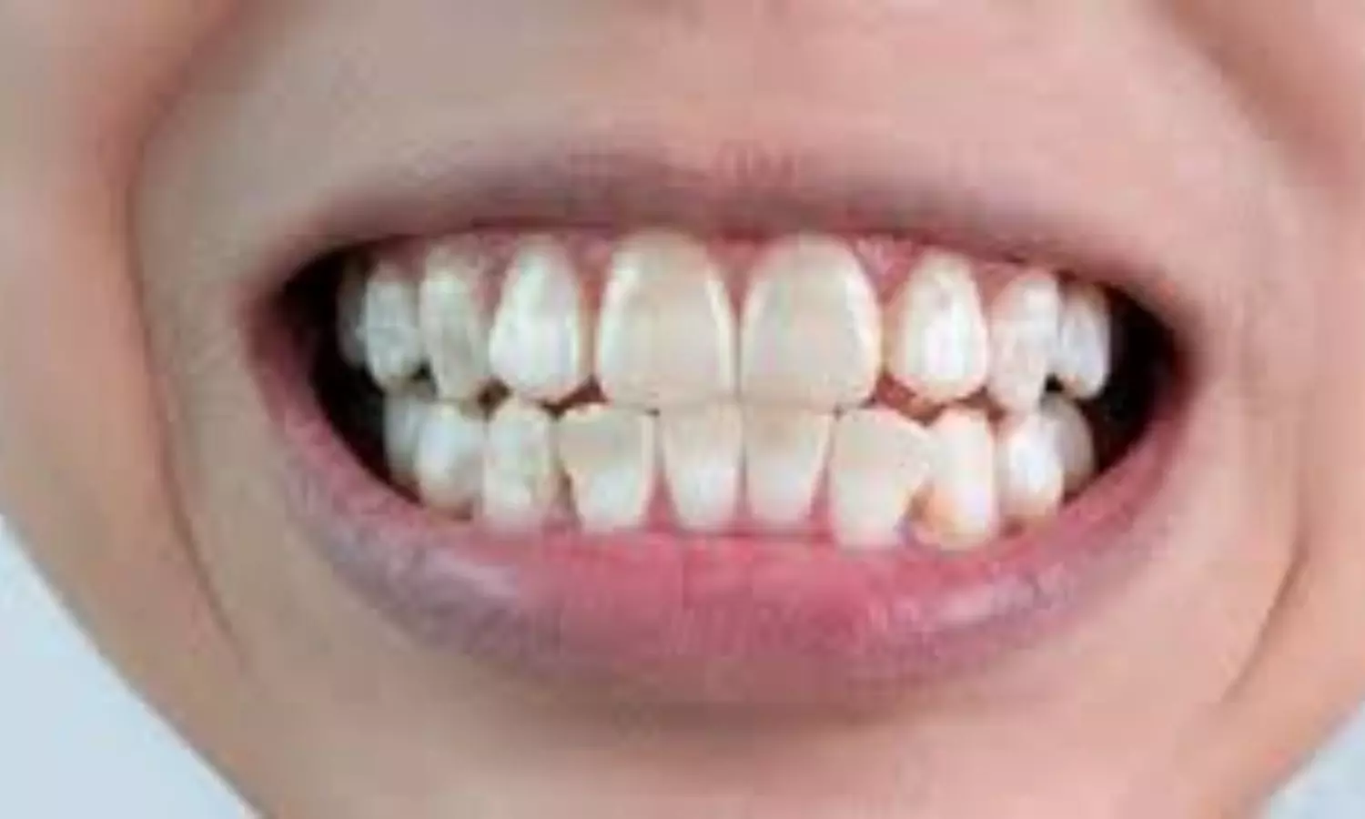
Dehydration affects shade of teeth and may look them whiter

Lebanon: A prospective in vivo study published in Cureus has shed light on the effect of dehydration on tooth colour.

The researchers showed that dehydration impacted the teeth’s shade: lightness increased, and therefore, the teeth appeared whiter.

“Dentists should consider the teeth’s hydration status when evaluating colour for treatments, as there is a significant impact of dehydration on shade matching: the more the dehydration time elapses, there is more increase in the colour difference compared to the baseline,” the researchers wrote.

In dentistry, the teeth’s shade is an essential factor that determines the aesthetics, and the patient’s satisfaction. Therefore, shade matching is a crucial step that reflects the success of the aesthetic restoration outcome. According to several technicians and dentists, this step represents the major challenge. Thus, particular importance must be given to it.

The impact of dehydration on tooth shade constitutes a significant, making it an important area of study, specifically within the field of restorative dentistry. Céline Alamé and Carina Mehanna Zogheib from Saint Joseph University in Beirut, LBN, and colleagues aimed to evaluate the effect of dehydration on the shade of the teeth using a spectrophotometer in a prospective study.

The following null hypotheses were tested: (1) The shade of the teeth does not vary according to their hydration degree, and (2) the ability of shade matching could be conducted at any given time.

For this purpose, the researchers recruited twenty-five participants; the study was conducted between October 2022 and January 2023. The study’s inclusion criteria primarily comprised four intact maxillary incisors. Cavities, damage, restorations, and staining served as exclusion criteria.

The measurements were recorded using the VITA Easyshade Advance spectrophotometer. The measures of L (representing darkness to lightness), a (representing greenness to redness), and b (representing blueness to yellowness) were registered at the centre of each tooth’s labial surface at baseline and then at 10-minute intervals for 30 minutes while the teeth were dehydrating due to rubber dam placement.

Calculation of the colour difference (ΔE) was done using L*a*b* measures, with the perceptibility threshold set at ΔE00=0.8. Statistical analyses were performed using the Bonferroni post hoc test, the Friedman test, and the Wilcoxon signed-rank test.

The study led to the following findings:

· The different colour changes represented by ΔE (ΔE1, ΔE2, and ΔE3) were significantly higher than the perceptibility threshold of 0.8.

· A statistically significant difference was found between ΔE1 and ΔE3.

· A statistically significant difference was found between L0, L10, L20, and L30.

· Statistically significant differences exist between the means of L0 and L20, the means of L0 and L30, the means of L10 and L20, and the means of L10 and L30.

The study concluded that there is a significant impact of teeth dehydration on teeth shade, particularly affecting the perception of whiteness. The dehydration time duration directly corresponds to the extent of colour difference from the baseline. This strongly refutes the initial hypotheses, asserting that teeth shade remains constant irrespective of hydration status and that shade matching can be indiscriminately performed at any time during a dental procedure.

“These findings emphasize the importance of carrying out shade matching before any procedure leading to teeth dehydration, such as rubber dam isolation,” the researchers wrote. “We hope that these insights will guide dental practitioners in augmenting their shade-matching practices, ultimately improving patient satisfaction.”

“Further research would be needed that might focus on identifying ways to reduce the impact of hydration on colour change or developing more adaptive shade-matching techniques that account for teeth hydration levels,” they concluded.

Reference:

Alamé C, Mehanna Zogheib C (November 02, 2023) The Effect of Dehydration on Tooth Color: A Prospective In Vivo Study. Cureus 15(11): e48140. doi:10.7759/cureus.48140

Powered by WPeMatico

Kidney not visualised after Hernia Operation: Hyderabad Hospital doctors slapped Rs 30 lakh compensation for allegedly removing kidney

Hyderabad: The Telangana State Consumer Disputes Redressal Commission recently directed two doctors from a private hospital in Secunderabad to pay Rs 30 lakh compensation to a patient for alleged wrongful removal of a kidney.

It was alleged by the Complainant that his left kidney was removed by them during the hernia operation in 2009. In this regard, the complainant referred to several investigation reports at the later stage mentioning that the left kidney was not visualized properly.

While considering the matter, the State consumer court applied the principle of res ipsa loquitor and noted that the treating hospital represented by its two doctors could not prove that the left kidney was not removed but had shrunk due to atrophy and the same is not visualized and in this regard, they could not submit any evidence also.

The case concerned the complainant who was diagnosed with stones in the bladder in the year 2007. Allegedly, the doctor at the hospital had left the operated portion open in order to ooze out the pus that was formed inside and subsequently, the outer portion was sutured, leaving open the inner portion, and after one month he was discharged.

Following this, in the year 2009, the patient developed hernia due to which he approached Poulomi Hospitals where he was admitted and underwent all the investigations done by the hospital and the ultrasound scan of the whole abdomen was also taken. As per the complainant’s submission, the report mentioned that both the kidneys of the complainant were normal in contour and echo texture, the size of the right kidney was also noted as 96 x 48 mm; left kidney size: 117 x 60 mm.

Consequently, on 20.07.2009, the doctors at the hospital performed the surgery for Hernia and on 31.07.2009, the patient was discharged from the hospital. However, the patient was in regular touch with the Urologist Dr Madhekar who had been prescribing the medicines regularly. Due to the ill health of the complainant, he was asked to rest for several days and was further advised not to take physical strain.

Meanwhile, his health condition was deteriorating and therefore in 2011, the complainant went to Calcutta to visit his father-in-law on 10.10.2011. However, he developed severe stomach pain and approached Dr. Sarkar at Arogya Maternity & Nursing Home. He was admitted to the hospital on 21.11.2011 and he was operated for hernia and a mesh was put in order to protect him from further pain, and during his stay, there were several investigations and scans done and the doctor had informed that his “left kidney was not well visualised”. 

At that time, the complainant allegedly realised that the treating hospital while conducting the hernia operation in 2009 had fraudulently in collusion with each other had removed his kidney without his knowledge, information and consent and the matter came to light only in 2011 when the doctor in Calcutta had informed him.

On 16.05.2022, the patient again developed severe stomach pain and thereafter he approached Kothagudem Area Hospital, who referred the patient to Medicare Diagnostic Centre for medical test, USG abdomen and pelvis. The concerned scanning report also revealed that the left kidney was not visualised. Again on 13.06.2012, the complainant visited Mamata Medical College Hospital, Khammam and the scan report there also revealed that the left kidney was not visible.

The Complainant alleged that he has been suffering since then due to fraudulent acts of the doctors at the treating hospital, who had fraudulently removed his left kidney.

Due to such culpable act of the opposite party, the complainant approached the AP State Human Rights Commission and registered a case which was referred to Kushaiguda Police Station, Cyberbad, who conducted investigations and inquiry and opined that the act committed by the doctors at the treating hospital was a deficiency of service and advised the complainant to approach Consumer Court and submitted the said report dated 4.2.2015.

It was alleged by the complainant that due to such fraudulent and deficient acts of the hospital doctors, the complainant had been suffering continuously since 2009. Due to this, the precious life of the complainant had been ruined, his life span was reduced and he developed several malfunctioning, his urinary system had also totally crippled his life due to which his marital status has also been affected and he has no children till date.

Approaching the consumer court bench, the complainant demanded a compensation of Rs 50 lakh with interest @ 24% p.a., from the date of complaint till the realization.

On the other hand, the doctors at the hospital pointed out that before coming to their hospital, the patient underwent treatment at Gandhi Hospital and underwent multiple surgeries. They also informed the consumer court bench that non-visualisation of the kidney does not necessarily mean that the kidney was removed. It was submitted that in case of atrophy of kidney, there is a possibility of non visualization of kidney and that there is no nexus between the surgery performed in 2009 and the alleged pain the complainant is suffering from.

Further, they pointed out that on the basis of the complaint made before the Human Rights Commission, the matter was referred to Kushaiguda police station and after conducting the inquiry, the concerned police found that there was no substance in the allegations made by the complainant.

It was further argued that to receive and kidney for transplantation, huge process and regulations are involved under the Special Act called “Organ Donation Act” and the donor of such kidney should be in a healthy condition whereas in the instant case, the complainant himself is suffering from stones in his kidneys and had underwent several surgeries for the same. Therefore, the question of removing the kidney does not arise.

Further, it was pointed out that to transplant the kidney from the donee to the donor both must be closely related and shall be in good health. The doctors further pleaded that after performing the PCNL for renal calculus and Hernia removal on 20.07.2009, an ultra scan was done on 28.07.2023 and the report categorically showed the presence of the left kidney admeasuring 123×63 mm and the same was allegedly shown to the complainant before his discharge.

It was further argued that since the complainant had not paid any consideration and availed the services under the Arogyasree Scheme, he does not fall into the category of Consumer and therefore, the complaint is not maintainable.

Referring to this contention, the consumer court noted that “It is a fact borne by record that the complainant has not paid any consideration to the opposite parties but the Government has paid the consideration on behalf of the complainant hence it implies that the complainant has paid the valid consideration and had availed the services of opposite party as a beneficiary as such he falls into the definition of ‘consumer’.”

While considering the allegations that the left kidney of the patient was fraudulently removed, the Consumer Court noted that when the complainant visited the hospital for treatment of hernia in 2009, both his kidneys were normal and the ultrasound scan dated 09.07.2009 revealed the same.

The Commission noted that the complainant’s case was based on the whole abdomen report dated 18.11.2011 of Medvue Medical Services, Kolkata and the USG Abdomen and Pelvis report dated 16.05.2012 of Medicare Diagnostics, Kothagudem, Ultra Sonography of Abdomen/Pelvis report dated 13.06.2012 of Mamata General & Super Speciality Hospital, Khammam and USG Abdomen and Pelvis report dated 18.07.2014 of Medicare Diagnostics, Kothagudem.

In all these reports, it was reported that the “left kidney not visualised”. Therefore, based on these documents, the complainant stated that when he visited the hospital, both his kidneys were normal and the doctors at the hospital while performing the Hernia operation had fraudulently removed his left kidney.

The Commission noted that the hospital doctors appeared and filed the written version but they failed to file any evidence affidavit nor any documents and had contended in the written version that in case of Atrophy of kidney, the kidneys will not be visible.

“In a medical negligence case, when negligence is alleged and the complainant has supported his allegations with documentary evidence, the burden shifts on the opposite party to prove that they were not wrong and have rendered their services diligently, but in the instant case the opposite party did not choose to file any evidence nor had put any efforts to prove that the kidney was not visualised due to atrophy,” noted the Commission.

Further, it noted that the doctors at the treating hospital had specifically stated that at the time of discharge from the hospital and ultrasound was done on 28..07.2009 to study the left lumbar region post-operation and the left kidney was well visible in the said report with a measurement of 123×62 mm. However, the Commission noted that “no such report is filed before this Commission”.

Observing that the Commission is not medical expert to analyse and conclude that in case of Atrophy, the kidney will shrink to the extent that it becomes invisible, the Commission noted that it was for the doctors at the treating hospital to furnish such proofs.

“A mere statement will not suffice, the same has to be established and proved with cogent evidence, the opposite party failed to substantiate their contention with sufficient evidence or medical literature. In the absence of the same an adverse inference can be drawn against the opposite parties that they might have certainly played some mischief with the complainant at the time of surgery for hernia in the year 2009,” mentioned the consumer court.

Meanwhile, the doctors at the hospital remained silent and to come to a truthful conclusion, the Commission sent the complainant for examination to the Gandhi Govt. Hospital at Secunderabad and instructed to examine the complainant and to report whether the left kidney of the complainant is visible or is removed by surgical intervention.

Accordingly, the Gandhi Hospital conducted the examination and sent a report on 19.07.2023. However, the Commission noted that the report was not conclusive in nature, because it was not supported with any pathology report nor the films of MRI etc. At one point the report mentioned that the left kidney “Not visualized” and at the same time it also mentioned that the left kidney has a calculus in it.

Therefore, the Commission held that the report by Gandhi Hospital cannot be relied upon. Meanwhile, the Commission noted that the doctors of the treating hospital, despite being aware of the fact that the complainant was sent for examination to Gandhi Hospital, did not choose to come forward and give any explanation.

At this outset, the Commission referred to the Supreme Court order in the case of CPL Ashish Kumar Chauhan Vs Commanding Officer and others, where the Apex Court relied on its earlier decisions invoked the principle of Res Ipsa loquitor. 

Applying the same principle, the Commission noted that the Complainant by producing the documents substantiated that his left kidney was not visualized and alleged that the same was removed by the doctors at the treating hospital without his consent during the surgery for hernia.

“These documents speak for themselves, in view of these documents the burden shifts on the opposite party to prove that the left kidney was not removed but had shrunk due to atrophy and the same is not visualized but no such evidence is forth coming from the opposite parties,” noted the Commission.

Holding them negligent, the Commission held,

“Therefore based on the above discussion and failure on the part of the opposite party to produce the crucial document ‘ultra scan’ dated 28.07.2009 and the silence of the opposite party in not coming forward with any sort of explanation supported with evidence and medical literature, all these acts and avoiding and escaping behaviour of opposite parties leads us to irresistibly conclude that the opposite parties under the guise of Hernia operation had illegally removed the left kidney of the complainant, the opposite party had taken undue advantage of the ignorance of the complainant and deceived him, thereby not only causing organ loss to the complainant but the complainant is forced to live with the fear, frustration and disappointment.”

While considering the issue of compensation, the Commission observed that “…strictly speaking the act committed by the opposite parties are criminal in nature.”

It noted that no amount of compensation will restore the loss suffered by the complainant,

“Since due to the act of the opposite parties, the life expectancy of the complainant has come down and he is forced to live with one kidney, what will the fate of the complainant be if unfortunately his right kidney fails.”

Therefore, the Commission ordered Poulomi Hospitals represented by Urologist Dr. Madhekar and another Dr. Behara to jointly and severally pay Rs 30 lakh as compensation and another Rs 25,000 as costs.

To read the order, click on the link below:

https://medicaldialogues.in/pdf_upload/telangana-state-commission-rs-30-lakh-compensation-228797.pdf

Also Read: Death after Anaesthesia administration during implant removal :Bengaluru Hospital, Doctors to Pay Rs 38 Lakh Compensation
Powered by WPeMatico

Owning pets helps to cope with loneliness and cognitive decline

In the past few decades, the number of individuals living alone has shown an upward trend. In 2021, the proportion of single-person households in the United Kingdom (UK) and the US reached 29.4% and 28.5%, respectively.

Loneliness is a potential mediator in the association of living alone with dementia among older adults. Contrary to living alone,10 pet ownership (eg, raising dogs and cats) is related to reduced loneliness.

To explore the association of pet ownership with cognitive decline, the interaction between pet ownership and living alone, and the extent to which pet ownership mitigates the association between living alone and cognitive decline in older adults.

A recent study in JAMA Network suggests that pet ownership can be associated with slower cognitive decline among older adults living alone. Slower rates of decline in verbal memory and verbal fluency among individuals living alone, but not among those living with others. Pet ownership offset the association between living alone and declining rates of verbal memory and verbal fluency.

Researchers designed a cohort study used data from waves 5 (June 2010 to July 2011) to 9 (from June 2018 to July 2019) in the English Longitudinal Study of Ageing. Participants included adults 50 years and older. Data were analyzed from April 1 to June 30, 2023. Pet ownership and living alone in wave 5. In waves 5 to 9, verbal memory and verbal fluency were assessed, and composite verbal cognition was further calculated.

The key findings of the study are

  • A total of 7945 participants included, the mean (SD) age was 66.3 (8.8) years, and 4446 (56.0%) were women. Pet ownership was associated with slower rates of decline in composite verbal cognition (β = 0.008 [95% CI, 0.002-0.014] SD/y), verbal memory (β = 0.006 [95% CI, 0.001-0.012] SD/y), and verbal fluency (β = 0.007 [95% CI, 0.001-0.013] SD/y).
  • Three-way interaction tests showed that living alone was a significant modifier in all 3 associations. Stratified analyses showed that pet ownership was associated with slower rates of decline in composite verbal cognition (β = 0.023 [95% CI, 0.011-0.035] SD/y), verbal memory (β = 0.021 [95% CI, 0.008-0.034] SD/y), and verbal fluency (β = 0.018 [95% CI, 0.005-0.030] SD/y) among individuals living alone, but not among those living with others.
  • Joint association analyses showed no significant difference in rates of decline in composite verbal cognition, verbal memory, or verbal fluency between pet owners living alone and pet owners living with others.

Researchers concluded that ” In this cohort study, pet ownership was associated with slower rates of decline in verbal memory and verbal fluency among older adults living alone, but not among those living with others, and pet ownership offset the associations between living alone and declining rates in verbal memory and verbal fluency. Further studies are needed to assess whether pet ownership slows the rate of cognitive decline in older adults living alone.”

Reference: Li Y, Wang W, Zhu L, et al. Pet Ownership, Living Alone, and Cognitive Decline Among Adults 50 Years and Older. JAMA Netw Open. 2023;6(12):e2349241. doi:10.1001/jamanetworkopen.2023.49241.

Powered by WPeMatico

Risk of young-onset dementia could be reduced by targeting health and lifestyle factors

Researchers have identified a wide range of risk factors for young-onset dementia. The findings challenge the notion that genetics are the sole cause of the condition, laying the groundwork for new prevention strategies.

The largescale study identified 15 risk factors, which are similar to those for late-onset dementia. For the first time, they indicate that it may be possible to reduce the risk of young-onset dementia by targeting health and lifestyle factors.

Relatively little research has been done on young-onset dementia, though globally there are around 370,000 new cases of young-onset dementia each year.

Published in JAMA Neurology, the new research by the University of Exeter and Maastricht University followed more than 350,000 participants younger than 65 across the United Kingdom from the UK Biobank study. The team evaluated a broad array of risk factors ranging from genetic predispositions to lifestyle and environmental influences. The study revealed that lower formal education, lower socioeconomic status, genetic variation, lifestyle factors such as alcohol use disorder and social isolation, and health issues including vitamin D deficiency, depression, stroke, hearing impairment and heart disease significantly elevate risk of young-onset dementia

Professor David Llewellyn of the University of Exeter emphasized the importance of the findings: “This breakthrough study illustrates the crucial role of international collaboration and big data in advancing our understanding of dementia. There’s still much to learn in our ongoing mission to prevent, identify, and treat dementia in all its forms in a more targeted way. This is the largest and most robust study of its kind ever conducted. Excitingly, for the first time it reveals that we may be able to take action to reduce risk of this debilitating condition, through targeting a range of different factors.

Dr Stevie Hendriks, Researcher at Maastricht University, said: “Young-onset dementia has a very serious impact, because the people affected usually still have a job, children, and a busy life. The cause is often assumed to be genetic, but for many people we don’t actually know exactly what the cause is. This is why we also wanted to investigate other risk factors in this study.”

Sebastian Köhler, Professor of Neuroepidemiology at Maastricht University, said: “We already knew from research on people who develop dementia at older age that there are a series of modifiable risk factors. In addition to physical factors, mental health also plays an important role, including avoiding chronic stress, loneliness and depression. The fact that this is also evident in young-onset dementia came as a surprise to me, and it may offer opportunities to reduce risk in this group too.”

Dr Janice Ranson, Senior Research Fellow at the University of Exeter, said: “Our research breaks new ground in identifying that the risk of young-onset dementia can be reduced. We think this could herald a new era in interventions to reduce new cases of this condition.”

Dr Leah Mursaleen, Head of Clinical Research at Alzheimer’s Research UK, which co-funded the study, said: “We’re witnessing a transformation in understanding of dementia risk and, potentially, how to reduce it on both an individual and societal level. In recent years, there’s been a growing consensus that dementia is linked to 12 specific modifiable risk factors such as smoking, blood pressure and hearing loss . It’s now accepted that up to four in 10 dementia cases worldwide are linked to these factors.

“This pioneering study shines important and much-needed light on factors that can influence the risk of young-onset dementia. This starts to fill in an important gap in our knowledge. It will be important to build on these findings in broader studies.’

Reference:

Hendriks S, Ranson JM, Peetoom K, et al. Risk Factors for Young-Onset Dementia in the UK Biobank. JAMA Neurol. Published online December 26, 2023. doi:10.1001/jamaneurol.2023.4929.

Powered by WPeMatico

High-sensitivity troponin HEART pathway allows earlier detection of acute myocardial infarction: JAMA

USA: A recent cohort study published in JAMA Network Open has suggested that the high-sensitivity troponin HEART pathway may allow earlier detection of acute myocardial infarction (AMI) and improve resource use.

The multicenter cohort study involved 17 384 emergency department (ED) patients with chest pain risk stratified with a HEART pathway incorporating conventional vs high-sensitivity (hs) troponin levels. The study revealed that the high-sensitivity troponin group demonstrated a higher rate of AMI diagnosis during the ED visit and a lower AMI diagnosis rate within 30 days after the visit, along with lower rates of revascularization, stress testing, and admission. Mortality was rare and similar across groups.

Patients presenting to the EDs with chest pain are routinely risk-stratified for major adverse cardiac events (MACEs) using the HEART (History, Electrocardiogram, Age, Risk factors, and Troponin) score pathway. The pathway incorporates risk factors, clinical features, initial serum troponin testing, and electrocardiography findings hs-troponin levels are suggested to improve risk stratification among patients with possible acute myocardial infarction.

Mackensie Yore, Veterans Affairs/University of California Los Angeles National Clinician Scholars Program, Los Angeles, and colleagues aimed to compare resource use and health outcomes among emergency department patients undergoing cardiac risk stratification with a HEART pathway using conventional vs high-sensitivity serum troponin.

The study was conducted at 16 Kaiser Permanente Southern California hospitals during hs-serum troponin assay uptake. It included 17 384 adult patients who presented to an emergency department with chest pain and were risk-stratified with a HEART pathway based on high-sensitivity troponin or conventional troponin.

The study’s main outcome was AMI detection in the ED and within 30 days.

The study led to the following findings:

  • Of the 17 384 patients (median age, 58 years; 56.2% were women), 12 440 were risk stratified with a HEART pathway based on conventional troponin, and 4944 were risk stratified with a HEART pathway based on high-sensitivity troponin.
  • Detection of AMI within 30 days was higher for the high-sensitivity troponin group than the conventional troponin group (5.8% versus 4.4%), while the 30-day all-cause mortality rate was unchanged (0.3% versus 0.4%).
  • In the emergency department, 4.6% of patients in the high-sensitivity troponin group received a diagnosis of AMI compared with 2.0% in the conventional troponin group.
  • Among those who did not receive a diagnosis of AMI in the emergency department, an additional 1.2% of patients in the high-sensitivity troponin group and 2.4% in the conventional troponin group received a diagnosis within 30 days.
  • Patients in the high-sensitivity troponin group had lower rates of healthcare use compared with the conventional troponin group, including admission (12.2% versus 15.0%), stress testing within 7 days (10.2% versus 12.8%), and coronary revascularization within 30 days (1.0% vs 2.0%).

“The findings indicate that the hsTn HEART pathway was linked with higher rates of ED AMI diagnoses and lower rates of AMI diagnoses after the index ED visit and within 30 days,” the researchers wrote.

The team added, “We also found less health service use for the hsTn group compared with the cTn group.”

“A high-sensitivity troponin algorithm may improve the ED evaluation of AMI, both catching AMI earlier and mitigating advanced testing and unnecessary admission,” they concluded.

Reference:

Yore M, Sharp A, Wu Y, et al. Emergency Department Cardiac Risk Stratification With High-Sensitivity vs Conventional Troponin HEART Pathway. JAMA Netw Open. 2023;6(12):e2348351. doi:10.1001/jamanetworkopen.2023.48351

Powered by WPeMatico

JIPMER Announces Conduction Of 1st Year Orientation Program For MD, MS, DM, MCH, MDS students

Puducherry: Jawaharlal Institute of Postgraduate Medical Education & Research (JIPMER) has announced the conduction of 1st year postgraduate orientation program for MD, MS, DM, MCH, and MDS courses for the January 2024 batch. 

The postgraduate orientation program for the first-year postgraduate degree students admitted in the January 2024 session has been scheduled for 2nd January and 5th – 6th February 2024 in the NTTC Hall-1, JIPMER Academic Centre, JIPMER. Registration for the Programme will start sharp at 8.30 am on 2nd January 2024.

All the Heads of Departments are requested to circulate this among the first-year MD, MS, DM, MCh & MDS students who have joined in the first and second round of seat allotment and permit them to attend the program. The Heads should not post them on night duty before and during the program days and inform all unit heads to relieve the students on time, which will enable them to attend all sessions on time.

The guide and PG dissertation topic details of the students as per the format given below to nttc.jipmer@gmail.com on or before 02.02.2024 need to be sent.

Sl. No.

Name of the resident

Name of the research guide

Proposed area of research/ topic (Optional)

The students are to come with a fully charged laptop, pen drive, and a hard copy of the PGRMC (Word Doc) form, which can be downloaded from the link mentioned in the notice below.

The Jawaharlal Institute of Postgraduate Medical Education & Research is a medical school located in Pondicherry. It is an institute of national importance (INI) and a tertiary care referral hospital. It is under the direct administrative control of the Ministry of Health and Family Welfare and the Indian Government, with autonomy to run its internal administration. The courses include undergraduate, postgraduate, super specialty, fellowships, PhD programs, post-basic diplomas, and certified courses.

To view the notice, click on the link below –

https://medicaldialogues.in/pdf_upload/circular-i-yr-pgo-for-md-ms-dm-mch-mds-jan-2024-batch-228823.pdf

Powered by WPeMatico