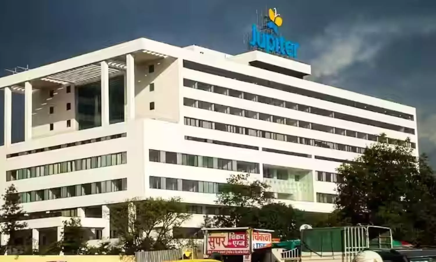
Jupiter Hospital Pune performs path-breaking bloodless living donor liver transplant on 52-year-old man

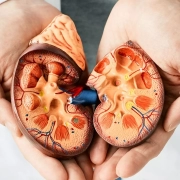
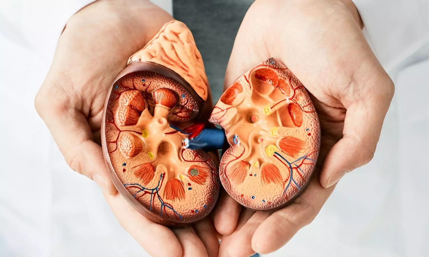
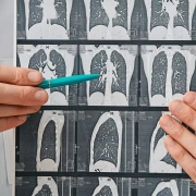
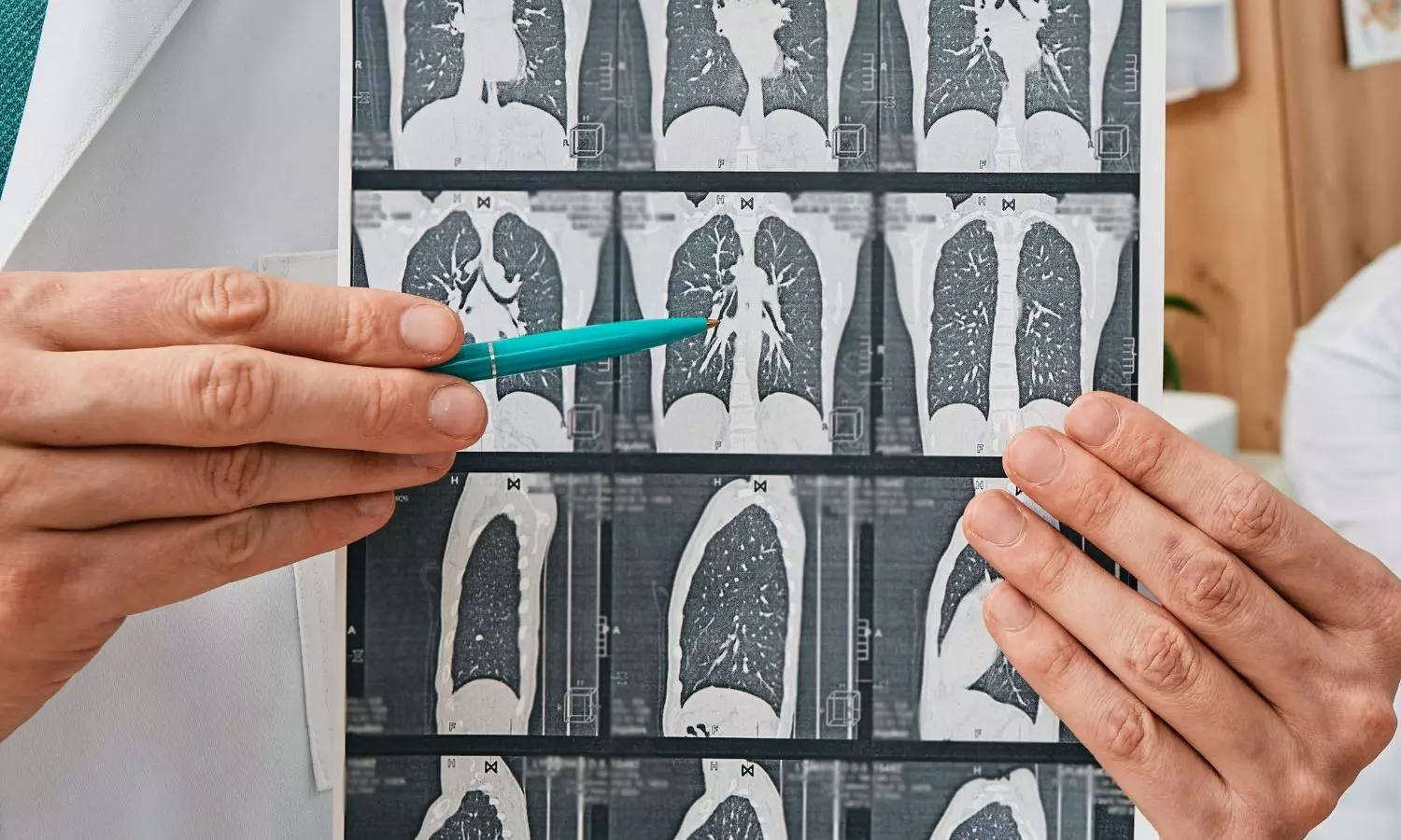
Pune: Living donor liver transplantation (LDLT) is considered one of the most intricate and demanding surgical procedures known to medical science. Traditionally, the procedure has been associated with substantial blood loss and the necessity for extensive blood and blood product transfusions.

However, in a significant medical breakthrough Jupiter Hospital, Pune successfully performed a ‘living donor liver transplantation’ without the need for any blood or blood product transfusion. 

The procedure became necessary when a 52-year-old man, diagnosed with end-stage liver disease, needed a living donor liver transplantation. His wife had donated a portion of her liver to save her husband’s life.

Also Read:Pune hospital performs live donor liver transplant without blood transfusion

Dr. Abhishek Yadav, Director HPB Surgery and Liver Transplant Surgeon, Jupiter Hospital, shedding light on the process said, “Living donor liver transplantation involves the removal of a portion of a healthy donor’s liver, which then regenerates in both the donor and the recipient. While this procedure has revolutionized the treatment of end-stage liver diseases, it has traditionally been associated with challenges such as significant blood loss and the subsequent need for blood transfusions.”

The breakthrough at Jupiter Hospital addresses these challenges and marks a transformative moment in the field.

Unlike conventional liver transplant procedures, the liver transplant team at Jupiter Hospital conducted the entire operation without resorting to blood transfusion. This successful operation showcased the exceptional skill and experience of the hospital’s liver transplant team, which includes proficient surgeons, Hepatologists, liver Anesthetists, and intensivists.

The success of this bloodless living donor liver transplantation underscores the pivotal role of a highly experienced medical team. Jupiter Hospital proudly has a very seasoned liver transplant team, which repeatedly navigates technical challenges with precision and expertise. The reduced reliance on blood transfusion aligns with improved patient outcomes and sets a new standard in liver transplant surgeries.

Following the 12-hour surgery, the patient emerged from the operation theatre with a smile. Remarkably, the patient did not require ventilation post-surgery, highlighting the hospital’s approach of minimizing the postoperative burden on patients.

Dr. Abhishek Yadav, Director HPB Surgery and Liver Transplant Surgeon, member of the liver transplant team at Jupiter Hospital, expressed his views on procedure. Says, “The successful completion of a living donor liver transplantation without the need for blood transfusion is a significant milestone in the field of transplant surgery. It is a result of the collaborative effort and expertise of our team. This breakthrough not only demonstrates the technical prowess of our surgeons but also emphasizes our commitment to advancing patient care standards.”

The patient’s experience, emerging from a 12-hour surgery without the need for ventilation, emphasizes the hospital’s commitment to patient-centred care. Beyond the medical achievement, the emphasis on a positive patient experience speaks about the holistic approach adopted by Jupiter Hospital, aiming for not only successful surgeries but also optimal postoperative outcomes.

This procedure also underscores a conscious effort to minimize the reliance on blood transfusions. This approach aligns with the hospital’s commitment to patient safety, reducing the risks associated with transfusions and promoting a smoother recovery process. It also marks a significant advancement in the overall safety and success rates of living donor liver transplantations.

Powered by WPeMatico

Resorbable scaffolds fare better than balloon angioplasty for infra-popliteal artery disease :NEJM

In patients experiencing chronic limb-threatening ischemia (CLTI) coupled with infrapopliteal artery disease, the utilization of angioplasty has been linked to frequent reinterventions and unfavorable limb outcomes due to restenosis. Presenting findings from the LIFE-BTK trial, researchers have demonstrated that employing an everolimus-eluting resorbable scaffold proves superior to angioplasty in achieving positive clinical outcomes, such as freedom from amputation or reintervention within a year. The results were recently published in NEJM.

Endovascular therapy applied to arterial vessels below the knees is known to have inherent limitations, including issues such as elastic recoil, dissection, and restenosis, which compromise the long-term success of the procedure. Atherosclerotic plaque below the knees typically contains higher levels of calcium and luminal thrombus compared to lesions above the knees, potentially influencing the response to therapies more commonly applied in treating upper limb arteries.

Varcoe et al conducted a rigorous single-blind, randomized, controlled trial (LIFE-BTK) to assess the safety and efficacy of a novel everolimus-eluting resorbable scaffold (Esprit BTK, Abbott Vascular) for treating infrapopliteal artery disease in CLTI patients.

During the trial, individuals with CLTI and infrapopliteal artery disease were randomly assigned in a 2:1 ratio to receive either the everolimus-eluting resorbable scaffold or balloon angioplasty. A pivotal eligibility criterion was Rutherford–Becker class 4 or 5 disease, excluding class 6. Additionally, arterial stenosis severity had to be at least 70% of the vessel diameter upon visual assessment.

The primary efficacy endpoint was freedom from specific events at the one-year mark, including amputation above the ankle, complete occlusion of the target vessel, clinically driven revascularization of the target lesion, and binary restenosis of the target lesion. Meanwhile, the primary safety endpoint was freedom from major adverse limb events at six months and perioperative death.

Following a median follow-up period of 390 days, the primary efficacy endpoint (i.e., no events occurred) was observed in 74% of the scaffold group and 44% of the angioplasty group. Subgroup analyses and assessments of other arterial patency endpoints consistently favored the scaffold treatment, serving as promising indicators of success for the evaluated therapy.

“Focus on clinical endpoints”

In an accompanying editorial, Joshua A. Beckman, M.D. has stressed that “…what truly marks this trial as important is not only the arterial patency data but also the clinical end points. The inclusion of clinical end points is less common in studies of new devices for the treatment of vascular disease of the leg than in studies of medications.”

The recently published literature supporting interventional management of peripheral artery disease includes PROMISE II and EMINENET trials. As the field moves forward with new treatments, the incorporation of clinical outcomes should be standard. Only then will clinicians be able to understand the true value of new tools and treatments.

Source: NEJM:

1. DOI: 10.1056/NEJMoa2305637

2. DOI: 10.1056/NEJMe2312167

Powered by WPeMatico

Residual Probing Depth and Periodontitis Grading Good Predictors of Tooth Loss Due to Periodontitis

Residual Probing Depth and Periodontitis Grading Good Predictors of Tooth Loss Due to Periodontitis suggests a new study published in the Journal of Periodontology.

Individuals enrolled in supportive periodontal therapy (SPT) can still present with tooth loss due to periodontitis (TLP). There is limited evidence on the influence of residual pockets (RPc) and a defined “threshold” at which a patient’s profile is set to be at high risk for tooth loss due to periodontitis in the literature. Therefore, this study aimed to assess the influence of residual pockets on tooth loss due to periodontitis and determine the predictive performance of residual pockets compared to the staging and grading of periodontitis on tooth loss due to periodontitis risk. Clinical data from 168 patients (3869 teeth) treated for periodontitis and receiving supportive periodontal therapy for at least 10 years were evaluated in this retrospective study. Tooth loss due to periodontitis and the percentage of sites with residual pockets ≥ 5 mm or ≥6 mm per patient were collected. The prognostic performance of residual pockets was compared to the staging and grading of the disease on tooth loss due to periodontitis using a multilevel Cox proportional hazard regression model. Results: Over a median follow-up of 25 years, 13.7% of teeth were lost, 4.6% of which were due to periodontitis. Most patients with tooth loss due to periodontitis had ≥1 site with residual pockets ≥5 mm (90.8%) or ≥6 mm (77.6%). Multivariate multilevel Cox regression revealed that patients with >15% of sites with residual pockets ≥5 mm had a hazard ratio of 2.34, and grade C had a hazard ratio of 4.6 for tooth loss due to periodontitis compared to residual pockets ≤4 mm/grade A. Grading exhibited the best discrimination and model fit. Patients with residual pockets ≥5 mm at >15% of the sites are at risk for tooth loss. Grading and residual pockets ≥5 mm displayed excellent predictive capability of tooth loss due to periodontitis.

Reference: Saleh MHA, Dias DR, Mandil O, et al. Influence of residual pockets on periodontal tooth loss: a retrospective analysis. J Periodontol. 2023; 1-12. https://doi.org/10.1002/JPER.23-0448

Keywords: Residual depth, Probing Depth, Periodontitis, perio Grading Predictors, Tooth Loss, Periodontitis, Saleh MHA, Dias DR, Mandil, Journal of Periodontology

Powered by WPeMatico

Delay in continuous renal replacement therapy initiation linked to worse major adverse kidney events among adolescents and adults

The early or accelerated CRRT initiation strategy among adults with a history of acute kidney injury or volume overload has not demonstrated a survival benefit. There needs to be more data on the association of the timing of CRRT initiation with outcomes among children and young adults.

An Original Investigation on Nephrology published in JAMA Network Open has concluded that delaying the initiation of continuous renal replacement therapy (CRRT) in critically ill children increases the risk of adverse events. This includes death, dialysis dependence, and persistent kidney dysfunction at 90 days.

In this cohort study of 969 patients with data for the primary outcome of MAKE-90, a propensity score–weighted analysis found that each 1-day delay in CRRT initiation was associated with 3% higher odds of MAKE-90.

This retrospective cohort study utilized data from the Worldwide Exploration of Renal Replacement Outcome Collaborative in Kidney Disease (WE-ROCK) registry, collected between 2015 and 2021 across 32 centres in 7 countries. The participants included children and young adults (birth to 25 years) diagnosed with acute kidney injury or VO. The primary exposure was the time from intensive care unit admission to CRRT initiation, and the primary outcome was MAKE-90 (death, dialysis dependence, or persistent kidney dysfunction).

Critical points in the study are:

A total of 996 patients were enrolled.

· MAKE-90 occurred in 65 % of patients (n=630).

· 368 patients, constituting 58.4 %, died.

· Out of 601 patients who survived, 262 patients had persistent kidney dysfunction. Out of these patients, 91 patients were dependent on dialysis.

· The time taken to initiate CRRT was about a day longer for those with MAKE-90 (three days vs two days.

· In the generalized propensity score-weighted regression, there were approximately 3% higher odds of MAKE-90 for every 1-day delay in CRRT initiation, with an odds ratio of 1.03.

In this study of children and young adults receiving CRRT, it was observed that a longer time in initiating CRRT is associated with a greater risk of MAKE-90 outcomes, particularly death. These results imply further research to determine the optimal time to initiate CRRT and its interaction with other factors like VO to improve survival rates and reduce complications in this category of patients.

Reference:

Gist KM, et al. Time to Continuous Renal Replacement Therapy Initiation and 90-Day Major Adverse Kidney Events in Children and Young Adults. JAMA Netw Open. 2024;7(1):e2349871. doi:10.1001/jamanetworkopen.2023.49871

Powered by WPeMatico

Peri operative antibiotic prophylaxis not necessary for patients undergoing orthopaedic surgery using K-wire fixation

Kirschner (K) wires are commonly used in orthopaedic surgery for fixation of unstable fractures such as supracondylar humerus fractures in paediatric patients, distal radius fractures in paediatric and adult patients and elective surgery of the foot. They can be inserted percutaneously with minimal damage to soft tissues such as tendons and neurovascular structures, followed by cutting and bending of the wires. The wires are then either left protruding outside the skin or buried.

Overall, there is no consensus or clear guidance on the administration of antibiotics for K-wire. Reducing the incidence of SSI needs to be balanced with potential risks of the unnecessary use of antibiotics, such as antibiotic resistance and allergic reaction. Currently, there are no systematic reviews or meta-analyses that investigate the use of prophylactic antibiotics for orthopaedic surgery using K-wire.

Ahmad Abul et al conducted a study to investigate incidence of SSI using prophylactic antibiotics compared to no antibiotics in the context of percutaneous fixation of fractures with K-wire.

Two authors independently searched the following electronic databases: MEDLINE, EMBASE, EMCARE, CINAHL, and the Cochrane Central Register of Controlled Trials (CENTRAL). The last search was run on 10th January 2022.

A systematic review and meta-analysis were performed as per the Preferred Reporting Items for Systematic Reviews and Meta-analyses (PRISMA) Guidelines and a search of electronic information was conducted to identify all randomised controlled trials (RCTs) and non-randomised studies comparing the outcomes of antibiotic prophylaxis group versus those without antibiotic in patients undergoing orthopaedic surgery in which K-wire fixation was used. Incidence of surgical site infection (SSI) was the primary outcome. Random effects modelling was used for the analysis. The study has been published in “Indian Journal of Orthopaedics.”

Key findings of the study were:

• Four retrospective cohort studies and one RCT were identified with a total of 2316 patients.

• There was no significant difference between the prophylactic antibiotic and no antibiotic groups in terms of incidence of SSI (odds ratio [OR] =0.72, P=0.18).

“In conclusion, based on the current available evidence there is no significant difference in administering perioperative antibiotics for patients undergoing fracture fixation with K-wire. Further high quality, prospective studies are required to improve the evidence base to influence guidelines” the authors commented.

Further reading:

Peri operative Antibiotic Prophylaxis in K Wire Fixation: A Systematic Review and Meta analysis Ahmad Abul, Mohammad Karam et al Indian Journal of Orthopaedics (2023) 57:1000–1007 https://doi.org/10.1007/s43465-023-00879-6

Powered by WPeMatico

Unsupervised home-use of transcranial direct current fails to improve major depression: JAMA

Transcranial direct current stimulation (tDCS) is moderately effective in treating depression when administered by trained professionals. However, it remains unclear whether self-administered tDCS, either alone or in combination with a digital psychological intervention, is also effective in treating depression. 

According to an Original Investigation published in JAMA Psychiatry, Unsupervised home-use transcranial direct current stimulation (tDCS) along with a digital psychological intervention or a digital placebo demonstrates no superiority compared to sham treating individuals with major depressive episodes.

In this randomized clinical trial involving 210 adults with a major depressive episode, no significant differences were found between home-use tDCS combined with either a digital psychological intervention or digital placebo and sham in reducing depressive symptoms after six weeks. The aim was to determine whether fully unsupervised home-use tDCS, combined with a digital psychological intervention or digital placebo, is effective for treating major depressive episodes.

The intervention involved 2-mA, 30-minute prefrontal tDCS sessions for 15 consecutive weekdays and twice-weekly sessions for three weeks. The digital intervention had 46 sessions based on behavioral therapy. Digital placebo included internet browsing. The main outcome measure was the change in HDRS-17 score at week 6.

Key findings from the study are:

  • Among the 210 participants, aged 38.9 years on average, 30 were men, and 180 were women. All of them had major depression, at least eight years of education, and access to a smartphone and home internet.
  • 64 participants were allocated to double active, 73 to tDCS only, or 73 to double sham. One hundred ninety-nine participants finished the trial.
  • No statistically significant differences were found in HDRS-17 scores between groups.
  • Skin redness and heat or burning sensations were more common in the double active and tDCS-only groups, and one nonfatal suicide attempt occurred in the tDCS-only group.

According to the results, it is not advisable to recommend unsupervised home use of tDCS for clinical practice.

Reference:

Borrione L et al. Home-Use Transcranial Direct Current Stimulation for the Treatment of a Major Depressive Episode: A Randomized Clinical Trial. JAMA Psychiatry.

doi:10.1001/jamapsychiatry.2023.4948

Powered by WPeMatico

Lung Biopsy Crucial in Diagnosing diffuse developmental disorder of lung in Pediatric Intensive Care

In a recent study published in the Pediatric Pulmonology found the critical role of lung biopsy in diagnosing critical lung conditions in pediatric intensive care units (ICUs). This investigation was conducted between 1995 and 2022 and majorly focused on the diagnostic yield of lung biopsies in 31 critically ill children. This provides valuable insights into the delicate balance between diagnostic necessity and the invasive nature of the procedure.

This study spanned over two decades involved children ranging from newborns to 10.8 years old who underwent lung biopsy in the ICU were examined thoroughly. The criteria for inclusion excluded biopsies conducted in operating rooms and post-mortem cases. Out of the 31 patients, majority (67.7%) were newborns who required invasive mechanical ventilation. Also, 70.9% were placed under extracorporeal membrane oxygenation (ECMO) and 89.7% had pulmonary hypertension.

The lung biopsy proved to be a diagnostic success in 81% of cases with reliability seemingly decreasing with age. The study identified diffuse developmental disorders of the lung as a primary diagnostic outcomes with alveolar capillary dysplasia and surfactant disorders. but, complications were observed in 29% of cases, with a significant association with ECMO usage, particularly showcasing a higher risk in cases extending beyond 10 days and in children over 2 years old.

The study underscores the significance of lung biopsy as a reliable diagnostic tool, specially in neonates who undergo critical situations with suspected diffuse developmental lung disorders. As the findings also highlight the complications linked to ECMO, this study calls paves way for a prospective evaluation of biopsy-related risks under ECMO, specially in prolonged cases and older children.

This research deepens the understanding of the diagnostic efficacy of lung biopsies in critical pediatric cases and also highlights the need for a nuanced approach that considers both diagnostic benefits and potential complications to optimize patient outcomes in intensive care settings.

Reference:

Levy, Y., Bitton, L., Sileo, C., Rambaud, J., Soreze, Y., Louvrier, C., Ducou le Pointe, H., Corvol, H., Hervieux, E., Irtan, S., Leger, P., Prévost, B., Coulomb L’Herminé, A., & Nathan, N. (2024). Lung biopsies in infants and children in critical care situation. In Pediatric Pulmonology. Wiley. https://doi.org/10.1002/ppul.26845

Powered by WPeMatico

Choroidal Microvasculature Changes Linked to Smoking in Glaucoma Patients

A recent study published by BMJ Open Ophthalmology clarified the effect of smoking on choroidal microvasculature dropout (MvD) in glaucoma patients. Based on a cross-sectional investigation, the results indicate that higher smoking intensity is linked to larger MvD regions, specially in individuals with more severe forms of the disease.

Undertaken at a tertiary glaucoma center, 223 eyes from 163 patients with primary open-angle glaucoma were included in this study. As part of the Diagnostic Innovations in Glaucoma Study, researchers used optical coherence tomography angiography and sent out smoking questionnaires to collect data.

The study revealed that smoking history was associated with MvD in 51.4% of eyes, while non-smokers exhibited MvD in 44.4% of cases. Though the overall prevalence of MvD didn’t significantly differ between the two groups (p=0.389), some important distinctions were observed.

Compared to non-smokers, smokers displayed larger MvD areas (p=0.068) and wider angular circumferences (p=0.046). Importantly, a multivariable model showed that smoking intensity had a significant link to MvD area (0.30; 95% CI 0.01 to 0.60) for each 0.01 mm² per 10 pack-years (p=0.044).

However, the impact of smoking on MvD wasn’t uniform across glaucoma severity levels. In individuals with moderate to severe glaucoma (MD < −6), smoking intensity was significantly associated with larger MvD areas (0.47; 95% CI 0.11 to 0.83) for each 0.01 mm² per 10 pack-years (p=0.011). This effect was not observed in individuals with early glaucoma (MD ≥ −6), where the association was not significant (−0.08; 95% CI −0.26 to 0.11, p=0.401).

These findings raise concerns about the potential impact of smoking on the eye health of glaucoma patients. The study suggests that smoking, particularly its intensity, may contribute to larger MvD areas, especially in individuals with moderate to severe forms of the condition. The study highlights the importance of addressing smoking cessation in glaucoma management and underscores the need for further research into the relationship between smoking and eye health. Understanding these connections can aid in the development of targeted interventions to improve the eye health and overall well-being of glaucoma patients, especially those who smoke.

Reference:

Nishida, T., Micheletti, E., Latif, K., Du, K. H., Weinreb, R. N., & Moghimi, S. (2023). Impact of smoking on choroidal microvasculature dropout in glaucoma: a cross-sectional study. In BMJ Open Ophthalmology (Vol. 8, Issue 1, p. e001421). BMJ. https://doi.org/10.1136/bmjophth-2023-001421

Powered by WPeMatico

Novel antibiotic class promising against invasive infections caused by carbapenem-resistant Acinetobacter: Study

Switzerland: A team of researchers from Swiss drugmaker Roche have identified a novel class of antibiotics that target a highly drug-resistant and deadly bacterial pathogen.

In the research published in Nature, Claudia Zampaloni, Roche Innovation Center Basel, F. Hoffmann-La Roche, Basel, Switzerland, and colleagues described the identification and optimization of tethered macrocyclic peptide (MCP) antibiotics. These antibiotics target carbapenem-resistant Acinetobacter baumannii (CRAB) by inhibiting one of the building blocks of the bacterium’s hard-to-penetrate outer wall.

They further found the high effectiveness of a clinical candidate from this class called zosurabalpin (RG6006) at treating CRAB isolates both in vitro and in mouse models of infection.

“This chemical class is a promising treatment paradigm for patients with invasive infections due to CRAB, for whom current treatment options are inadequate,” the researchers wrote. They additionally identified LptB2FGC as a tractable target for antimicrobial drug development.

Carbapenem-resistant Acinetobacter baumannii has emerged as a major global pathogen having limited treatment options. In patients over 50 years old, no new antibiotic chemical class with activity against A. baumannii has been reached. Dr. Zampaloni and colleagues report the identification and optimization of tethered macrocyclic peptide antibiotics with potent antibacterial activity against CRAB.

CRAB is considered one of the leading antibiotic resistance threats by both the Centers for Disease Control and Prevention and the World Health Organization and a priority for antibiotic development, with mortality estimates for invasive infections ranging from 40% to 60% and limited treatment options.

Beyond carbapenems, CRAB infections are often resistant to multiple other antibiotics, leaving patients with limited treatment options. However, A baumannii has a tough outer membrane (like other gram-negative bacterial pathogens) blocking the entry of most antibiotics, making it a difficult target for antibiotic developers.

Collectively, these data support the hypothesis that antibacterial activity is mediated through a new target.

“MCP displayed a highly similar phenotypic profile across several tested compounds while differentiating from other known antibiotic classes,” they wrote. “Taken together, these data support the hypothesis that antibacterial activity is mediated through a new target.”

According to the researchers, tethered MCPs, identified through the screening of about 45,000 compounds, solve the problem by blocking the transport of lipopolysaccharide (LPS) from the inner membrane to the outer membrane, which is requisite for the formation of an outer membrane and antibiotic resistance.

A process called bacterial phenotypic fingerprint profiling revealed that a cluster of the identified MCP shared this mode of action.

“Collectively, these data demonstrate the zosurabalpin’s potential as an antibiotic and human clinical trials have been initiated to further develop this compound to provide a treatment option for invasive infections caused by CRAB,” the researchers wrote.

Reference:

Zampaloni, C., Mattei, P., Bleicher, K., Winther, L., Thäte, C., Bucher, C., Adam, J., Alanine, A., Amrein, K. E., Baidin, V., Bieniossek, C., Bissantz, C., Boess, F., Cantrill, C., Clairfeuille, T., Dey, F., Di Giorgio, P., Du Castel, P., Dylus, D., . . . Bradley, K. A. (2024). A novel antibiotic class targeting the lipopolysaccharide transporter. Nature, 1-6. https://doi.org/10.1038/s41586-023-06873-0

Powered by WPeMatico

18F-FDG-PET/CT helps in prediction of brain metastasis in melanoma patients

Malignant melanoma is known for its aggressive nature and disproportionately high mortality rate among skin-related malignancies and is the third most common cancer to metastasize to the brain. A recent retrospective study published in the Journal of Cancers unveiled the potential breakthrough in prediction of brain metastases in melanoma patients.

18F-FDG PET/CT is a powerful imaging modality for detecting melanoma metastases, this study evaluated its prognostic value in the context of brain metastases. The limitation of 18F-FDG PET/CT, an unfavorable tumor-to-background uptake ratio in the brain, prompted researchers to explore its potential as a prognostic tool.

This retrospective analysis included a total of 201 melanoma patients with pathology-proven cases between 2008 and 2021. The baseline assessments included histopathology, 18F-FDG PET/CT, and brain MRI. A median follow-up of six years was conducted that revealed intriguing associations.

Among the clinical variables, the initial M-stage and TNM-stage were significantly associated to the occurrence of brain metastasis.18F-FDG PET/CT parameters, specially regional metastatic lymph node uptake values, prominent SULmax (pSULmax) and prominent SUVmean (pSUVmean), demonstrated significant links with the outcome.

The cumulative incidences observed following 1, 5, and 10 years were 10%, 31%, and 35.2%, respectively. The notable significance were the correlations between pSULmax and pSUVmean and the occurrence of brain metastases, where higher values corresponded to a swifter development of brain metastases.

This outcomes not only highlight the potential of baseline 18F-FDG PET/CT in predicting brain metastasis but also emphasize the importance of follow-up and complementary evaluations, like the MRI brain for melanoma patients with high total metabolic activity.

This study deepens the understanding of melanoma progression and also improves possibilities of targeted and timely interventions by potentially transforming the melanoma management.

Source:

Kalantari, F., Mirshahvalad, S. A., Hoellwerth, M., Schweighofer-Zwink, G., Huber-Schönauer, U., Hitzl, W., Rendl, G., Koelblinger, P., Pirich, C., & Beheshti, M. (2023). Prognostic Value of Baseline 18F-FDG PET/CT to Predict Brain Metastasis Development in Melanoma Patients. In Cancers (Vol. 16, Issue 1, p. 127). MDPI AG. https://doi.org/10.3390/cancers16010127

Powered by WPeMatico