Tai Chi Emerges as Powerful Ally in Battle Against Prehypertension

In a pivotal study conducted across two tertiary public
hospitals in China, researchers have unearthed a groundbreaking discovery in
the realm of cardiovascular health. The investigation, spanning from July 2019
to January 2022, focused on evaluating the effectiveness of Tai Chi versus
aerobic exercise in individuals grappling with prehypertension and found that a
12-month Tai Chi intervention is more effective in reducing SBP than aerobic
exercise for individuals with prehypertension.


The study results were published in the journal JAMA Network
Open.

Prehypertension, characterized by elevated blood pressure
falling just below the hypertension threshold, poses a significant health risk.
Recognizing the urgency for effective interventions, researchers aimed to
compare the impact of Tai Chi and aerobic exercise on blood pressure levels
among adults aged 18 to 65 with prehypertension.


This single-blinded randomized clinical trial enrolled 342
participants and set out to assess the efficacy of Tai Chi and aerobic
exercise. The participants, selected from two public hospitals in China, were
randomized in a 1:1 ratio into either the Tai Chi group (n=173) or the aerobic
exercise group (n=169). Both groups underwent four 60-minute supervised
sessions per week for a span of 12 months.


Key Findings:


  • At the heart of this groundbreaking revelation is the
    notable superiority of Tai Chi in reducing blood pressure levels.
  • After the 12-month intervention, the Tai Chi group
    demonstrated a statistically significant reduction in office systolic blood
    pressure (SBP) compared to the aerobic exercise group.

  • The mean change in SBP for the Tai Chi group was -7.01 mm
    Hg, while the aerobic exercise group exhibited a change of -4.61 mm Hg.

  • This trend was consistent at the 6-month mark, emphasizing
    the enduring impact of Tai Chi over time.


  • Beyond office-based measurements, the study delved into
    24-hour ambulatory blood pressure readings.
  • The Tai Chi group exhibited substantial reductions in both
    24-hour ambulatory SBP and nighttime ambulatory SBP compared to their
    counterparts engaging in aerobic exercise.


The implications of these findings are profound, suggesting
that Tai Chi could be a transformative element in cardiovascular health
interventions. The accessibility and holistic nature of Tai Chi make it a
promising avenue for individuals at risk of developing hypertension. This
discovery heralds a new era in preventive healthcare, advocating for the
integration of mindful practices like Tai Chi into broader strategies for
cardiovascular wellness.

Further reading: Li X, Chang P, Wu M, et al. Effect of Tai Chi vs Aerobic Exercise on Blood Pressure in Patients With Prehypertension: A Randomized Clinical Trial. JAMA Netw Open. 2024;7(2):e2354937. doi:10.1001/jamanetworkopen.2023.54937

Powered by WPeMatico

A new drug candidate can shrink kidney cysts

Autosomal dominant polycystic kidney disease (ADPKD), the most common form of polycystic kidney disease, can lead to kidney enlargement and eventual loss of function. The disease affects more than 12 million people worldwide, and many patients end up needing dialysis or a kidney transplant by the time they reach their 60s.

Researchers at MIT and Yale University School of Medicine have now found that a compound originally developed as a potential cancer treatment holds promise for treating ADPKD. The drug works by exploiting kidney cyst cells’ vulnerability to oxidative stress-a state of imbalance between damaging free radicals and beneficial antioxidants.

In a study employing two mouse models of the disease, the researchers found that the drug dramatically shrank kidney cysts without harming healthy kidney cells.

“We really believe this has potential to impact the field and provide a different treatment paradigm for this important disease,” says Bogdan Fedeles, a research scientist and program manager in MIT’s Center for Environmental Health Sciences and the lead author of the study, which appears this week in the Proceedings of the National Academy of Sciences.

John Essigmann, the William R. and Betsy P. Leitch Professor of Biological Engineering and Chemistry at MIT; Sorin Fedeles, executive director of the Polycystic Kidney Disease Outcomes Consortium and assistant professor (adjunct) at Yale University School of Medicine; and Stefan Somlo, the C.N.H. Long Professor of Medicine and Genetics and chief of nephrology at Yale University School of Medicine, are the senior authors of the paper.

Cells under stress

ADPKD typically progresses slowly. Often diagnosed when patients are in their 30s, it usually doesn’t cause serious impairment of kidney function until patients reach their 60s. The only drug that is FDA-approved to treat the disease, tolvaptan, slows growth of the cysts but has side effects that include frequent urination and possible liver damage.

Essigmann’s lab did not originally set out to study PKD; the new study grew out of work on potential new drugs for cancer. Nearly 25 years ago, MIT research scientist Robert Croy, also an author of the new PNAS study, designed compounds that contain a DNA-damaging agent known as an aniline mustard, which can induce cell death in cancer cells.

In the mid 2000s, Fedeles, then a grad student in Essigmann’s lab, along with Essigmann and Croy, discovered that in addition to damaging DNA, these compounds also induce oxidative stress by interfering with mitochondria-the organelles that generate energy for cells.

Tumor cells are already under oxidative stress because of their abnormal metabolism. When they are treated with these compounds, known as 11beta compounds, the additional disruption helps to kill the cells. In a study published in 2011, Fedeles reported that treatment with 11beta compounds significantly suppressed the growth of prostate tumors implanted in mice.

A conversation with his brother, Sorin Fedeles, who studies polycystic kidney disease, led the pair to theorize that these compounds might also be good candidates for treating kidney cysts. At the time, research in ADPKD was beginning to suggest that kidney cyst cells also experience oxidative stress, due to an abnormal metabolism that resembles that of cancer cells.

“We were talking about a mechanism of what would be a good drug for polycystic kidney disease, and we had this intuition that the compounds that I was working with might actually have an impact in ADPKD,” Bogdan Fedeles says.

The 11beta compounds work by disrupting the mitochondria’s ability to generate ATP (the molecules that cells use to store energy), as well as a cofactor known as NADPH, which can act as an antioxidant to help cells neutralize damaging free radicals. Tumor cells and kidney cyst cells tend to produce increased levels of free radicals because of the oxidative stress they’re under. When these cells are treated with 11beta compounds, the extra oxidative stress, including the further depletion of NADPH, pushes the cells over the edge.

“A little bit of oxidative stress is OK, but the cystic cells have a low threshold for tolerating it. Whereas normal cells survive treatment, the cystic cells will die because they exceed the threshold,” Essigmann says.

Shrinking cysts

Using two different mouse models of ADPKD, the researchers showed that 11beta-dichloro could significantly reduce the size of kidney cysts and improve kidney function.

The researchers also synthesized a “defanged” version of the compound called 11beta-dipropyl, which does not include any direct DNA-damaging ability and could potentially be safer for use in humans. They tested this compound in the early-onset model of PKD and found that it was as effective as 11beta-dichloro.

In all of the experiments, healthy kidney cells did not appear to be affected by the treatment. That’s because healthy cells are able to withstand a small increase in oxidative stress, unlike the diseased cells, which are highly susceptible to any new disturbances, the researchers say. In addition to restoring kidney function, the treatment also ameliorated other clinical features of ADPKD; biomarkers for tissue inflammation and fibrosis were decreased in the treated mice compared to the control animals.

The results also suggest that in patients, treatment with 11beta compounds once every few months, or even once a year, could significantly delay disease progression, and thus avoid the need for continuous, burdensome antiproliferative therapies such as tolvaptan.

“Based on what we know about the cyst growth paradigm, you could in theory treat patients in a pulsatile manner-once a year, or perhaps even less often-and have a meaningful impact on total kidney volume and kidney function,” Sorin Fedeles says.

The researchers now hope to run further tests on 11beta-dipropyl, as well as develop ways to produce it on a larger scale. They also plan to explore related compounds that could be good drug candidates for PKD.

Reference:

Bogdan I. Fedeles, Rishi Bhardwaj, Yasunobu Ishikawa, Stefan Somlo, A synthetic agent ameliorates polycystic kidney disease by promoting apoptosis of cystic cells through increased oxidative stress, Proceedings of the National Academy of Sciences, https://doi.org/10.1073/pnas.2317344121.

Powered by WPeMatico

Low-Dose Positron Emission Mammography Shows Promise in Breast Cancer Detection

In a groundbreaking prospective study approved by the
research ethics board, scientists have explored the feasibility of utilizing
low-dose positron emission mammography (PEM) concurrently with magnetic
resonance imaging (MRI) to identify breast cancer and assess its local extent.


The trial results were published in the journal Radiology:
Imaging Cancer.


The early detection of breast cancer is crucial for
improving overall survival rates. While mammography is the primary imaging
modality, challenges persist in detecting nonpalpable lesions, especially in
patients with dense breast tissue. Advances in modalities like MRI,
contrast-enhanced mammography, and molecular breast imaging (MBI) show promise
in overcoming these limitations. This study explores the feasibility of
low-dose positron emission mammography (PEM) alongside MRI for effective breast
cancer identification and localization.


The research, conducted with newly diagnosed breast cancer
patients undergoing concurrent breast MRI acquisitions, included participants
independent of breast density, tumor size, and histopathologic cancer subtype.
Low-dose PEM, utilizing up to 185 MBq of fluorine 18–labeled fluorodeoxyglucose
(18F-FDG), was administered. PEM images acquired 1- and 4 hours post-injection,
were reviewed by two breast radiologists unaware of cancer locations. The
findings were then correlated with histopathologic results. Logistic regression
and summary statistics were employed for detection accuracy and participant
details, and a comparative analysis with MRI assessed the efficacy of PEM in
detecting additional lesions.


Results:


The cohort consisted of 25 female participants with a median
age of 52 years.

Remarkably, low-dose PEM identified 96% (24 out of 25) of
cancers, including 19 invasive cancers and five in situ diseases, from 100 sets
of bilateral images.

This performance was maintained even after 3 hours of
radiotracer uptake.

The median size of invasive cancers detected was 31 mm,
ranging from 10 to 120 mm.

Notably, three additional in situ grade 2 lesions were
missed at PEM, though the study did not find a statistically significant
difference.

False-positive additional lesions were less frequent with
PEM compared to MRI (16% vs. 62%; P = .14), highlighting the potential of PEM
in minimizing false-positive results.

The study presents compelling evidence supporting the
feasibility of a low-dose PEM system in the detection of invasive breast
cancer. While acknowledging the need for large-scale clinical trials to
validate these initial findings, this research underscores the promising role
of low-dose PEM as an imaging tool in the diagnosis of breast cancer. The
potential implications of this innovative approach could significantly
contribute to enhancing accuracy and reducing false positives in breast cancer
detection, ultimately improving patient outcomes.

Further reading: Freitas V, Li X, Scaranelo A, et al. Breast Cancer Detection Using a Low-Dose Positron Emission Digital Mammography System. Radiol Imaging Cancer. 2024;6(2):e230020. doi:10.1148/rycan.230020

Powered by WPeMatico

Limiting screen time and returning to school early following a concussion may speed up recovery among kids

Researchers at Nationwide Children’s Hospital have found that when it comes to concussion recovery, activity type matters. In a study published today in British Journal of Sports Medicine, researchers found that limiting screen time and returning to school early following a concussion may speed up recovery.

“Increased time spent in the classroom, participating in some after-school activities or working a job was associated in our study with faster symptom resolution, especially for participants with lower post-acute symptom scores,” said lead author Jingzhen Ginger Yang, PhD, MPH,  principal investigator in the Center for Injury Research and Policy of the Abigail Wexner Research Institute at Nationwide Children’s. “However, when activities involved significant screen time – like surfing the internet or playing video/computer games – during the first week post-injury, symptoms resolved more slowly.”

While some research on pediatric concussion has suggested an earlier introduction of physical activity promotes recovery, little is known about the intensity and duration of cognitive activity post-concussion and its potential influence on concussion recovery outcomes.

This new study, conducted by experts in Nationwide Children’s Center for Injury Research and Policy, division of Sports Medicine, Center for Biobehavioral Health, and division of Emergency Medicine evaluated the intensity and duration of daily cognitive activity reported by adolescents (age 11-17 years) following concussion and examined the connections between these activities and symptom duration. Participants reported increases in low-intensity cognitive activities -such as listening to music, reading, watching television, and making or receiving phone calls-and total minutes of overall cognitive activities as their symptoms resolved.

Previous clinical guidelines for youth with concussions have recommended complete physical and mental rest until symptoms resolve to avoid re-injury and reduce the demands on injured tissue. However, recent clinical and experimental data have demonstrated that prolonged physical rest does little to improve functional outcomes, and the resulting isolation and withdrawal can have unintended physical, social and educational consequences.

“Children and teens should be encouraged to get back to their routines and take part in activities like clubs, jobs and homework after experiencing concussion, but keep in mind that for some patients, prolonged screen time in non-school activities such as gaming or internet scrolling can affect recovery if it is worsening their symptoms,” said co-author Thomas Pommering, DO, division chief and director of concussion clinics of Nationwide Children’s division of Sports Medicine. “We recommend concussion patients pick their screen activity wisely, focusing on school demands first.”

According to the study, the average time children returned to school after a concussion was almost one week. Symptoms resolved more slowly when returning to school was delayed. Additionally, participating in club activities was associated with faster symptom resolution.

“Having a better understanding of the appropriate ‘dosing’ and timing of introducing cognitive activity is critically needed for our patients,” explains co-author James MacDonald, MD, MPH, sports medicine physician at Nationwide Children’s. “Increased engagement in the classroom during the first week post-concussion, especially for youth with lower post-concussion symptom scores, can mean symptoms resolve faster and teens get back to normal life.”

The relationship between level of cognitive activity and symptom resolution may be bidirectional, with improving post-concussion symptoms prompting higher levels of cognitive activity and vice versa. Thus, controlling for current symptom levels when assessing associations between cognitive activity and symptom resolution, as this study did, is crucial.

This study has several important clinical implications:

  • Adolescents with concussion, especially those with relatively less severe post-acute symptoms, may benefit from moderate- to high-intensity cognitive activity as early as the first week post-injury.
  • Limiting (but not prohibiting) moderate-intensity activities involving significant screen time (e.g., surfing the internet or video/computer gaming) during the first-week post-injury may help speed recovery.
  • Early introduction of returning to school following concussion is an important consideration that may hasten timelines for recovery.

Families should collaborate with their child’s care team to develop a plan based on injury severity and recovery milestones.

Reference:

Yang J, Alshaikh E, Asa N, et alExploring the association between cognitive activity and symptom resolution following concussion in adolescents aged 11–17 yearsBritish Journal of Sports Medicine Published Online First: 11 February 2024. doi: 10.1136/bjsports-2023-107601

Powered by WPeMatico

Thoracic Epidural Anaesthesia as effective as Rectus Sheath Catheter Analgesia for pain reduction after major abdominal operations

Although it has long been a problem, postoperative pain management is a basic human right. Because of the size of the abdominal wall incision, laparotomies, which entail considerable levels of postoperative discomfort, are often associated with postoperative pain. With an emphasis on early mobilisation and enteral or oral feeding, enhanced recovery procedures seek to reduce morbidity and hasten functional recovery. To provide the best possible surgical recovery, effective analgesia is essential for lowering stress, enabling patient mobility, enhancing respiratory function, and preventing gastrointestinal issues. Inadequate pain management has the potential to worsen physical function, raise morbidity, lower quality of life, and delay healing. Pain may impair the functioning of many organ systems, change the immune system, and interfere with the healing of wounds. Inadequate pain management may lead to longer hospital stays, greater readmission rates, and higher overall healthcare expenses.

Overuse of systemic opioid analgesia is linked to a high rate of postoperative adverse effects, including constipation, urine retention, itching, pseudo-obstruction or ileus, excessive sedation, and reduced respiratory function. Rectus sheath catheters (RSC) relieve anterior abdominal wall tissues’ discomfort during treatments that call for a midline incision. Recently published research evaluated how well thoracic epidural analgesia (TEA) and rectal sheath catheter (RSC) relieve pain after major abdominal surgery.

The chi-squared test and I2 test were utilised in the study’s statistical analysis, which was conducted using the RevMan 5.3 software programme. The random effect model analysis was performed using the Mantel-Haenszel technique to determine the relative risk. Postoperative pain ratings at rest and during movement after 24 hours were the main goal, while postoperative pain scores at rest and during movement after 48 hours after the laparotomy were the secondary endpoint.

Four RCTs with 351 patients, carried out between 2017 and 2022, were included in the meta-analysis. The patients’ ages varied from 47.53 ± 9.43 to 67 years old on average. Table 1 displays the key features of the included research, and Table 2 displays the analgesic method used in each study.

Four RCTs on rectal sheath catheter (RSC) surgery and thoracic epidural analgesia (TEA) were included in the research. The analgesic approach, the kind of operation, and the surgery itself are the primary features of the studies that are included. Also included was the analgesic method used in each experiment.

The study’s conclusion was that TEA was superior than RSC in terms of minimising pain after laparotomy; also, the analgesic method used in each experiment was examined.

In this systematic review, four Randomised Controlled Trials (RCTs) involving 351 patients undergoing open major abdominal surgery were examined. The results showed that there was no significant difference in VAS pain scores at rest and during movement between thoracic epidural analgesia (TEA) and rectal sheath catheter (RSC) after 24 and 48 hours. There was no discernible change in pain levels between the two groups, according to the random effect model analysis. Significant heterogeneity, on the other hand, was seen throughout the trials that were included, suggesting that the included research are inconsistent for reasons other than chance.

The research has a few limitations, but its conclusions are consistent with those of other meta-analyses on the topic. There are just four RCTs on 351 individuals in the study, which is regarded as a modest number. To adequately evaluate the success of any analgesic treatment, other factors including the requirement for further analgesics and the impact on the patient’s shoulder’s functional condition must be taken into account. When comparing analgesic modalities, it is important to take into account the safety of each one as well as any potential adverse effects.

Conclusion: Based on almost identical VAS pain ratings at 24 and 48 hours during both rest and movement stages, there is no discernible difference between the use of TEA and RSC for pain reduction after major abdominal operations. However, the safety of these approaches was not investigated because of the study’s limitations. To ascertain whether mode of analgesia is safer and more effective for postoperative pain management in patients after major abdominal surgery, a multicenter randomised controlled trial (RCT) with a large sample population is recommended. This RCT compares all components of a successful mode of analgesia.

Reference –

Nour H M, Elmansi Abdalla H E, Abogabal S, et al. (November 15, 2023) Comparing Thoracic Epidural Anaesthesia to Rectus Sheath Catheter Analgesia for Postoperative Pain After Major Abdominal Surgeries: A Systematic Review. Cureus 15(11): e48842. doi:10.7759/cureus.48842

Powered by WPeMatico

Graves’ disease patients with thymic hyperplasia may exhibit higher serum calcium levels

Graves’ disease patients with thymic hyperplasia may exhibit higher serum calcium levels suggests a new study published in the BMC Endocrine Disorders.

Graves’ disease increases bone resorption in hyperthyroidism, leading to elevated serum calcium levels and a negative bone balance. Thymic hyperplasia is observed in some Graves’ disease patients. What’s more, there have been a few reports of increased serum calcium and severe osteoporosis induced by Graves’ disease with thymic hyperplasia. It remains unclear whether Graves’ disease with thymic hyperplasia is associated with higher serum calcium levels. Our study aimed to investigate the possibility of elevated serum calcium levels and aggravated bone mobilization in Graves’ disease patients with thymic hyperplasia. Newly diagnosed and untreated patients with Graves’ disease (n = 96) were enrolled. They were divided into two groups based on the incidental detection of thymic hyperplasia during imaging. Albumin, alkaline phosphatase, calcium, free triiodothyronine, free thyroxine, thyroid-stimulating hormone, and thyrotrophin receptor antibody (TRAb) were measured, and a computerized tomography of the chest was obtained. Results: Patients with Graves’ disease who had thymic hyperplasia were notably younger (P=0.018) and exhibited higher serum calcium levels (P=0.001) compared to those with Graves’ disease without thymic hyperplasia. In the multiple regression analysis, thymic hyperplasia, TRAb, and female gender were significant variables associated with elevated serum calcium levels in patients with Graves’ disease, collectively accounting for 31.7% of the variation in serum calcium. Graves’ disease patients with thymic hyperplasia showed higher serum calcium levels. thymic hyperplasia, TRAb, and female gender were found to be correlated with increased serum calcium levels in Graves’ disease, suggesting a potential association between thymic hyperplasia and bone mobilization in Graves’ disease.

Reference:

Zeng, J., Li, L. & Wei, D. Association between thymic hyperplasia and serum calcium level in Graves’ disease. BMC Endocr Disord 24, 15 (2024). https://doi.org/10.1186/s12902-024-01541-4

Keywords:

Graves’ disease, thymic hyperplasia, serum calcium levels, Zeng, J., Li, L. & Wei, D, Graves’ disease, Thymic hyperplasia, Calcium, BMC Endocrine Disorders

Powered by WPeMatico

Saffron Promising in Reducing the Symptoms of Ulcerative Colitis: Study

A study conducted at Yazd University explored the therapeutic benefits of saffron as an additional intervention to improve clinical manifestations in the patients with Ulcerative Colitis (UC). The findings were published in the journal of Inflammatory Bowel Diseases.

The study involved a total of 30 UC patients who were randomly assigned to receive a placebo, a low dose (25 mg), or a high dose (50 mg) of saffron twice daily for 8 weeks. The key markers including hemoglobin, platelet levels, CRP, ESR and fecal calprotectin were recorded along with clinical data such as the Simple Clinical Colitis Activity Index (SCCAI), Partial Mayo Score and Hamilton Depression Anxiety Score (HDRS).

The results indicated that the high dose saffron group experienced significant improvements in HDRS score, SCCAI, Partial Mayo score, fecal calprotectin, and CRP compared to baseline. To validate the findings, the outcomes collaborated with Howard University, where three UC patients received 50mg of saffron twice daily for 8 weeks. The inflammatory markers and cytokines were compared before and after the saffron treatment which revealed significant improvements in Partial Mayo Score, health-related quality of life and HDRS. The pro-inflammatory cytokines decreased, while the anti-inflammatory markers increased.

Also, 16S rDNA analysis on stool DNA for gut microbiome showed a decrease in gamma Proteobacteria and an enrichment in Ruminococcaceae. The study demonstrated that saffron led to a reduction in gamma Proteobacteria and enrichment in Ruminococcaceae by indicating its potential impact on the gut microbiome composition.

The study highlighted that all participants completed the study successfully with no adverse events reported which emphasized the safety profile of saffron supplementation. The findings suggest that incorporating saffron supplements along with standard treatment protocols may yield beneficial effects for individuals with UC which could offer improved clinical responses and a better quality of life.

Reference:

Ashktorab, H., Roghani, R. S., Rohgani, H., Chirumamilla, L., Farjana, N., Rashid, M., Aduli, F., Kibreab, A., Laiyemo, A., Challa, S. R., Oppong-Twene, P., & Brim, H. (2024). Protective role of saffron to reduce inflammation and improve clinical manifestations in Ulcerative colitis patients. Inflammatory Bowel Diseases, 30(Supplement_1), S00–S00. https://doi.org/10.1093/ibd/izae020.021

Powered by WPeMatico

Maternal autistic traits during pregnancy tied to elevated risk of preterm birth: JAMA

Japan: A recent study published in JAMA Network Open revealed an increased risk of adverse birth outcomes, particularly very preterm births in women with higher levels of autistic traits.

The cohort study of 87 687 women showed an association of higher levels of maternal autistic traits with increased risk of preterm birth, particularly very preterm birth and small for gestational age. These risks were most noticeable for women within the clinical range, although most of them lacked a formal diagnosis of autism spectrum disorder (ASD).

“Acknowledging the risks and providing timely and tailored antenatal care support to women having high levels of autistic traits in the general population, specifically women with autistic traits within the clinical range, irrespective of formal diagnosis, is warranted,” the researchers suggested.

Previous studies have shown that women with high autistic trait levels in the general population may experience larger health disparities during pregnancy, specifically, women diagnosed with ASD, which in turn may be tied to an increased risk of adverse birth outcomes. Mariko Hosozawa, National Center for Global Health and Medicine, Tokyo, Japan, and colleagues aimed to determine the association between maternal autistic traits and the general population’s risk of adverse birth outcomes.

The researchers included women from the Japan Environmental Children’s Study, a nationwide, multicenter prospective birth cohort. Expecting mothers were recruited between 2011 and 2014.

Using the short form of the Autism-Spectrum Quotient Japanese version (AQ-J10) (score range, 0-10; clinical range, ≥7), autistic traits were self-reported during the second and third trimesters.

Data on neonates born small for gestational age (SGA) and preterm birth (<37 weeks’ gestation) were transcribed from medical records. The research group also analysed gestational age groups (very preterm birth, <32 weeks’ gestation; moderate-to-late preterm birth, 32-36 weeks’ gestation).

The study led to the following findings:

  • 87 687 women (mean age, 31.2 years) were included in the study, of which 2.7% had AQ-J10 scores within the clinical range, yet only 0.02% were diagnosed with autism spectrum disorder.
  • A higher AQ-J10 score was associated with an increased risk of all birth outcomes, including preterm births (relative risk [RR] per 1-SD increase, 1.06), moderate-to-late preterm births (RR per 1-SD increase, 1.05), very preterm births (RR per 1-SD increase, 1.16), and child born SGA (RR per 1-SD increase, 1.04) after adjusting for maternal and pregnancy-related factors.
  • The risks of all outcomes increased with higher AQ-J10 scores; compared with women below the clinical range, women within the clinical range had a greater risk of preterm births (RR, 1.16), very preterm births (RR, 1.49), moderate-to-late preterm births (RR, 1.12), and a child born SGA (RR, 1.11).

The researchers suggest that “healthcare practitioners should acknowledge the significant perinatal health disparity experienced by women with a high level of autistic traits, particularly those with autistic traits in the clinical range.”

They further suggest providing inclusive comprehensive antenatal care, including the assessment of antenatal psychological distress and offering timely and tailored support that meets the needs of women, irrespective of whether a formal diagnosis of ASD is received.

“If provided, these approaches may improve maternal well-being with the potential to reduce adverse birth outcomes risk and have a downstream effect on child development and health,” they concluded.

Reference:

Hosozawa M, Cable N, Ikehara S, et al. Maternal Autistic Traits and Adverse Birth Outcomes. JAMA Netw Open. 2024;7(1):e2352809. doi:10.1001/jamanetworkopen.2023.52809

Powered by WPeMatico

Consumer Court disregards medical council order, slaps compensation on Sir Ganga Ram Hospital for medical negligence

New Delhi: The State Consumer Disputes Redressal Commission Delhi recently held Sir Ganga Ram Hospital and its five doctors guilty of medical negligence while providing treatment to a patient who underwent splenectomy procedure.

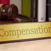
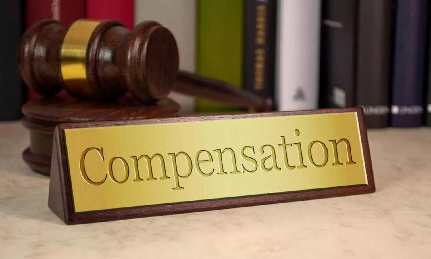
Holding them negligent and deficient in providing their services pertaining to accurate diagnosis and operative care of the patient, the consumer court ordered them to pay Rs 5,10,000 as damages, Rs 1,20,000 as mental agony and Rs 90,000 as litigation charges.

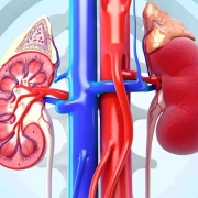
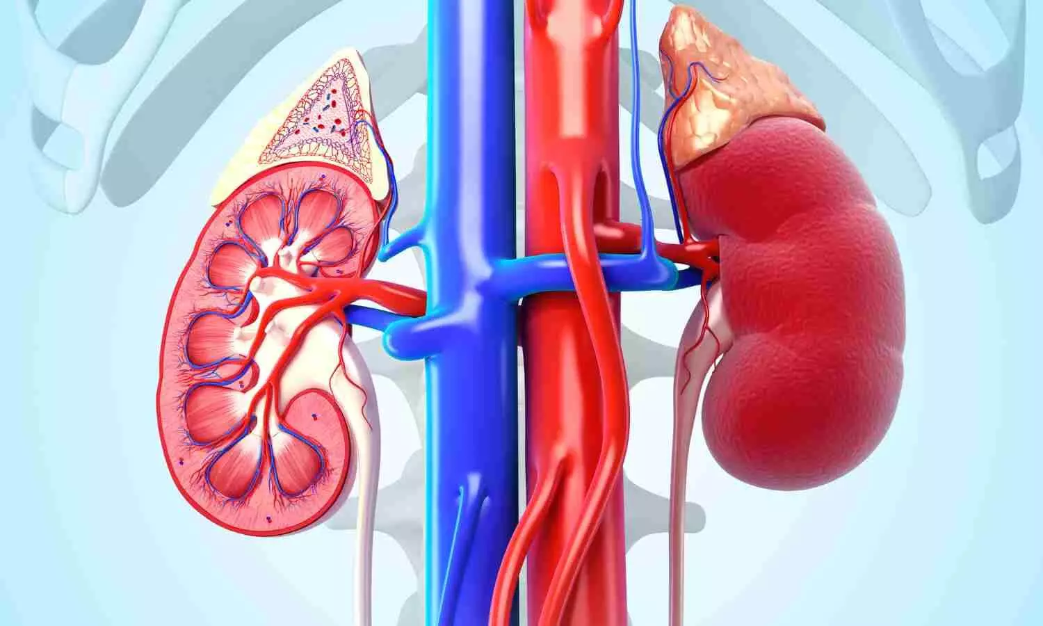
Such a decision was taken by the Commission after noting that the abdominal ultrasound report, conducted after the surgery showed that the “Spleen is normal in size and echo texture.”

The matter goes back to 2014 when the complainant’s wife was admitted to Sir Ganga Ram Hospital and was diagnosed for Non-Hodkin Lymphoma by Dr. Aggarwal. Later, she was advised splenectomy by him and consequently, the patient underwent the said procedure by Dr Kalhan.

Allegedly, the complainant was informed that the surgery was uneventful and the spleen was successfully removed from the patient’s body. Consequently, the complainant requested Dr. Kalhan to show the specimen of the removed spleen. However, the doctor allegedly refused to show the removed spleen. A few months later, the patient was again admitted to the hospital with complaints of dehydration and associated symptoms.

The complainant alleged that the treating doctors and the hospital conducted Ultra Sound and allied tests and started the treatment of the patient relying on the reports of the said tests. However, the patient finally succumbed to her ailments a few days later.

It was submitted by the complainant that even though he made the whole payment for the treatment, he was not given the medical records of the patient and therefore, he sent several letters to the hospital and other authorities such as the Medical Council of India (MCI), Directorate of Health Services (DGHS), Delhi requesting for the requisition of the complete medical record and the original documents such as the Discharge summary, total bills, bone marrow reports, ultrasound films, spleen specimen etc and other allied reports.

Further, it was alleged that Dr. Garg prepared a forged and baseless discharge summary to shield the hospital. After complaining before various authorities, the complainant, with the aid of DGHS, got three sets of medical treatment papers including the Discharge Summary and bills. However, he submitted that the documents pertaining to the status of the spleen, films of the ultrasound, bone marrow reports etc. were not provided to him even though charges against Bone Marrow Biopsy and allied tests were paid to the hospital.

Apart from this, the Complainant submitted that there was a great contradiction between the ultrasound report and the discharge summary. Besides, the abdominal ultrasound report showed the spleen to be normal in size and echo texture, though the spleen was already removed through splenectomy performed on 23.02.2015.

It was also submitted that till date, despite having made several requests/complaints, the complainant did not receive the status of Spleen which was allegedly removed. Therefore, the complainant alleged that the hospital and the doctors committed gross errors in diagnosis and post-operative treatment and therefore, they are liable for medical negligence and professional misconduct.

On the other hand, the doctors and the hospital filed a joint reply and stated that the entire record of the case sheets, investigations, bills, death summary etc. were provided to the complainant on 07.07.2015 and the spleen of the patient was handed as biomedical waste as per Biomedical Waste Rules. It was further submitted that in the present case, no CCTV recording was done and a reply was sent to the complainant mentioning that not all surgical procedures are routinely recorded.

It was also highlighted that even though the complainant made a complaint to the Delhi Medical Council alleging medical negligence, the Council in its orders observed that no medical negligence was made on the part of the doctors and the hospital. The hospital and the doctors claimed that the patient was provided with the standard level of treatment.

While considering the matter, the State Consumer Court noted that even though the complainant was provided the medical record of the patient, it did not contain documents about the Bone Marrow Reports, Spleen Status etc. 

“It is not denied by the Complainant that he did not receive any documents. It is further clear that, though 3 sets of documents as per the direction of the DGHS were supplied to the Complainant vide speed post… yet the same did not contain the documents pertaining to the spleen status as demanded by the Complainant,” noted the consumer court, adding, “…it is clear that the Complainant had only asked for the reports of the test conducted prior to the death of the patient, which were already prepared beforehand, yet the Opposite Party-hospital made inordinate delays in supplying the same until the intervention of DGHS. Therefore, it is clear that prima facie that Opposite Parties kept the Complainant dangling in the air for no reason whatsoever.”

To adjudicate the issue of medical negligence, if any, the consumer court noted that after consulting Dr Aggarwal, the patient was advised PET Scan for clinical evaluation which showed active uptake of FDG in spleen. Thereafter, another Dr. Aggarwal treated the patient and referred her to Dr. Kalhan for laparoscopic splenectomy. Subsequently, the patient was admitted as a case of Non-Hodgkin’s Lymphoma Post Chemotherapy with Splenic Abscess for Laparoscopic Splenectomy on 21.02.2015. The surgery was allegedly performed under General Anesthesia on 23.02.2015.

At this outset, the consumer court also referred to the details of the abdominal ultrasound report dated 09.06.2015, which mentioned, “Spleen is normal in size and echo texture.”

Referring to this, the State Consumer Court observed,

“A perusal of the aforesaid report clearly establishes that spleen was normal in size and echotexture…it is abysmally surprising to note that the ultra sound report dated 09.06.2015 shows that the spleen is intact and normal in size. It is implausible as to how could the spleen be spotted normal in size and texture when the same was removed on 21.02.2015 through Laparoscopic Splenectomy procedure. It is further inexplicable as to how could the Opposite Party-hospital charge for a surgery, which as per test reports, was never performed, or to the contrary, even if performed, constitutes a total failure as is negated by the ultra sound report which shows the spleen intact.”

“It is to be noted further that it is a standard medical practice to show the patient or his/her relatives the specimen of the removed organ after an organ removal surgery. However, in the present case, it is not in dispute that neither the Complainant nor the patient or any other relative thereof was shown the removed spleen after surgery and the same was treated as biomedical waste,” it further noted.

Further, the consumer court referred to another anomaly that no CCTV footage of the operation was recorded/preserved in the first place and the doctors and the hospital also clearly stated in their joint reply that no CCTV recording was done in the ICU.

Holding the hospital and its doctors liable, the commission noted,

“Here, it is pertinent to remark that the aforesaid findings /discrepancies in the line of treatment, highly reek of an unprofessional and heedless attitude of the Opposite Parties towards the patient, thus rendering the present case absolutely fit to fall in the domain of the doctrine of res ipsa loquitor. Here, the principle of res ipsa loquitor very well comes into play, as prima facie, the conduct of the Opposite Parties tantamounts to negligent conduct…The aforesaid findings independently make way for raising an adverse presumption against the Opposite Parties that either the Spleen was not removed at all and the Complainant was wrongly charged for the said operation, or to the contrary, even if it is assumed that the spleen was removed, the post-operative treatment was erroneous as it was based on a faulty test report/diagnosis which showed the spleen to be present intact within the patient’s body. It is crucial to remark here that none of the Opposite Parties ever reflected on the aforesaid discrepancy and continued to provide treatment based on a faulty line of diagnosis/ test reports. Therefore, either way, the Opposite Parties cannot shrug off their liability in so far so the Opposite Parties failed to exercise reasonable care and diligence in extending post operative care to the patient.”

Although the doctors and the hospital argued that the scan was sub-optimal and the Delhi Medical Council had absolved them of all charges, the State Commission opined that the hospital and the doctors owed a duty of care towards the patient and the whole post-operative treatment of the patient was based on the test reports which turned out to be faulty. 

“This Commission is of the view that even if it is assumed that the scan was sub-optimal, the Opposite Parties ought to have referred the patient for a second scan and ought not to have proceeded with further course of treatment till an optimal scan was obtained. The Opposite Parties have prima facie failed in exercising reasonable care towards the patient in so much so that all the treating doctors proceeded with further treatment based on an erroneous report which shows the spleen to be intact when the same was already removed,” the Commission noted.

The Consumer Court also referred to the Delhi Medical Council’s order to note,

“A perusal of the aforesaid order makes it abundantly clear that Dr. *** misunderstood the liver to be spleen. His explanation that when the spleen has been excised or is small due to other reasons, the left lobe of liver grows to fill the left subdiaphragmatic space and because of similar echotexture can frequently mimic like spleen, is not a medically tenable explanation. The aforesaid finding prima facie gives rise to an adverse presumption against the competency of the treating doctors in being well conversant with the anatomy of the human body, failing to clinically correlate the findings in the reports for diagnosing the disease and to later provide treatment accordingly.”

Further, the Commission noted that the post operative PET NCCT scan duplicate report dated 16.06.2015 also gave a clear-cut finding that “the spleen is not visualized consistent with post operative status”. Thus, the Commission opined that it confirmed there being no spleen in the body of the patient and the ultra sound report was an erroneous one which wrongly reported the spleen to be normal in size.

“Therefore, the aforesaid events prima facie tantamount to seemingly evident negligence and are sufficient proof to carve out a case of medical negligence,” opined the State Consumer Court.

It also observed that there was a glaring discrepancy in the abdominal ultra sound report and death summary for the period of 08.06.2015 and 18.06.2015. “A juxtaposition of the ultra sound report and the clinical summary as recorded in the death summary project a contradictory picture. The clinical summary states that the liver is observed to be enlarged than its normal size. However, the ultra sound report makes it clear that the liver is normal in size. The aforesaid observations again indicate towards a confused state of conduct, raising an adverse inference against the level of skill and competence of the Opposite Parties,” the Commission noted.

Holding the hospital and its doctors liable, the Commission observed,

“Therefore, in view of the aforesaid discussion, it can be concluded beyond doubt that the conduct of the Opposite Parties fell below that of the standards of a reasonably competent practitioner in exercising skill and competence and the Opposite Parties conjointly failed to take reasonable care of the patient.”

Opining that they were negligent and deficient in providing their services pertaining to accurate diagnosis and operative care of the patient, the Commission ordered, “the Opposite Parties No.1-6 to pay the Complainant a sum of Rs. 85,000/- each to the Complainant totalling to Rs.5,10,000/- as damages towards the physical agony suffered by the patient including Rs.1,97,900/- being the charges of surgical procedure.”

“…the Opposite Parties No.1-6 to pay a sum of Rs.20,000/- each to the Complainant as mental agony,” further granted the Commission, adding “the Opposite Parties No.1-6 to pay a sum of Rs. 15,000/- each to the Complainant as litigation charges.”

To view the order, click on the link below:

https://medicaldialogues.in/pdf_upload/sir-ganga-ram-hospital-negligence-232130.pdf

Also Read: Delhi-based Private hospital, doctors directed to pay compensation for failing to provide appendix treatment to minor patient despite diagnosis

Powered by WPeMatico

PGI Dr Parul Gupta bags Sushruta Award 2024 for contributions in organ donation

Chandigarh: In a significant achievement, Dr Parul Gupta, the Transplant Coordinator at the Postgraduate Institute of Medical Education and Research (PGIMER), Chandigarh received the esteemed Sushruta Award 2024 on Thursday. 

The award was presented to her in recognition of her contributions to the field of organ donation by facilitating over 150 organ and tissue donations at PGIMER Chandigarh since 2019.

Union Minister for Health and Family Welfare, Mansukh Mandaviya handed the award to Dr Gupta at a televised health conclave organised by Medically Speaking The awards ceremony, held at Bharat Mandapam, Pragati Maidan, New Delhi, on February 8, 2024, celebrated the exceptional achievements of healthcare professionals.

Also read- Renowned Interventional Cardiologist Dr Tejas Patel Wins Padma Bhushan, Expresses Gratitude

National Oral Healthcare Sushruta Awards are conceptualized by the IDA Foundation a philanthropic organization to address the educational and humanitarian commitments of the Indian Dental Association (IDA).

It recognizes the highest standards of excellence in oral healthcare, honouring individuals in respective fields for their outstanding achievements. These awards are instrumental in motivating other professionals to strive for greater heights and to contribute more actively towards promoting the profession’s intellectual, creative and ethical value systems.

During the conclave, Gupta participated in a panel discussion on the topic ‘Donate organs and save lives’, and shared about the role of transplant coordinators in deceased organ donation. She emphasised the right approach to meet potential donor families, HT reports. 

She has been involved in initiatives like organ donation after cardiac death (DCD), and organ donation in very young children, including a 39-day-old infant, which was mentioned by PM Narendra Modi, in his 99th episode of “Mann Ki Baat” and the successful organ donation of a child aged just 70 hours at PGIMER Chandigarh in February 2020.

Also read- Padma Awards In Medicine 2024: 3 Doctors Get Padma Bhushan, 10 Conferred Padma Shri

Powered by WPeMatico