PM Modi to inaugurate Rs 114 crore ESI Hospital at Kakinada on February 25

Kakinada: Prime Minister Narendra Modi will inaugurate the ESI hospital constructed at a cost of Rs. 114 crores in the Sambamurtynagar area here on February 25 in virtual mode. 

Kakinada MP Vanga Geeta Viswanath told media persons at the hospital here on Thursday that the construction of the hospital was started in 2020 and completed on schedule despite the COVID epidemic and heavy rains lashing the area.

Also Read:Nine new ESIC Hospitals, 17 dispensaries approved: Bhupender Yadav

City MLA Dwarampudi Chandrasekhara Reddy, who also inspected various wings of the hospital along with the MP, has attributed the speedy completion of the ESI hospital to the personal interest taken by Chief Minister Jaganmohan Reddy. He said, but for CM’s personal intervention, the ESI hospital would have been shifted from Kakinada.

He said the hospital was provided with modern equipment and has four operating theatres.

Additional Municipal Commissioner Ch Naga Narasimharao, Municipal Health Officer Dr. D Prudhvicharan, Incharge Superintendent K Srinivasarao, and SE P. Satyakumari were among those who attended.

Medical Dialogues team had earlier reported that Dr Mansukh Mandaviya, Union Minister of Health and Family Welfare laid the foundation stone for 2 Critical Care Blocks and a BSL-3 Laboratory in Vijayawada, Andhra Pradesh. He also inaugurated 7 Integrated Public Health Laboratories to be set up in various districts of the state. Smt. Vidadala Rajini, Minister of Health, Government of Andhra Pradesh was also present. These new facilities will give a boost to the healthcare infrastructure of Andhra Pradesh and help in addressing the challenges of any health emergency.

Expressing his elation at the event, Dr Mandaviya said the BSL laboratory, seven Integrated Public Health laboratories and the two Critical Care Blocks, once operational, will play a pivotal role in providing quality medical services to the people of Andhra Pradesh.

Powered by WPeMatico

Air Pollution Increase Mortality Risk in Rheumatoid Arthritis Patients: BMJ

A recent study published in the BMJ Rheumatic & Musculoskeletal Diseases highlights the potential impact of air pollution on the prognosis of Rheumatoid Arthritis-associated Interstitial Lung Disease (RA-ILD). This study aimed to assess the effects of long-term exposure to particulate matter with an aerodynamic diameter of ≤10 µm (PM10) and nitrogen dioxide (NO2) on mortality among patients with RA-ILD.

This research incorporating data from 309 patients with RA-ILD, with a mean age of 61.7 years and a male population of 44.3% and analyzed individual-level long-term exposure to PM10 and NO2 using a national-scale exposure prediction model. The research spanned a median follow-up period of 4.8 years and observed that 40.8% of patients either passed away or underwent lung transplantation during the study period.

The findings revealed that while there was no significant association between air pollutant concentration and mortality when pollutant levels were stratified by quartiles, a significant correlation emerged when stratified by high exposure (top 25th percentile) against low exposure (bottom 75th percentile). Also, a significant association was identified between high PM10 exposure and mortality, with a hazard ratio (HR) of 1.68 and a confidence interval (CI) of 1.11 to 2.52.

Further analysis in subgroup demographics revealed that the effect of high PM10 exposure on mortality was particularly pronounced in patients under the age of 65, with an HR of 1.98 and a CI of 1.02 to 3.85. These findings underline the potential health implications of air pollution, particularly PM10, on patients with RA-ILD. While NO2 exposure did not express a significant association with mortality in this study, the implications of prolonged exposure to PM10 on the prognosis of RA-ILD require further attention and research.

Source:

Kim, S. H., Kim, S.-Y., Yoon, H.-Y., & Song, J. W. (2024). PM10increases mortality risk in rheumatoid arthritis-associated interstitial lung disease. In RMD Open (Vol. 10, Issue 1, p. e003680). BMJ. https://doi.org/10.1136/rmdopen-2023-003680

Powered by WPeMatico

Doctor duped of Rs 56 Lakh under pretext of building hospital

New Delhi: A Nellore-based doctor has been duped of Rs 56 lakh under the pretext of constructing a hospital. The Delhi Police’s Crime Branch has initiated an investigation into a doctor’s complaint who informed the police about his plan to establish a dental hospital in his hometown.

According to the doctor, a family friend promised to facilitate a government grant for a 100-bed multispecialty hospital. However, the friend insisted on a payment of Rs 60 lakh to government officials to secure the grant.
As per a recent media report by the Times of India, the doctor, unable to arrange the full amount, was then offered the option to pay in installments by the friend and his wife. The friend’s wife claimed to have connections with several ministers, adding an air of legitimacy to the scheme. As the doctor agreed to the proposed payment plan and paid his friend Rs 56 lakhs. “The doctor has paid Rs 56 lakh to the accused duo.
During Covid-19, he suffered a financial loss and even had to sell a piece of land, which he had kept for his dental hospital. After waiting for years and finding that nothing was happening, he asked his friend to return the money. But he refused and the doctor then approached police,” an officer said.

The fraudulent scheme underscores the vulnerability of individuals seeking to establish medical facilities, especially when promised government assistance. The Delhi Police’s Crime Branch is now actively investigating the matter, aiming to uncover the details of the scam and identify those responsible for the financial deceit. 

Frauds targeting individuals with aspirations for healthcare ventures are not uncommon, emphasizing the need for increased vigilance and scrutiny in such business dealings. A similar incident took place a few years back, where police registered a case against four persons, including two doctors, for allegedly cheating a woman doctor of Rs 1.54 crore under the pretext of arranging funds for a hospital in Thane district of Maharashtra. 

Powered by WPeMatico

High prevalence and persistence of vitamin D deficiency revealed over time in patients with acute MI: Study

Italy: A recent study published in Nutrition, Metabolism and Cardiovascular Diseases has shed light on the persistence of vitamin D deficiency among Italian patients with acute myocardial infarction (AMI).

The researchers revealed a high prevalence and persistence of vitamin D deficiency (Hypovitaminosis D) in AMI patients despite a trend towards increasing 25(OH)D concentrations over the years. The COVID-19 pandemic and lockdowns did not affect vitamin D levels.

Previous studies have shown vitamin D deficiency is a common cardiovascular (CV) risk factor linked with atherosclerosis development. Aneta Aleksova, University of Trieste in Trieste, Italy, and colleagues evaluated changes in 25(OH)D concentrations in 1510 patients with acute myocardial infarction over a long observation period, including the COVID-19 pandemic.

Patients were divided into four groups according to the year of enrolment, group 1 (2009-2010), group 2 (2014-2016), group 3 (2017-2019), and group 4 (2020-2022).

The key findings of the study were as follows:

· The median 25(OH)D concentration in the overall cohort was 17.15 ng/mL. The median plasma concentrations of 25(OH)D for groups 1, 2, 3, and 4 were 14.45 ng/mL, 17.3 ng/mL, 18.95 ng/mL and 19.05 ng/mL, respectively.

· Although 25(OH)D levels increased over the years, the prevalence of vitamin D deficiency remained high in each group (68.4%, 61.4%, 53.8%, and 52%, respectively).

· Hypovitaminosis D was predicted by the season influence (OR:2.03), higher body mass index (OR:1.25), smoking (OR:1.47), diabetes mellitus (OR:1.54), higher triglycerides levels (OR:1.02), older age (OR:1.07), and female gender (OR:1.3).

· After multivariable adjustment, vitamin D≤20 ng/mL was an independent predictor of mortality.

The findings showed that Hypovitaminosis D is highly prevalent and persistent over time in patients with AMI despite increasing vitamin D levels. The frequent lockdowns did not reduce 25(OH)D levels in the fourth group (2020-2022).

Vitamin D deficiency is highly prevalent and persistent in patients with AMI despite a trend towards increasing 25(OH)D concentrations over the years. The frequent lockdowns did not reduce the levels of 25(OH)D in the fourth group (2020-2022). However, the lowest incidence of hypovitaminosis D was observed between 2020 and 2022.

“Low levels of 25(OH)D are an independent predictor of mortality,” the researchers concluded.

Reference:

DOI: https://doi.org/10.1016/j.numecd.2024.02.007

Powered by WPeMatico

Increased use of Paxlovid could cut hospitalizations, deaths and costs

Increased use of Paxlovid, the antiviral drug used to treat COVID-19, could prevent hundreds of thousands of hospitalizations and save tens of billions of dollars a year, according to a new epidemiological model published by researchers at The University of Texas at Austin. In fact, epidemiologists found that treating even 20% of symptomatic cases would save lives and improve public health.

A 2023 National Institutes of Health study found that only about 15% of high-risk patients take Paxlovid when infected with COVID-19. Using a multiscale mathematical model based on conditions seen over 300 days beginning in January 2022, the researchers found that using Paxlovid on 20% of symptomatic COVID-19 patients during the omicron wave would have resulted in up to 850,000 fewer hospitalizations and saved up to $170 billion. Even with lower transmission levels of the virus, the researchers estimate that an expanded use of Paxlovid could save approximately 30,000 lives during an outbreak.

The findings appear in the February issue of Emerging Infectious Diseases.

“This model shows us there are real benefits to using Paxlovid, not just for the patients receiving treatment, but for the people around them,” said Lauren Ancel Meyers, UT professor of integrative biology and statistics and data sciences, director of the Center for Pandemic Decision Science and corresponding author of the paper. “Not only does this drug help keep high-risk patients out of the hospital, but it can substantially decrease the chance that a treated patient will infect other people.”

The team of researchers assumed patients would take Paxlovid within five days of symptom onset, which is recommended, and estimated different outcomes based on different potential levels of viral transmission, which can vary in communities and with the variant of the virus. If each symptomatic person was assumed to go on to infect about one other person, giving Paxlovid to even 1 out of every 5 of all symptomatic patients could result in 280,000 fewer hospitalizations and save nearly $57 billion. If the virus were to lead the average symptomatic patients to go on to infect closer to three people, as some research has found with the omicron variant, using Paxlovid in 20% of patients would be predicted to result in 850,000 fewer hospitalizations and save more than $170 billion.

“We conducted this analysis to help doctors and policymakers make good decisions about using Paxlovid to combat future waves of COVID,” Meyers said. “A lot of our work is aimed at improving global preparedness for future pandemics. These kinds of models can help to ensure that the U.S. has enough antivirals stockpiled and to design playbooks for using vaccine, drugs and other measures in the heat of threat to slow viral spread and save as many lives as possible.”

Reference:

Bai Y, Du Z, Wang L, Lau E, Fung I, Holme P, et al. Public Health Impact of Paxlovid as Treatment for COVID-19, United States. Emerg Infect Dis. 2024;30(2):262-269. https://doi.org/10.3201/eid3002.230835.

Powered by WPeMatico

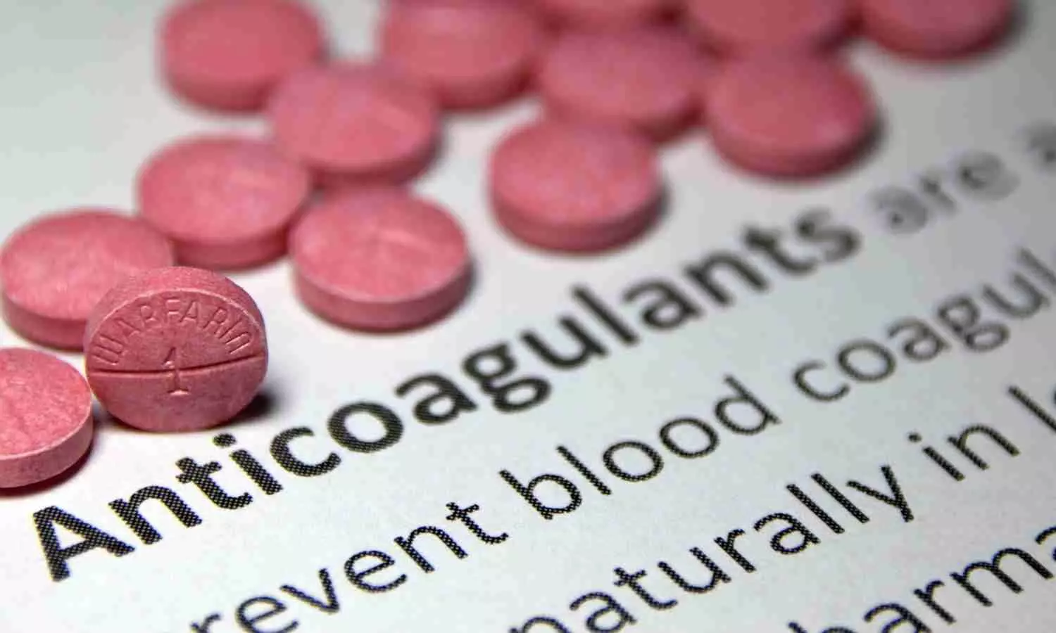
Do’s and don’ts with direct oral anticoagulants

Direct oral anticoagulants (DOACs) are a common treatment for patients with a wide variety of cardiovascular conditions. DOACs are the preferred treatment over vitamin K antagonists (VKAs) for many patients with atrial fibrillation or venous thromboembolism, since the latter would have a higher risk of intracranial bleeding and more complex dosing routine. However, new research suggests that DOACs should not be the first line of treatment for every patient who need to treat or prevent blood clots. A systematic overview from researchers at Brigham and Women’s Hospital, a founding member of Mass General Brigham, discusses the efficacy of DOACs compared to other treatment methods.

This review utilized data from randomized controlled trials to compare DOACs with other treatment methods for various cardiovascular conditions. Although there is merit to using DOACs in many common conditions, the manuscript provides a robust summary of clinical trials indicating that DOACs fare worse in patients with mechanical heart valves, thrombotic antiphospholipid syndrome, atrial fibrillation associated with rheumatic heart disease, and patients with embolic stroke of unclear source. The authors also highlight clinical scenarios in which there is uncertainty, with a look toward future for better evidence generation.

“The results we reviewed here have significant implications for optimizing anticoagulation therapy and improving patient outcomes in clinical practice,” said Behnood Bikdeli, MD, MS, of the Brigham’s Heart and Vascular Center. “There is a critical need for further research regarding why DOACs are less efficacious or safe than the standard of care in certain scenarios.”

Reference:

Antoine Bejjani, Candrika D. Khairani, Ali Assi, Gregory Piazza, Parham Sadeghipour, Azita H. Talasaz, John Fanikos, Jean M. Connors, Deborah M. Siegal, Geoffrey D. Barnes, Karlyn A. Martin, Dominick J. Angiolillo, Dawn Kleindorfer, Manuel Monreal, David Jimenez, Saskia Middeldorp, Mitchell S.V. Elkind, Christian T. Ruff, Samuel Z. Goldhaber, Harlan M. Krumholz, Roxana Mehran, Mary Cushman, John W. Eikelboom, Gregory Y.H. Lip, Jeffrey I. Weitz, Renato D. Lopes, Behnood Bikdeli, When Direct Oral Anticoagulants Should Not Be Standard Treatment: JACC State-of-the-Art Review, Journal of the American College of Cardiology, https://doi.org/10.1016/j.jacc.2023.10.038.

Powered by WPeMatico

Latest research redefines neurodevelopmental risks, outcomes for congenital heart disease

More than a decade of new knowledge about neurodevelopmental risk in people with congenital heart disease has changed the thinking about who is most at risk and the factors that impact neurological development, learning, emotions and behaviors, according to a new American Heart Association scientific statement published today in the Association’s flagship, peer-reviewed journal Circulation.

Congenital heart disease, defined as structural abnormalities in the heart or nearby blood vessels that arise before birth, is the most common birth defect. While advances in treatment have helped more than 90% of people with congenital heart disease in developed countries live to adulthood, the risk of neurodevelopmental issues when individuals have a more severe form of congenital heart disease has not meaningfully improved.

The new scientific statement, “Neurodevelopmental Outcomes for Individuals With Congenital Heart Disease: Updates in Neuroprotection, Risk-Stratification, Evaluation, and Management,” describes the significant advancement in understanding the impact of congenital heart disease on an individual’s development, learning, emotions and behaviors throughout childhood and adulthood.

“Neurodevelopmental difficulties are among the most common and enduring complications faced by people with congenital heart disease. These difficulties can affect a person’s ability to function well at school, at work or with peers, and can affect health-related quality of life throughout childhood and into adulthood,” said Vice Chair of the writing statement group Erica Sood, Ph.D., a senior research scientist and pediatric psychologist at Nemours Children’s Health, Delaware Valley. “It is important for health care professionals and individuals with congenital heart disease and their families to understand how common neurodevelopmental difficulties are. It is also important to understand what places a person with congenital heart disease at high-risk for these difficulties, as well as how these difficulties can be prevented or managed.”

The statement includes updated guidance for health care professionals on how to identify which patients are at high-risk for neurodevelopmental difficulties and what type of evaluations may be helpful to better understand these difficulties. Optimizing neurodevelopmental outcomes through clinical care and research has become increasingly critical since more patients are surviving into adulthood.

The key findings of the statement include:

  • The algorithm for risk stratification of people with congenital heart disease into high or low risk for developmental delays or disorders has been revised to reflect the latest research.
  • The statement suggests health care professionals sequentially review three risk categories: Risk Category 1 includes patients with a history of cardiac surgery with cardiopulmonary bypass during infancy. Risk Category 2 is people with a history of chronic cyanosis, those with blue or purple discoloration due to low blood-oxygen levels, who did not undergo cardiac surgery with cardiopulmonary bypass during infancy. Risk Category 3 has two criteria. The first criterion for Risk Category 3 is a history of an intervention or hospitalization secondary to congenital heart disease in infancy, childhood or adolescence. The second criterion is the presence of one or more factors known to increase neurodevelopmental risk.
  • The statement features an updated list of factors known to increase neurodevelopmental risk, including genetic, fetal and perinatal impact, surgical aspects of treatment and care, socioeconomic and family influences and factors related to growth and development. For example, genetic variants that may alter fetal development of the heart, brain and other organs cause up to nearly a third of congenital heart disease cases.
  • There is a new section on emerging risk factors, such as abnormal placental development, prolonged or repeated anesthetic exposure and exposure to neurotoxic chemicals.
  • In addition, there is a new section on neuroprotective strategies, including detection of congenital heart disease before birth, monitoring of brain blood flow and the delivery of oxygen, and functional support care, such as physical therapy, occupational therapy and speech-language pathology.
  • The statement provides updated information about referral to age-based evaluation of people with congenital heart disease at high risk for developmental delay or disorder. The statement refers to guidance from the Cardiac Neurodevelopmental Outcome Collaborative, which recommends that children with congenital heart disease at high risk for developmental delay or disorders have neurodevelopmental assessments throughout infancy, childhood and adolescence.
  • The statement also provides updated information about management of developmental delay or disorder in infants, children and adolescents, and a new section on management of neuropsychological deficits in adults.

“Reducing barriers that people with congenital heart disease and their families often face when trying to access neurodevelopmental supports and services, and ensuring sufficient research funding are priority areas for future policies,” said Chair of the statement writing group Bradley S. Marino, M.D., M.P.P., M.S.C.E., M.B.A., FAHA, chief of cardiology and cardiovascular medicine at Cleveland Clinic Children’s. “More research will result in a better understanding of how to prevent and manage neurodevelopmental conditions related to congenital heart disease, which will ultimately improve neurodevelopmental outcomes and health-related quality of life for people with congenital heart disease across their life span.”

Reference:

Erica Sood, Jane W. Newburger, Julia S. Anixt, Adam R. Cassidy, Jamie L. Jackson, Richard A. Jonas, Amy J. Lisanti, Keila N. Lopez, Shabnam Peyvandi, Bradley S. Marino, Neurodevelopmental Outcomes for Individuals With Congenital Heart Disease: Updates in Neuroprotection, Risk-Stratification, Evaluation, and Management: A Scientific Statement From the American Heart Association, Circulation, https://doi.org/10.1161/CIR.0000000000001211.

Powered by WPeMatico

Feeling depressed after menopause? Hormone therapy may help

Hormone therapy (HT) is already recognized as the most effective treatment option for physiologic menopause symptoms such as hot flashes, but a new study is one of the first to suggest that it might also help manage depressive symptoms. It further documented a high rate of depression during menopause.

Results of the study have been published online in Menopause, the journal of The Menopause Society.

Depression and other mood-related symptoms can strike at any time during a woman’s life but have been shown to be especially likely during and immediately after the menopause transition. In a new study involving 170 women who were patients at a menopause clinic in Ontario, Canada, there was a high prevalenc-62%-of participants who scored as being “depressed.” Although these symptoms may not necessarily reach the severity of a major depressive disorder, they still could significantly affect quality of life and relationships.

To date, the effectiveness of HT to effectively manage hot flashes (technically known as vasomotor symptoms) has been well documented. However, its ability to address mood-related symptoms is less established. This new study from Canada sought to correct that problem.

Hormone influences on neurotransmitter-receptor pathways, which regulate mood during and after the menopause transition, are thought to play a role in depressive symptoms. In addition, the domino theory suggests that the toll of physiologic symptoms experienced during menopause, such as hot flashes and an array of genitourinary problems, could also contribute to adverse moods.

As part of their study, researchers attempted to determine whether the stage of menopause contributed to an increased risk of depression, but no association was found, although a lower education level (high school or less) and younger age did show a direct correlation. The addition of progestin to the HT regimen did not have a significant effect on overall effectiveness. Additionally, although women who went through natural menopause experienced significant improvements with regard to their depressive symptoms, women who experienced iatrogenic menopause (surgical or chemotherapy based) did not have similar improvements.

On the basis of these results, the researchers concluded that HT-whether used alone or in conjunction with antidepressants-can improve not only hot flashes but also depressive symptoms commonly associated with menopause.

Survey results are published in the article “Does menopause hormone therapy improve symptoms of depression? Findings from a specialized menopause clinic.”

“This study showed a beneficial effect of hormone therapy on mood symptoms during menopause when used alone and a synergistic effect when used in combination with antidepressants. It also highlights the high prevalence of mood symptoms during this transition and the need to address women’s symptoms holistically rather than having a singular focus on hot flash management,” says Dr. Stephanie Faubion, medical director for The Menopause Society.

Reference:

Gnanasegar R, Wolfman W, Galan LH, Cullimore A, Shea AK. Does menopause hormone therapy improve symptoms of depression? Findings from a specialized menopause clinic. Menopause. 2024 Feb 20. doi: 10.1097/GME.0000000000002325.  

Powered by WPeMatico

Hepatitis E Vaccine Provides Long-term Protection

Hepatitis E virus (HEV) is a common cause of acute hepatitis, yet its prevention often receives less attention compared to other hepatitis viruses. Understanding the long-term effectiveness of hepatitis E vaccines is crucial for disease control and prevention efforts.

A recent study conducted in Dongtai County, China, extended a phase-3 clinical trial to evaluate the durability of protection provided by a hepatitis E vaccine over a 10-year period. The initial trial enrolled over 112,000 healthy adults and randomly assigned them to receive either the hepatitis E vaccine or a placebo. This study was published in the journal The Lancet by Shoujie H. and colleagues.

Participants received three doses of the vaccine or placebo intramuscularly at months 0, 1, and 6. A comprehensive surveillance system was established to monitor hepatitis E cases in the study region over the following 10 years. The primary outcome was the efficacy of the vaccine in preventing confirmed cases of hepatitis E occurring at least 30 days after the third dose.

The key findings of the study were:

  • Over the 10-year study period, 90 cases of hepatitis E were identified, with significantly fewer cases in the vaccine group compared to the placebo group.

  • The vaccine demonstrated an efficacy of 83.1% in the modified intention-to-treat analysis and 86.6% in the per-protocol analysis.

  • Moreover, antibody persistence was assessed in subsets of participants, showing that a significant proportion maintained detectable antibody levels even after several years.

The study findings indicate that immunization with the hepatitis E vaccine provides long-lasting protection against hepatitis E, with vaccine-induced antibodies persisting for at least 8.5 years. This underscores the importance of vaccination programs in preventing hepatitis E infections and highlights the effectiveness of the vaccine in reducing disease burden over the long term.

Reference:

Huang, S., Zhang, X., Su, Y., Zhuang, C., Tang, Z., Huang, X., Chen, Q., Zhu, K., Hu, X., Ying, D., Liu, X., Jiang, H., Zang, X., Wang, Z., Yang, C., Liu, D., Wang, Y., Tang, Q., Shen, W., … Xia, N. Long-term efficacy of a recombinant hepatitis E vaccine in adults: 10-year results from a randomised, double-blind, placebo-controlled, phase 3 trial. Lancet,2024. https://doi.org/10.1016/s0140-6736(23)02234-1

Powered by WPeMatico

Vision impairment in older adults tied to symptoms of depression, anxiety and social isolation: JAMA

USA: A cross-sectional study comprising 2822 US adults revealed that worse examination-based and self-reported vision impairment is associated with anxiety and depressive symptoms, and worse examination-based vision impairment is linked with severe social isolation.

These findings, published in JAMA Ophthalmology, provide evidence to support prioritizing research aimed at enhancing the health and inclusion of people with vision impairment.

Vision impairment and psychosocial function, including symptoms of anxiety, depression and social isolation, are a major cause of morbidity in the US. However, there is a lack of nationally representative studies evaluating associations between subjective and objective vision impairment with psychosocial function following the COVID-19 pandemic.

To address this knowledge gap, Louay Almidani, Wilmer Eye Institute, Johns Hopkins University School of Medicine, Baltimore, Maryland, and colleagues provided updated national estimates on the associations of vision impairment with depressive and anxiety symptoms and social isolation in US adults 65 years and older.

The study was a cross-sectional analysis of the National Health and Aging Trends Study rounds 9 (2019) and 11 (2021), a nationally representative sample of Medicare beneficiaries aged 65 years and above. Binocular distance visual acuity, contrast sensitivity, and near visual acuity were tested.

Objectively measured vision impairment was defined as vision impairment in either distance visual acuity (worse than 20/40), near visual acuity (worse than 20/40), or contrast sensitivity (worse than 1.55 logCS). Self-reported vision impairment was defined based on participants’ reports on their vision status.

Depression and anxiety symptoms were assessed via the Patient Health Questionnaire. Social isolation was defined based on communication frequency, living arrangement, and activity participation responses.

The researchers reported the following findings:

  • Among 2822 community-dwelling adults sampled from a population of 26 182 090, the mean age was 78.5 years; 54.7% were female; 32.3% had objectively measured vision impairment, and 6.4% had self-reported vision impairment.
  • In adjusted models, all outcomes were significantly associated with objectively measured vision impairment, including depressive symptoms (odds ratio [OR], 1.81), anxiety symptoms (OR, 1.74), and severe social isolation (OR, 2.01).
  • Depressive symptoms (OR, 2.37) and anxiety symptoms (OR, 2.10) but not severe social isolation symptoms (OR, 2.07) were significantly associated with self-reported vision impairment.

In the study, vision impairment was associated with several psychosocial outcomes, including symptoms of depression, anxiety and social isolation.

Reference:

Almidani L, Miller R, Varadaraj V, Mihailovic A, Swenor BK, Ramulu PY. Vision Impairment and Psychosocial Function in US Adults. JAMA Ophthalmol. Published online February 22, 2024. doi:10.1001/jamaophthalmol.2023.6943

Powered by WPeMatico