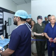
Max Healthcare and Medtronic launch advanced surgical skill lab

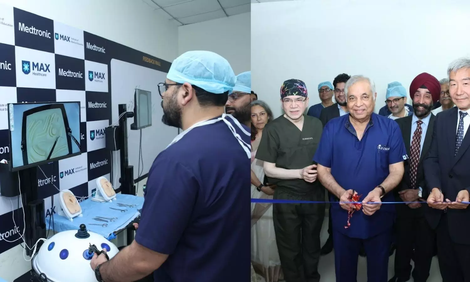
New Delhi: Max Healthcare, one of the largest private sector healthcare services companies in India, and Medtronic, a global leader in healthcare technology, announced the inauguration of the Max-Medtronic Skill Lab at Max Super Specialty Hospital, Saket, New Delhi.

The facility is designed to advance training in minimally invasive surgical techniques and marks a significant step in their shared commitment to clinical excellence and education. 

This strategic collaboration aims to advance medical education and clinical training in laparoscopic and minimally invasive surgical techniques. In addition to training, Max-Medtronic Skill Lab will also prioritize educating patients and caregivers about the benefits of these advanced minimally invasive procedures, such as faster recovery, smaller incisions, reduced infection risk, and less tissue trauma compared to traditional surgeries .

Also Read:Max Healthcare, Global Health Alliance UK partner for Medical Innovation

The Lab will offer comprehensive, structured training modules for a wide range of medical professionals, including surgeons, nurses, OT technicians, and paramedics, ranging from hands-on workshops to advanced upskilling sessions.

Spearheaded by Max Healthcare’s experienced in-house faculty and facilitated by Medtronic, these programs aim to provide a robust, practical, and impactful learning experience for all medical professionals.

“At Max Healthcare, we are committed to fostering a culture of continuous learning and clinical excellence. The Max-Medtronic Skill Lab is a significant step toward creating a structured, high-impact training ecosystem for our medical professionals. This initiative not only aims to improve clinical competencies but also empowers patients and caregivers through increased awareness of advanced surgical techniques”, said Dr Sandeep Budhiraja, Group Medical Director, Max Healthcare.

“This collaboration with Max Healthcare reaffirms our unwavering commitment to advancing access to high-quality healthcare through education, innovation, and strategic partnerships. Max-Medtronic Skill Lab is designed to provide hands-on training and foster continuous learning in laparoscopic and minimally invasive surgical techniques.

Through this initiative, Medtronic seeks to empower healthcare professionals with the tools and knowledge needed to deliver better outcomes and transform patient care”, said Mandeep Singh Kumar, Managing Director and Vice President of Medtronic India.

As part of this collaboration, both partners will also focus on regular upskilling programs for paramedical staff, an often overlooked yet critical component of the surgical care team.

The Max-Medtronic Skill Lab is set to become a benchmark in clinical education and innovation, aligning with the Government’s vision for a skilled and empowered healthcare workforce.

At the inaugural event, present were key leaders from Max Healthcare and Medtronic, including Dr. Pradeep Chowbey, Chairman – Max Institute of Laparoscopic, Robotic and Bariatric Surgery; Dr Vinitaa Malhotra Jha Executive Vice President – Research & Academics Clinical Directorate, Max Healthcare; Dr Subhash Gupta, Chairman – Max Centre for Liver and Biliary Sciences, Dr. Manish Baijal, Senior Director – Max Institute of Laparoscopic, Robotic and Bariatric Surgery. From Medtronic, attendees included Feng Dong, Vice President, Asia Region-Led Markets, EurAsia; Mandeep Singh Kumar, Managing Director and Vice President; and Abhishek Bhargava, Senior Director, Medical Surgical, Medtronic India.

Also Read:Max Healthcare opens 300-bed hospital in Dwarka, plans 3,700 more beds by 2028

Powered by WPeMatico

Tirzepatide Improves Blood Sugar Control in Children With Type 2 Diabetes: SURPASS-PEDS Trial

New research shows that that the diabetes/obesity medication tirzepatide can cause clinically meaningful improvements in blood sugar control and weight loss in children and adolescents with type 2 diabetes aged 10-17 years whose diabetes and weight are inadequately controlled with an existing treatment regimen of metformin, insulin, or both.

The study (the SURPASS-PEDS trial), by Dr Tamara Hannon, Division of Pediatric Endocrinology and Diabetology, Indiana University School of Medicine, Indiana University School of Medicine, Indianapolis, USA, and colleagues is presented at this year’s Annual Meeting of the European Association for the Study of Diabetes (EASD) in Vienna, Austria (15-19 September) and published in The Lancet. The study is sponsored by Eli Lilly and company, the manufacturer of tirzepatide.

Youth-onset type 2 diabetes (YT2D) is a rapidly progressing disease with rising incidence in recent years, primarily driven by the increase in the global prevalence of obesity. The overall incidence of type 2 diabetes in children and adolescents in USA has nearly doubled across 15 years, going from 9.0 to 17.9 cases per 100,000 persons per year between 2002-03 and 2017-18.

There are limited treatment options to improve blood sugar (glycaemic) control in YT2D. Existing therapeutics have generally demonstrated lower glycaemic efficacy in YT2D compared to adults with T2D, without clinically meaningful impact on weight as measured by body mass index (BMI). Tirzepatide is a once weekly GIP/GLP-1 receptor agonist approved for the treatment of adults with T2D, obesity, and obstructive sleep apnoea (in the USA, and approved for treating T2D and obesity in many other countries). The safety and efficacy of tirzepatide in YT2D is yet to be reported.

In this phase 3 trial, 99 young people aged 10-17 years with YT2D with inadequate glycaemic control with metformin, basal insulin, or both, were randomized in a 1:1:1 ratio to receive blinded treatment with tirzepatide (5 mg or 10 mg) or placebo (PBO) once weekly for 30 weeks followed by a 22-week open-label extension*. The primary aim was to demonstrate superiority of tirzepatide (looking at the pooled results of both doses combined) versus placebo for change in glycated haemoglobin (HbA1c – a measure of blood sugar control) at 30 weeks. Analyses included all participants who received at least 1 dose of study drug, excluding data after discontinuation of study drug or initiation of glycaemic rescue therapy (this is when a person’s blood sugar is so high they need intensive medical treatment to recover).

At baseline, the mean age was 14.7 years, mean duration of diabetes was 2.4 years, and participants were treated with metformin (68.7%), basal insulin (8.1%), or both (23.2%). At 30 weeks, tirzepatide was superior to placebo for improving HbA1c, fasting serum glucose, BMI, and incidence of HbA1c of 6.5% or less (below the range for diabetes) and 5.7% of less (below the range for prediabetes). In the placebo group, these indicators barely changed or did not change across the 30 weeks of the study.

At the start of the study all of the children had HbA1c above 6.5%, classifying them as having type 2 diabetes. At 30 weeks, the pooled tirzepatide results shows that more than three quarters (79%) of children taking tirzepatide had HbA1c of less than 6.5%, and more than half (53%) had and HbA1c of less than 5.7%, compared to 29% and 14% respectively for those taking placebo

And while mean BMI fell just 0.4 points across the 30 weeks in the placebo group, from 34.7 to 34.3 kg/m2, in the pooled tirzepatide group mean BMI fell 9.3 units from 35.6 to 26.3 kg/m2. Fasting glucose levels fell by around 6 times more in the pooled tirzepatide group (2.46 units) than the placebo group (0.44 units)

The estimated mean treatment difference (between tirzepatide pooled results and placebo) for change from baseline were: HbA1c -24.9 mmol/mol (-2.3%), FSG -2.0 mmol/L (-36.3 mg/dL) and BMI -8.9%. At 52 weeks, estimated mean change from baseline in HbA1c was -24.2 mmol/mol (-2.21%) and percent change in BMI from baseline was -12.0%, for TZP pooled.

The most common adverse events on tirzepatide were gastrointestinal, mild to moderate in severity, occurred mostly during dose escalation, and generally decreased over time (consistent with trials in adults). Treatment discontinuation due to adverse events occurred in 6.3% of participants in tirzepatide 5 mg and 0% in tirzepatide 10 mg and placebo. No severe hypoglycaemia episodes were reported during the study. Glycaemic rescue therapy was initiated by 18% of participants in the placebo group and 0 participants in the tirzepatide pooled group.

The authors conclude “Tirzepatide demonstrated significant and clinically meaningful improvements in blood sugar control and BMI in youth with type 2 diabetes. The impact on blood sugar control was sustained over the one year trial period and improvements in BMI continued through the year and did not plateau.”

“Tirzepatide is the first drug used for type 2 diabetes in this age group that has shown sustained clinically, meaningful BMI lowering effects…These results support tirzepatide as a potential safe and efficacious treatment option for youth-onset type 2 diabetes.”

Reference:

Prof Tamara S Hannon, Lily C Chao, Margarita Barrientos-Pérez, Karthik Chandrasekhar Pamidipati, Efficacy and safety of tirzepatide in children and adolescents with type 2 diabetes (SURPASS-PEDS): a randomised, double-blind, placebo-controlled, phase 3 trial, The Lancet.

Powered by WPeMatico

Deep learning model helps in identifying the risk of complete heart block, finds JAMA

A new study published in the Journal of American Medical Association showed that patients at risk for complete heart block (CHB) may be risk-stratified using the AI-ECG model known as AIRE-CHB, which might inform therapy choices such empirical pacemaker insertion or rhythm monitoring.

Ventricular standstill, syncopal damage, and abrupt cardiac death are all possible outcomes of complete heart block, a potentially fatal condition brought on by severe conduction system dysfunction. 

When there are no reversible reasons of CHB, permanent pacemaker (PPM) implantation is the therapy of choice. The gold standard for diagnosing CHB is an electrocardiogram (ECG). The difficulty in diagnosing CHB is that the conduction system illness may be intermittent, and the ECG may not exhibit signs of high-grade atrioventricular (AV) block at the time of recording.

Recent research has demonstrated the great potential of artificial intelligence-enhanced ECG (AI-ECG) to identify concealed cardiovascular illness and forecast future disease risk. The goal of this work was to create an AI-ECG risk estimator for CHB (AIRE-CHB) that could forecast incident CHB.

The UK Biobank volunteer cohort served as the external validation for this cohort research, which was a development and external validation prognostic study carried out at Beth Israel Deaconess Medical Center. more than 31 days following the ECG, a new CHB diagnosis. AIRE-CHB was trained to predict incident CHB using a residual convolutional neural network architecture with a discrete-time survival loss function.

The Beth Israel Deaconess Medical Center cohort comprised 189,539 patients’ 1,163,401 ECGs. With an area under the receiver operating characteristics curve (AUROC) of 0.889 (95% CI, 0.863-0.916) and a C index of 0.836 (95% CI, 0.819-0.534), AIRE-CHB forecasted incidence CHB within a year.

By contrast, bifascicular block was associated with an AUROC of 0.594 (95% CI, 0.567-0.620). When compared to the low-risk group, the adjusted hazard ratio (aHR) for the development of incident CHB was 11.6 (95% CI, 7.62-17.7; P <.001) for participants in the high-risk quartile.

The C index for incident CHB prediction in the UKB UK Biobank cohort, which included 50,641 ECGs from 189,539 patients, was 0.936 (95% CI, 0.900-0.972), and the aHR was 7.17 (95% CI, 1.67-30.81; P <.001). Overall, an unprecedented deep learning algorithm determined the likelihood of event CHB. The model’s credibility was established through thorough evaluations of explainability and biological plausibility. 

Reference:

Sau, A., Zhang, H., Barker, J., Pastika, L., Patlatzoglou, K., Zeidaabadi, B., El-Medany, A., Khattak, G. R., McGurk, K. A., Sieliwonczyk, E., Ware, J. S., Peters, N. S., Kramer, D. B., Waks, J. W., & Ng, F. S. (2025). Artificial intelligence–enhanced electrocardiography for complete heart block risk stratification. JAMA Cardiology. https://doi.org/10.1001/jamacardio.2025.2522

Powered by WPeMatico

Pneumococcal polysaccharide vaccine Found Ineffective in Reducing Cardiovascular Events, reports JAMA

A new study published in the Journal of American Medical Association found that pneumococcal polysaccharide vaccine (PPV23) vaccination did not reduce rates of fatal and nonfatal acute coronary syndrome or ischemic stroke.

The trial was conducted at 6 centers across Australia investigated the vaccine’s cardiovascular impact. Between 2016 and 2017, this research recruited 4,725 community-dwelling adults aged 55 to 60 who had no prior history of cardiovascular disease but did carry at least 2 risk factors such as obesity, high blood pressure, or elevated cholesterol.

The participants were randomly assigned to receive either the 23-valent pneumococcal polysaccharide vaccine (PPV23) or a saline placebo, with neither patients nor clinicians aware of the allocation. Over an average follow-up of 7 years, where this study tracked participants through electronic health records covering hospital admissions, emergency visits, and mortality data. The primary measure was a combined endpoint of fatal and nonfatal acute coronary syndrome (including heart attacks) and ischemic stroke.

The results showed no statistically significant difference between the 2 groups. In the PPV23 arm, 58 participants experienced a primary outcome event, when compared with 64 in the placebo arm. This translated to a hazard ratio of 0.90 (95% confidence interval, 0.63–1.28; P = .57), which suggested no meaningful reduction in risk. Exploratory analyses of all-cause mortality, hospitalizations for any reason, and cardiovascular-related procedures also showed no significant differences.

The overall number of cardiovascular events was lower than anticipated. This low event rate reduced the statistical power of the trial, making it difficult to detect modest protective effects if they exist. While the vaccine was safe and well tolerated, the data do not support its use as a strategy for preventing cardiovascular disease.

Previous studies had suggested that pneumococcal vaccination might play a role in reducing atherosclerosis through immune-mediated mechanisms. However, without randomized trial evidence, those signals remained speculative. Overall, this trial highlights the need for adequately powered studies before vaccines can be repurposed for heart disease prevention. While PPV23 remains an essential tool for preventing pneumococcal infections in older adults and vulnerable populations, its role in cardiovascular protection appears limited. 

Source:

Hure, A., Peel, R., D’Este, C., Abhayaratna, W. P., Tonkin, A., Hopper, I., Thrift, A. G., Levi, C., Sturm, J., Durrheim, D., Hung, J., Briffa, T., Chew, D. P., Ren, S., McEvoy, M., Hansbro, P., Newby, D., Szwec, S., Chiu, S., & Attia, J. (2025). Prevention of Adverse Cardiovascular Events Using the 23-Valent Pneumococcal Polysaccharide Vaccine. JAMA Cardiology. https://doi.org/10.1001/jamacardio.2025.3043

Powered by WPeMatico

Emphysema at CT lung screening increases death risk in asymptomatic adults: Study

Emphysema detected on baseline low-dose chest CT (LDCT) in the lung cancer screening cohort of more than 9,000 asymptomatic adults was associated with death from all causes, chronic obstructive pulmonary disease (COPD), and cardiovascular disease within a 25-year follow-up period in a new study published today in Radiology, a journal of the Radiological Society of North America (RSNA).

Emphysema is a permanent and progressive lung disease in which air sacs in the lungs become damaged, making breathing difficult. It is primarily caused by long-term exposure to irritants like cigarette smoke and air pollution.

“Until now, we didn’t know if baseline visual emphysema scoring on LDCT in the lung cancer screening setting had any prognostic value,” said Claudia I. Henschke, Ph.D., M.D., a radiologist and professor of radiology in the Department of Diagnostic, Molecular, and Interventional Radiology at Icahn School of Medicine at Mount Sinai, in New York. “Our study stands out for its long follow-up and comprehensive analysis of the causes of death in a large lung cancer screening cohort.”

In the study, a lung cancer screening cohort of 9,047 asymptomatic adults (ages 40-85 at enrollment; 4,614 female) with a smoking history underwent baseline LDCT in New York and were followed for up to 25 years as part of the International Early Lung Cancer Action Program (I-ELCAP).

Participants (median age 65 years, median pack-years 43, and median follow-up 23.3 years) underwent baseline LDCT between 2000 and 2008 and were followed when possible until death or December 31, 2024. Pack-years is a measure of lifetime exposure to cigarette smoke calculated by multiplying the number of years spent smoking by the number of cigarette packs smoked per day.

An experienced chest radiologist assessed each LDCT and assigned a score reflecting the level of emphysema present from 0 (none) to 3 (severe).

“Lung cancer screening shouldn’t just be looking for nodules,” said Dr. Henschke, principal investigator for the I-ELCAP. “That’s a small part of what we see on the CT scan. As radiologists, we’re responsible for the entire image.”

Among the study’s participants, 70.9% had no evidence of emphysema. The percentage of mild, moderate and severe emphysema was 21.1%, 5.7%, and 2.4%, respectively. Nearly 80% of participants identified with emphysema on their baseline LDCT had not been previously diagnosed, including five percent of participants with moderate or severe emphysema.

Slightly more males than women were diagnosed with emphysema (30.1% versus 28.2%). Evidence of the disease increased with advanced age and higher cumulative smoking exposure.

By year-end 2024, 3,738 participants (41.3%) had died, most commonly from cardiovascular disease (12.7%) and COPD (3.3%). The median age at the time of death from all causes was 81, and from COPD, cardiovascular disease, and other causes, it was 81, 82, and 81 years, respectively.

A statistical analysis of associations between emphysema and mortality demonstrated that the lung disease was associated with COPD mortality but not cardiovascular disease mortality.

“Clinically, these findings suggest emphysema is not merely an incidental CT finding, but a distinct disease entity associated with worst outcomes and increased mortality, not only from lung cancer but also from respiratory and cardiovascular diseases,” Dr. Henschke said. “The findings show an increased risk of all causes of death by the presence of emphysema and its severity, ranging from a 1.15-fold increase for mild disease and a 2.28-fold increase for severe emphysema. For deaths due to COPD, the increased risk ranged from a 2.07-fold for mild disease to 12.06-fold increase for severe emphysema.”

The study’s results will enable healthcare providers to tailor risk-based treatment to prevent the progression of the disease. Dr. Henschke said using a visual assessment of emphysema as a predictor of health two decades into the future is part of a new era of preventive health.

“The amount of information you get and the ability to act on it in a meaningful way is something preventive health only dreamed of being just a few years ago,” she said.

Dr. Henschke envisions a comprehensive lung cancer screening program that also assesses COPD and cardiovascular disease risk to identify individuals who may benefit from interventions to improve outcomes.

“Pulmonologists, cardiologists and radiologists need to work together, because one influences the other,” she said. “We have to work towards solutions holistically.”

Although the United States Preventive Services Task Force recommends annual lung cancer screening with LDCT for adults aged 50 to 80 who have a 20 pack-year smoking history and currently smoke or quit within the past 15 years, Dr. Henschke said she would also like to see screening benefits extended to individuals who have never smoked.

“In the U.S., about 30,000 to 40,000 deaths each year are found in non-smokers,” she said. “That’s about a third of the total deaths due to lung cancer annually.”

Reference:

Jessica González Gutiérrez, Rowena Yip, Javier J. Zulueta, Samuel M. Aguayo, Daniel M. Libby, Mark W. Pasmantier, Emphysema at Baseline Low-Dose CT Lung Cancer Screening Predicts Death from Chronic Obstructive Pulmonary Disease and Cardiovascular Disease Up to 25 Years Later, Radiology, https://doi.org/10.1148/radiol.250949

Powered by WPeMatico

Vitamin B3 Supplement Shows Promise in Cutting Skin Cancer Risk: JAMA

USA: A large Veterans Affairs (VA) cohort study has found that taking nicotinamide—a vitamin B3 derivative sold over the counter—significantly lowers the risk of developing skin cancer.

The protective effect was strongest when the supplement was started soon after a patient’s first skin cancer diagnosis, according to findings published in JAMA Dermatology by Dr. Kimberly F. Breglio and colleagues from the Durham VA Medical Center, North Carolina.
Nicotinamide, a form of vitamin B3, is inexpensive, widely available without prescription, and generally well tolerated. Previous small trials hinted at its protective potential, but large-scale evidence had been limited until now. This VA study adds weight to the idea that nicotinamide could be a practical chemopreventive option for patients with a history of skin cancer, especially when started early in the disease course.
The retrospective analysis explored whether regular nicotinamide supplementation could help prevent new skin cancers in a real-world population. Using Veterans Affairs electronic health records from October 1999 through December 2024, researchers reviewed data on 33,822 veterans. Among them, 12,287 patients took oral nicotinamide 500 mg twice daily for at least 30 days, while 21,479 served as matched controls who did not use the supplement. Participants were matched on factors such as age, sex, race, history of prior skin cancers, and other treatments, allowing a balanced comparison.
The study led to the following notable findings:
  • Nicotinamide use was linked to a 14% lower overall risk of developing new skin cancers compared to those not taking the supplement.
  • Starting the supplement soon after the first skin cancer reduced risk by 54%, showing the greatest benefit with early initiation.
  • The protective effect covered both basal cell carcinoma and cutaneous squamous cell carcinoma (cSCC), with the strongest impact on cSCC.
  • When nicotinamide was started after multiple skin cancers, its preventive benefit gradually declined.
  • Among solid organ transplant recipients—who face higher skin cancer risk due to immunosuppressive therapy—overall risk reduction was not statistically significant.
  • Early use of nicotinamide after a first cancer episode in transplant recipients still showed a notable decrease in cSCC risk, underscoring the importance of timing.
The authors emphasized that while the findings are promising, further prospective trials could help refine guidance on dosage, timing, and patient selection. Still, for many individuals at risk of recurring non-melanoma skin cancers, this easily accessible vitamin supplement may offer an effective and low-cost strategy to reduce future disease burden.
Reference:
Breglio KF, Knox KM, Hwang J, et al. Nicotinamide for Skin Cancer Chemoprevention. JAMA Dermatol. Published online September 17, 2025. doi:10.1001/jamadermatol.2025.3238

Powered by WPeMatico

Radioiodine Ablation Therapy Feasible in Hemodialysis-Dependent ESKD Patients with Low-Risk Thyroid Cancer: Case Series

Australia: In a recently published case series in BMC Nephrology, researchers from the University of Sydney, led by Dr. Raymond Lin, have demonstrated that with tailored protocols and safety precautions, radioiodine (I-131) therapy can be safely administered to patients with end-stage kidney disease (ESKD) on haemodialysis (HD) who are being treated for thyroid cancer.    

Radioiodine therapy is a well-established adjuvant treatment for differentiated thyroid cancer (DTC), particularly following thyroidectomy. However, its use in patients with impaired renal function poses a significant challenge. In those with ESKD, the clearance of I-131 is severely reduced, increasing the risk of prolonged radiation exposure and myelotoxicity. Currently, there is no universally accepted protocol for administering radioiodine in patients on dialysis, making treatment planning complex.

To address this, Dr. Lin and colleagues reported on two ESKD patients undergoing chronic haemodialysis who received I-131 therapy for low-risk thyroid cancer. The team adapted its institutional approach by implementing modifications to infrastructure, scheduling, and radiation safety protocols. This included pre-treatment patient training, altered dialysis timing, and close monitoring of serum radioactivity to ensure patient and staff safety.

Patient 1, who had undergone bilateral nephrectomy, was trained to self-cannulate in preparation for therapy, while Patient 2 had several comorbidities, including morbid obesity and a colostomy. Both patients underwent haemodialysis at 24, 72, and 144 hours post-I-131 administration. This schedule helped achieve radiation retention profiles comparable to those seen in individuals with normal kidney function.
Radiation exposure to the bone marrow—a primary concern in patients with impaired clearance—remained within acceptable safety margins (<0.3 Gy in both cases). The majority of radiation exposure to bone marrow occurred before the initial dialysis session, accounting for 60% in Patient 1 and 47% in Patient 2. Additionally, cumulative radiation exposure to dialysis staff remained well below the local annual safety threshold, measuring only 7 and 23 μSv, respectively.
At 24 months of follow-up, both patients showed undetectable thyroglobulin levels, indicating effective disease control. There were no significant complications during or after therapy.
The findings from this case series reinforce that radioiodine therapy can be administered safely in dialysis-dependent patients with proper planning. The study highlights the importance of adapting existing treatment pathways and monitoring protocols to address the unique risks posed by renal impairment.
While the small number of cases limits generalizability, the authors stress the value of sharing such clinical experiences. As standardization remains lacking in this area, collaborative efforts and pooled data from multiple centers could support the development of practical, flexible guidelines for managing radioiodine therapy in ESKD patients.
Reference:
Lin, R., Malaroda, A., Ryder, W. et al. Management of radioiodine ablation therapy in haemodialysis patients with thyroid cancer: a case series of two patients. BMC Nephrol 26, 420 (2025). https://doi.org/10.1186/s12882-025-04348-0

Powered by WPeMatico

New tracer could enable surgeons to see and hear prostate cancer: Study

A preclinical evaluation of a new ’dual-mode’ tracer agent shows promise in not only helping surgeons image and plan prostate cancer procedures, but also provide them with much more consistent and targeted guidance during surgery.

The agent uses a single tracer molecule labeled with Fluorine-18-a common isotope used in Positron Emission Tomography (PET) scans-for diagnostic imaging. It also provides a one-step, widely accessible solution that would enable combined fluorescence-guided and radio-guided surgery.

“Precision medicine is increasingly being practiced and developed to address the sophisticated treatment methods for diseases like cancer,” says Dr. David M Perrin, a University of British Columbia chemist and senior author on the paper, published in advance in the Journal of Medical Chemistry.

“Our tracer provides high-resolution visual guidance, but would also allow a surgeon to use a hand-held Geiger counter probes to ‘hear’ areas of high radiation density that would accumulate in cancerous tissue not immediately visible-whether it’s a lymph node, or distant metastasis, or local invasion in the like the bowel or the gut.”

The tracer targets and binds to PSMA-prostate-specific membrane antigen-a protein that is highly expressed on the surface of prostate cancer cells. It not only has a high uptake by the tumour for PET images, but high optical brightness in the fluorescent mode without requiring special visual equipment.

“There’s a real lack of good clinical options when it comes to dual-mode PSMA tracers,” adds Dr. Perrin. “So we feel this could fill an incredibly useful function in the treatment spectrum for prostate cancer, and potentially other diseases like larynx and ovarian cancer if the same approach can be applied to these.”

Dr. Perrin’s team and colleagues with the Department of Molecular Oncology at BC Cancer tested the tracer on mice with human tumours implanted in them. The next steps include Good Manufacturing Practices assessments, toxicity testing, and validation runs.

“By combining the technology of 18F-organotrifluoroborates with fluorescein, we have a very bright future in bringing dual-mode tracers closer to clinical applications,” says radiochemist Jerome Lozada, first author on the paper who conducted the experiments while at UBC. “The tracer is highly translatable to a larger variety of healthcare settings and smaller hospitals that typically have access to more standard suites of equipment.”

According to the Canadian Cancer Society, about one in eight Canadian men will develop prostate cancer during their lifetime-one in 30 will die from it. Treatment often involves trade-offs between complete tumour removal and preserving critical structures like nerves, the seminal vesicle, bowel, and bladder, particularly in cases of advanced localized disease.

“The implementation of dual mode fluorescent-PET racers in the surgical field is an exciting new approach to maximize benefit and minimize harm associated with more extended lymph node removal as well as to decrease the rate of positive surgical margins of a radical prostatectomy,” says Dr Larry Goldenberg, associate director of development and supportive care at the Vancouver Prostate Centre and a professor with the department of Urologic Sciences at UBC, who was not involved in the study.

“This novel approach has the potential to maximize local disease control and theoretically improve oncologic outcomes.”

“We already have similar approaches in breast cancer treatment using a radioactive tracer and methylene blue given as a separate injection,” explains Dr. Philip F Cohen, division head of nuclear medicine at Lions Gate Hospital, who was not involved in the research. “The surgeon uses a radioactive probe to detect the radioactivity and then sees if there is blue dye when they try to identify the lymph node visually. This new dual tracer does the same thing, but with potentially just one injection.”

Reference:

Jerome Lozada,Helen Merkens, Synthesis and Preclinical Evaluation of Dual-Mode Fluorescent 18F-PET Tracers Targeting PSMA, Journal of Medicinal Chemistry, DOI: 10.1021/acs.jmedchem.5c01480 

Powered by WPeMatico

Living in a food desert doubles stroke risk for patients with atrial fibrillation, study finds

Patients with atrial fibrillation who live in neighborhoods with poor access to full-service grocery stores face sharply higher odds of stroke and death, according to a new study from Tulane University.

Researchers at Tulane University School of Medicine found that, compared with similar patients in better-served areas, those in food deserts had more than double the risk of ischemic stroke and nearly four times the risk of death.

The study, published in the journal JACC: Advances, analyzed electronic health records for 1,553 patients treated for atrial fibrillation (an irregular heartbeat) in the New Orleans area between 2010 and 2019. Using federal maps that flag “food deserts,” defined as places where many residents live more than a mile from a supermarket, the team sorted patients by ZIP code into two groups: 1,115 living inside food deserts and 438 living outside them.

Researchers then compared patients with similar medical profiles but different levels of neighborhood food access. They tracked who was hospitalized, suffered stroke or died and adjusted for age, sex, body mass index, common health conditions (such as hypertension and diabetes) and medications, including blood thinners.

The differences were stark. After accounting for other risks, food-desert residence was linked to a 2.21-times higher risk of ischemic stroke and a 3.84-times higher risk of death over five years. A combined measure of “any bad outcome” (hospitalization, stroke or death) was 42% higher. Researchers believe it is likely that residents living in food deserts nationwide experience similar increased risks.

The findings demonstrate an urgent need to increase cardiovascular screenings for conditions such as atrial fibrillation, particularly in New Orleans and communities with similar socioeconomic profiles, said corresponding author Dr. Nassir Marrouche, director of the Tulane University Heart and Vascular Institute.

“This research shows that for patients with AF, the environment they live in, the basic infrastructure of their neighborhood, can be just as important as the care they receive in the clinic,” Dr. Marrouche said. “Something as fundamental as access to healthy food could literally save lives.”

Researchers used the REACHnet clinical research database to identify local patients and matched their ZIP codes to the U.S. Department of Agriculture’s Food Access Research Atlas. They then used standard survival curves and risk models to compare outcomes while controlling for other factors.

To help reduce risks for patients, the authors suggest clinicians ask simple screening questions about food access and connect at-risk patients to nutrition programs or social services. Policymakers and health systems could target nutrition support and grocery access in neighborhoods where medically vulnerable residents live.

“At the Tulane Research Innovation for Arrhythmia Discovery (TRIAD) Center, our research team is committed to addressing the specific needs of the New Orleans community,” Marrouche said. “Early detection through expanded screening efforts can save lives in these vulnerable communities where we’ve unearthed these striking disparities. By focusing on local data and real-world health disparities, we’re working to create more inclusive models of care and improve cardiovascular outcomes where it’s needed most.”

Reference: 

Christianson, E, Liu, Y, Dahl, A. et al. Impact of Food Desert Residence on Ischemic Stroke and Hospitalization Risk in Atrial Fibrillation Patients. JACC Adv. 2025 Oct, 4 https://doi.org/10.1016/j.jacadv.2025.102083

Powered by WPeMatico

AI could soon detect early voice box cancer from the sound of your voice, suggests study

Cancer of the voice box or larynx is an important public health burden. In 2021, there were an estimated 1.1 million cases of laryngeal cancer worldwide, and approximately 100,000 people died from it. Risk factors include smoking, alcohol abuse, and infection with human papillomavirus. The prognosis for laryngeal cancer ranges from 35% to 78% survival over five years when treated, depending on the tumor’s stage and its location within the voice box.

Catching cancer early is key for a patient’s prospects. At present, laryngeal cancers are diagnosed through video nasal endoscopy and biopsies – onerous, invasive procedures. Getting to a specialist who can perform these procedures can take time, causing delays in diagnosis. But now, researchers have shown in Frontiers in Digital Health that abnormalities of the vocal folds can be detected from the sound of the voice. Such ‘vocal fold lesions’ can be benign, like nodules or polyps, but may also represent the early stages of laryngeal cancer. These proof-of-principle results open the door for a new application of AI: namely, to recognize the early warning stages of laryngeal cancer from voice recordings.

“Here we show that with this dataset we could use vocal biomarkers to distinguish voices from patients with vocal fold lesions from those without such lesions,” said Dr Phillip Jenkins, a postdoctoral fellow in clinical informatics at Oregon Health & Science University, and the study’s corresponding author.

Voice messages

Jenkins and his colleagues are members of the ‘Bridge2AI-Voice’ project within the US National Institute of Health’s ‘Bridge to Artificial Intelligence’ (Bridge2AI) consortium, a nationwide endeavor to apply AI to complex biomedical challenges. Here, they analyzed variations in tone, pitch, volume, and clarity within the first version of the public Bridge2AI-Voice dataset, with 12,523 voice recordings of 306 participants from across North America.

A minority were from patients with known laryngeal cancer, benign vocal fold lesions, or two other conditions of the voice box: spasmodic dysphonia and unilateral vocal fold paralysis.

The researchers focused on differences in a number of acoustic features of the voice: for example, the mean fundamental frequency (pitch); jitter, variation in pitch within speech; shimmer, variation of the amplitude; and the harmonic-to-noise ratio, a measure of the relation between harmonic and noise components of speech.

The researchers found marked differences in the harmonic-to-noise ratio and fundamental frequency between men without any voice disorder, men with benign vocal fold lesions, and men with laryngeal cancer. They didn’t find any informative acoustic features among women, but it is possible that a larger dataset would reveal such differences.

The authors concluded that especially variation in the harmonic-to-noise ratio can be helpful to monitor the clinical evolution of vocal fold lesions, and to detect laryngeal cancer at an early stage, at least in men.

“Our results suggest that ethically sourced, large, multi‑institutional datasets like Bridge2AI‑Voice could soon help make our voice a practical biomarker for cancer risk in clinical care,” said Jenkins.

Building a bridge to AI

Now that the proof-of-principle has been established, the next step is to use these algorithms on more data and test them in clinical settings on patient voices.

“To move from this study to an AI tool that recognizes vocal fold lesions, we would train models using an even larger dataset of voice recordings, labeled by professionals. We then need to test the system to make sure it works equally well for women and men,” said Jenkins.

“Voice-based health tools are already being piloted. Building on our findings, I estimate that with larger datasets and clinical validation, similar tools to detect vocal fold lesions might enter pilot testing in the next couple of years,” predicted Jenkins.

Reference:

Phillip Jenkins, Rylan Harrison, Steven Bedrick, Voice as a biomarker: exploratory analysis for benign and malignant vocal fold lesions, Frontiers in Digital Health, https://doi.org/10.3389/fdgth.2025.1609811

Powered by WPeMatico