Patients with OSA at Greater risk of Gout, finds study

Patients with OSA are at Greater Gout Risk suggests a new study published in the Cureus.

Obstructive sleep apnea (OSA) is a comorbidity, which has shared risk factors with gout as well as causes pathophysiological mechanisms causing hyperuricemia. The relationship remains contentious. TrinetX, a global federated research network that provides a dataset of electronic medical records from different healthcare organizations (HCOs). We utilized this network to query patients who had a BMI greater than 30 and then two subgroups were made based on the presence or absence of OSA. Furthermore, propensity score matching (PSM) was carried out to match age, sex, race, chronic kidney disease (CKD), heart failure, and the use of diuretics. Compare outcome analytic function was utilized to map the co-relation with Gout. Results: A total of 3541566 patients who had a BMI >30 were identified, out of which 817638 (23.09%) patients had OSA. 7.19% of patients with OSA had gout while 2.84% without OSA had gout (p<0.0001). The odds of having gout are 2.65 times higher in patients with OSA than patients without OSA (hazard ratio is 2.393, 95% confidence interval (CI) 2.367-2.419, p<0.0001). After PSM, both the groups of obese patients with and without International Classification of Diseases, 10th Revision (ICD-10) diagnosis of OSA included 801526 patients, within which 6.93% of patients with OSA had gout while 4.63% of patients without OSA had gout (p<0.0001). The odds ratio was 1.533 (95% CI 1.512-1.554, p<0.0001) and the hazard ratio was 1.404 (95% CI 1.386-1.423).The study demonstrated that there is a strong correlation between gout and OSA. Chronic hypoxia-induced hyperuricemia is the most widespread explanation. OSA is a treatable condition with timely diagnosis and proper treatment. Prospective cohort studies are required to further test the strength of the relationship between OSA and gout.

Reference:

Khandwala P, Desai D, Sen M. Obstructive Sleep Apnea: A Contributing Factor in Gout. Cureus. 2024 Jan 11;16(1):e52115. doi: 10.7759/cureus.52115. PMID: 38344618; PMCID: PMC10858747.

Keywords:

Cureus, obstructive sleep apnea, gout risk, big data analysis; clinical rheumatology; gout; obstructive sleep apnea (osa); risk factor analysis.

Powered by WPeMatico

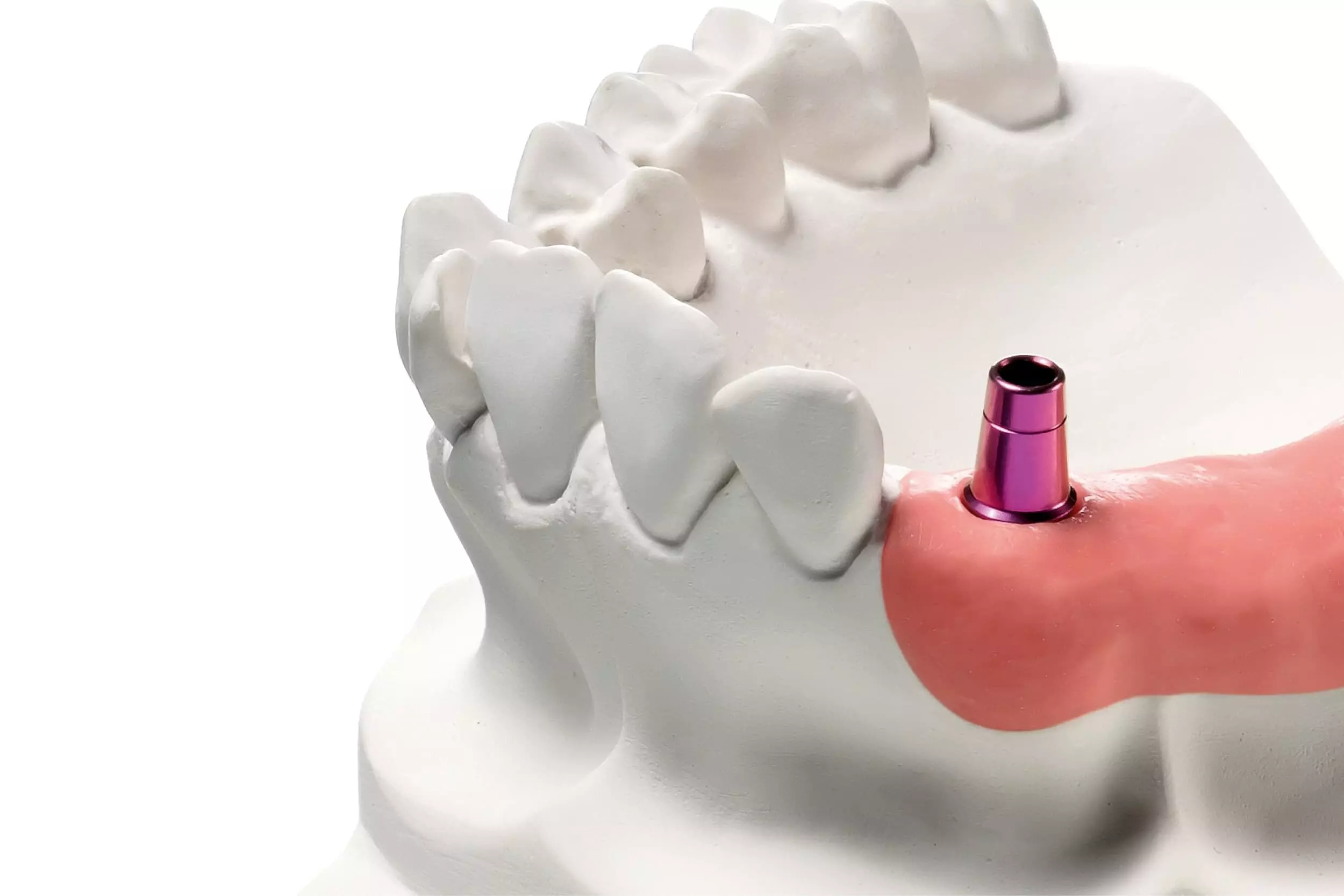
CAPS system may streamline fabrication process of implant supported fixed complete dental prostheses in one single visit: Study

CAPS system may streamline the fabrication process of implant-supported fixed complete dental prostheses in one single visit suggests a new study published in the Journal of Dentistry.

This article presents a novel complete-arch pillar system (CAPS) to register implant position and maxillomandibular relationship in one single visit for implant-supported fixed complete dental prostheses (IFCDPs). The novel system presents a 3-unit toolset comprising intraoral scan bodies (ISBs), lateral pillar attachments (LPAs) and occlusal pillar attachments (OPAs). A 2-stage single visit workflow by an intraoral scanner (Trios 5) was introduced. The first stage “Screw-Scan-Done” was used to describe complete-arch intraoral implant scanning using LPAs. The second stage “Screw-Occlude-Done” involved virtual occlusal recording using OPAs. Two patients with one single edentulous arch were selected for this study. In the first patient, 6 bone level implants (Bone Level Tapered, Straumann) were placed in the edentulous maxilla at positions 12, 14, 16, 22, 24 and 26. In the second patient, 4 bone level implants (NobelActive CC, Nobel Biocare) were placed in the edentulous mandible at positions 32, 35, 42 and 45. A CAD-CAM procedure was initiated with the acquired IOS data to fabricate an interim IFCDP at the same day. Periapical radiographs were obtained of the implant-prosthetic connection of the definitive IFCDPs to verify the passive fit. Metrology software (Geomagic Qualify, 3D Systems – Matlab, Mathworks) assessed the implant analog position in the 3D-printed casts used for fabricating the definitive IFCDPs. A quantitative occlusal relationship analysis was performed with IOS. Results: Radiographic examination revealed no gaps at the implant-prosthetic connection of the definitive IFCDPs. The 3D-printed casts showed an overall average distance deviation within the clinically acceptable range of errors of 150 µm. Quantitative occlusal relationship analysis with IOS showed well-distributed contacts. Within the limitations of this study, the following conclusions can be drawn: (1) A 3-unit toolset with ISBs, LPAs and OPAs allows to register the implant position and maxillomandibular relationship in one single visit; (2) the 2-stage clinical workflow with the CAPS system facilitates the IOS data acquisition for fabrication of an interim IFCDP at the same day; (3) a passive fit was demonstrated for the interim and the definitive IFCDPs. The CAPS system can help clinicians register the implant position and the maxillomandibular relationship in one visit to fabricate an IFCDP.

Keywords: Scan body; Complete-arch; Intraoral scanner; Pillar; Scan body attachment; Occlusal pillar

Reference:

Philippe Nuytens, Francesco Grande, Rani D’haese, Ziad Salameh, Luca Lepidi,

Novel complete-arch pillar system (CAPS) to register implant position and maxillomandibular relationship in one single visit, Journal of Dentistry, Volume 143, 2024, 104885, ISSN 0300-5712. https://doi.org/10.1016/j.jdent.2024.104885.

(https://www.sciencedirect.com/science/article/pii/S0300571224000551)

Keywords:

CAPS system, streamlined fabrication process, implant supported, fixed complete dental prostheses, one single visit, Journal of Dentistry, Philippe Nuytens, Francesco Grande, Rani D’haese, Ziad Salameh, Luca Lepidi

Powered by WPeMatico

Antisperm Antibodies After Vasectomy Reversal Not Linked to Pregnancy Rates or contraception method: Study

Following vasectomy reversal, patients often have concerns about whether antisperm antibodies (ASAs) will affect their ability to conceive. However, existing research on the relationship between ASA levels and pregnancy rates after vasectomy reversal is limited. A recent retrospective study aimed to examine this relationship and provide insights for patient counseling. This study was published in the journal of Urology by Catherine s. And colleagues.

While vasectomy reversal is a common procedure for restoring fertility in men who have undergone vasectomy, questions remain regarding the impact of ASAs on pregnancy outcomes post-reversal. The American Urological Association vasectomy guidelines highlight the need for further research to address these uncertainties.

The study conducted a retrospective chart review and phone interviews with patients who underwent vasectomy reversal between 2000 and 2018. Patients were categorized based on their ASA levels, and pregnancy rates and methods of conception were analyzed as primary outcomes.

Key Findings:

  • A total of 204 patients were included in the study, with a median age of 40 years and a median obstruction interval of 7.3 years.

  • 85 patients (42%) had low ASA levels (<50%), while 119 (58%) had high ASA levels (≥50%).

  • Among the patients with low ASA levels, 70% achieved pregnancy, with 59% resulting from spontaneous conception.

  • For patients with high ASA levels, 75% achieved pregnancy, with 40% resulting from spontaneous conception.

  • Fisher’s exact test p-value for the difference in pregnancy rates based on ASA levels was 0.2, indicating no significant association.

The study found no association between ASA levels and pregnancy rates or method of conception following vasectomy reversal. These findings provide valuable insights for patient counseling, alleviating concerns regarding the potential impact of ASAs on fertility outcomes post-reversal. As such, clinicians can better inform patients about their likelihood of achieving pregnancy after vasectomy reversal, regardless of ASA levels.

Reference:

Nam, C. S., Tooke, B. P., Strasser, O., Hameed, M. A., Chinnusamy, S., Van Til, M., Daignault-Newton, S., & Dupree, J. M. Antisperm antibody levels after vasectomy reversal are not associated with pregnancy rates or method of conception. Urology,2024. https://doi.org/10.1016/j.urology.2024.02.028

Powered by WPeMatico

Lower risk of death, major adverse cardiovascular events in hyperthyroid patients treated with surgery than with medication

Hyperthyroidism, or overactive thyroid, happens when your thyroid gland makes more thyroid hormones than your body needs. Excessive thyroid hormones from hyperthyroidism elevate cardiovascular risks. While hyperthyroidism can be treated with anti-thyroid drugs, radioactive iodine ablation or surgery, evidence comparing long-term benefits and risks of the three treatment modalities presents a challenge for informed decision-making and likely contributes to variations in treatment.

In a new study from Boston University Chobanian & Avedisian School of Medicine, researchers have found that hyperthyroid patients treated with surgery had a 47% lower risk of death and a 24% lower risk of major adverse cardiovascular events than patients treated with medication. They also found that patients treated with radioactive iodine had a 55% lower likelihood of major adverse cardiovascular events compared to the patients treated with medication.

“While there are three main treatment options for this condition, the selection of therapy needs to be individualized-this is not one-size-fits-all,” explains co-corresponding author Elizabeth Pearce, MD, MSc, professor of medicine at the school. “Our study suggests that it may be important to consider an individual’s cardiovascular risk in making decisions about hyperthyroidism treatment.”

The researchers used data from 114,062 patients from Taiwan’s National Health Insurance Research Database who were diagnosed with hyperthyroidism between 2011 and 2020. They compared rates of major cardiovascular events (acute myocardial infarction, stroke, heart failure, and death from cardiovascular disease) and death from all causes in patients treated with each of the three types of hyperthyroidism treatment.

According to the researchers, thyroid surgery or radioactive iodine treatment may be better treatment options than long-term anti-thyroid drug use in patients with hyperthyroidism who also are at high risk for cardiovascular diseases. “The improved outcomes observed in our study with surgery or radioactive iodine may have been related to more prompt and complete resolution of hyperthyroidism than was achieved with medication. However, future prospective studies will be needed to better understand these risks,” added Pearce who also is an endocrinologist at Boston Medical Center.

These finding appear online in JAMA Network Open.

Powered by WPeMatico

Smiling is secret to seeing happiness, new research reveals

Smiling for just a split second makes people more likely to see happiness in expressionless faces, new University of Essex research has revealed.

The study led by Dr Sebastian Korb, from the Department of Psychology, shows that even a brief weak grin makes faces appear more joyful.

The pioneering experiment used electrical stimulation to spark smiles and was inspired by photographs made famous by Charles Darwin.

A painless current manipulated muscles momentarily into action — creating a short uncontrollable smile.

This is the first time facial electrical stimulation has been shown to affect emotional perception.

Dr Korb hopes the research can explore potential treatments for depression or disorders that affect expression, like Parkinson’s and autism.

He said: “The finding that a controlled, brief and weak activation of facial muscles can literally create the illusion of happiness in an otherwise neutral or even slightly sad looking face, is ground-breaking.

“It is relevant for theoretical debates about the role of facial feedback in emotion perception and has potential for future clinical applications.”

Dr Korb used a modernised version of a technique first developed in the 19th century by the French physician Duchenne de Boulogne.

Darwin published drawings of Duchenne’s work in The Expression of the Emotions in Man and Animals — his third major work on evolution.

However, the voltage was dialled down for the new experiments to ensure the safety of participants and better control the smiles.

By using computers, the team were able to control the onset of smiles with millisecond precision.

In total 47 people took part in the Essex study which was published in Social Cognitive and Affective Neuroscience.

They were shown digital avatars and asked to assess whether they looked happy or sad.

In half the trials, smiling muscles were activated at the onset of the face.

It emerged that producing a weak smile for 500 milliseconds was enough to induce the perception of happiness.

Dr Korb says the results help us understand facial feedback and he hopes to expand the study.

He said: “We are currently conducting more al research to further explore the phenomenon in healthy participants.

“In the future, however, we hope to apply this technique to explore facial emotion recognition, for people with conditions like Parkinson’s, who are known to have reduced spontaneous facial mimicry and impaired facial emotion recognition.

“Moreover, we have published guidelines to allow other researchers to safely start using electrical facial muscle stimulation.”

Reference:

Themis Nikolas Efthimiou, Joshua Baker, Alasdair Clarke, Arthur Elsenaar, Marc Mehu, Sebastian Korb. Zygomaticus activation through facial neuromuscular electrical stimulation (fNMES) induces happiness perception in ambiguous facial expressions and affects neural correlates of face processing. Social Cognitive and Affective Neuroscience, 2024; 19 (1) DOI: 10.1093/scan/nsae013.

Powered by WPeMatico

Use of ultrahigh spatial-resolution photon-counting detector CT improves assessment of calcified stenoses: Study

Germany: A recent study published in the journal Radiology showed improvement in vivo and in vitro coronary stenosis assessment for calcified stenoses using ultrahigh spatial-resolution photon-counting detector (PCD) computed tomography (CT) reconstructions. This leads to a lower percentage of diameter stenosis (DS) compared with standard resolution and clinically relevant reclassification rates.

“Ultrahigh-spatial-resolution reconstructions led to the reclassification of 54.4% patients to lower CAD-RADS (Coronary Artery Disease Reporting and Data System) category than that assigned using standard resolution,” the researchers reported.

Coronary CT angiography is a first-line test in coronary artery disease (CAD) but is limited by severe calcifications. Photon-counting–detector CT improves spatial resolution. Therefore, Moritz C. Halfmann, University Medical Center of the Johannes Gutenberg-University, Langenbeckstr 1, Mainz, Germany, and colleagues aimed to investigate the effect of improved spatial resolution on coronary stenosis assessment and reclassification.

For this purpose, the researchers performed a prospective evaluation of coronary stenoses in a vessel phantom (in vitro) containing two stenoses (25%, 50%), and coronary stenoses were evaluated retrospectively in patients (in vivo) who underwent ultrahigh-spatial-resolution cardiac PCD CT (from July 2022 to April 2023).

Image reconstruction was done at standard resolution (section thickness, 0.6 mm; increment, 0.4 mm; Bv44 kernel), high spatial resolution (section thickness, 0.4 mm; increment, 0.2 mm; Bv44 kernel), and ultrahigh spatial resolution (section thickness, 0.2; increment, 0.1 mm; Bv64 kernel). Percentages of DS were compared between reconstructions.

In vitro values were compared with the manufacturer specifications of the phantom, and patient results were assessed regarding the impact on CAD-RADS reclassification.

The in vivo sample included 114 patients (mean age, 68 years; 71 male patients).

The study led to the following findings:

  • In vitro, percentage DS measurements were more accurate with increasing spatial resolution for 25% and 50% stenoses (mean bias for standard resolution, high spatial resolution, and ultrahigh spatial resolution, respectively: 10.1%, 8.0%, and 2.3%).
  • In vivo, results confirmed decreasing median percentage DS with increasing spatial resolution for calcified stenoses (n = 161) (standard resolution, high spatial resolution, and ultrahigh spatial resolution, respectively: 41.5%, 34.8%, and 26.7%), whereas noncalcified (n = 13) and mixed plaques (n = 19) did not show evidence of a difference.
  • Ultrahigh-spatial-resolution reconstructions led to a reclassification of 54.4% of patients to a lower CAD-RADS category than that assigned using standard resolution.

“Ultrahigh spatial resolution at photon-counting detector coronary CT angiography (CCTA) results in relevant rates of stenosis reclassification, which may impact the rate of downstream testing in patients with stable chest pain and may improve the role of CCTA as a gatekeeper for follow-up studies,” the researchers concluded.

Reference:

Halfmann MC, Bockius S, Emrich T, Hell M, Schoepf UJ, Laux GS, Kavermann L, Graafen D, Gori T, Yang Y, Klöckner R, Maurovich-Horvat P, Ricke J, Müller L, Varga-Szemes A, Fink N. Ultrahigh-Spatial-Resolution Photon-counting Detector CT Angiography of Coronary Artery Disease for Stenosis Assessment. Radiology. 2024 Feb;310(2):e231956. doi: 10.1148/radiol.231956. PMID: 38376407.

Powered by WPeMatico

Left Main Coronary Artery Calcium and Diabetes Confer Very-High-Risk Equivalence in CAC more than 1,000: Study

USA: Among asymptomatic patients with coronary artery calcium (CAC) score > 1,000, those with severe calcium in the left main coronary artery and diabetes have more than a sevenfold greater risk of death from atherosclerotic cardiovascular disease (ASCVD) versus those who have a high CAC score alone, a recent study has shown.

Not all patients with very high levels of CAC are created equal when it comes to CV risk, with the research published in JACC: Cardiovascular Imaging providing some clues on how to identify a particularly high-risk patient who could benefit from aggressive primary prevention strategies across several domains, including low-density lipoprotein-cholesterol lowering.

Although coronary artery calcium of ≥1,000 is a subclinical atherosclerosis threshold to consider combination lipid-lowering therapy, differentiating very high from high ASCVD risk in this patient population is not well-defined. Therefore, Alexander C. Razavi, Emory Clinical Cardiovascular Research Institute, Emory University School of Medicine, Atlanta, Georgia, USA, and colleagues aimed to identify risk factors equating with very high-risk ASCVD mortality rates among people with a CAC of ≥1,000.

For this purpose, the researchers studied 2,246 asymptomatic patients with a CAC of ≥1,000 from the CAC Consortium without a prior ASCVD event. The mean age was 66.6 years; 14% were female, and 10% were non-White. During a median follow-up of 11.3 years, Cox proportional hazards regression modelling was performed for ASCVD mortality.

Crude ASCVD mortality rates were compared with those reported for secondary prevention trial patients classified as very high risk, defined by ≥2 major ASCVD events or 1 major event and ≥2 high-risk conditions (1.4 per 100 person-years).

The study led to the following findings:

  • The median CAC score was 1,592, and 6% had severe left main (LM) CAC (vessel-specific CAC ≥300).
  • Diabetes (HR: 2.04) and severe LM CAC (HR: 2.32) were associated with ASCVD mortality.
  • The ASCVD mortality per 100 person-years for all patients was 0.8, although higher rates were observed for diabetes (1.4), severe LM CAC (1.3), and both diabetes and severe LM CAC (7.1).

“Among asymptomatic patients with a CAC of ≥1,000 without a prior index event, diabetes, and severe LM CAC define very high-risk atherosclerotic cardiovascular disease, identifying individuals who may benefit from more intensive prevention therapies across several domains, including LDL-cholesterol lowering,” the researchers wrote.

Reference:

Razavi AC, Shaw LJ, Berman DS, Budoff MJ, Wong ND, Vaccarino V, van Assen M, De Cecco CN, Quyyumi AA, Mehta A, Muntner P, Miedema MD, Rozanski A, Rumberger JA, Nasir K, Blumenthal RS, Sperling LS, Mortensen MB, Whelton SP, Blaha MJ, Dzaye O. Left Main Coronary Artery Calcium and Diabetes Confer Very-High-Risk Equivalence in Coronary Artery Calcium >1,000. JACC Cardiovasc Imaging. 2024 Feb 12:S1936-878X(24)00026-3. doi: 10.1016/j.jcmg.2023.12.006. Epub ahead of print. PMID: 38385932.

Powered by WPeMatico

Dysfunctional blood monocytes may predict CVD risk in patients with type 2 diabetes: Study

France: A recent study published in Circulation Research has revealed that frequency and monocyte phenotypic profile are closely associated with cardiovascular (CV) risk in patients with type 2 diabetes (T2D).

The findings imply that assessing monocyte count and frequency is a valuable predictive marker for the risk of CV events in diabetes patients.

Previous studies have shown diabetes to be a major risk factor for atherosclerotic cardiovascular diseases (ASCVD) with a 2-fold higher risk of CV events in patients with diabetes compared with those without. However, there is cardiovascular risk heterogeneity among patients with T2D that cardiovascular risk scores fail to detect. None of these cardiovascular risk scores considers proatherogenic immune effector, monocyte blood count.

Circulating monocytes are inflammatory effector cells involved in type 2 diabetes and atherogenesis. Nicolas Venteclef, IMMEDIAB Laboratory, Paris, France, and colleagues investigated the relationship between circulating monocytes and CV risk progression in people with T2D, using phenotypic, metabolomic, and transcriptomic analyses in a cross-sectional study population of 672 participants with T2D.

The study led to the following findings:

· Coronary artery calcium score was positively correlated with blood monocyte count and frequency of the classical monocyte subtype.

· Unsupervised k-means clustering based on monocyte subtype profiles revealed 3 main endotypes of people with type 2 diabetes at varying risk of cardiovascular events. These observations were confirmed in a validation cohort of 279 T2D participants.

· The predictive association between monocyte count and major adverse cardiovascular events was validated through an independent prospective cohort of 757 patients with T2D.

· Integration of monocyte transcriptome analyses and plasma metabolomes showed a disruption of mitochondrial pathways (tricarboxylic acid cycle, oxidative phosphorylation pathway) that underlined a proatherogenic phenotype.

The findings reveal an association between routine white blood cell-derived monocyte counts and CV risk among patients with T2D. By combining monocyte immunophenotypic, immunophenotypic, and plasma metabolomic analyses, the researchers showed an oxidative phosphorylation and mitochondrial modulation in a specific monocyte subtype, linked to an endotype of type 2 diabetes patients that are at higher cardiovascular risk.

We show an association of 2 phenomena: an increase in the monocyte pool, resulting in more ready infiltration of atherosclerotic plaques promoting progression,” the researchers wrote. “Mitochondrial dysfunction was apparent in these monocytes, providing a potential explanation for the increased CV risk associated with T2D.”

“Our findings present monocytes as good candidates for predicting cardiovascular complication development among type 2 diabetes patients and highlight the relevance of phenotyping circulating monocytes to assess CV risk in T2D,” they concluded.

Reference:

Julla JB, Girard D, Diedisheim M, Saulnier PJ, Tran Vuong B, Blériot C, Carcarino E, De Keizer J, Orliaguet L, Nemazanyy I, Potier C, Khider K, Tonui DC, Ejlalmanesh T, Ballaire R, Mambu Mambueni H, Germain S, Gaborit B, Vidal-Trécan T, Riveline JP, Garchon HJ, Fenaille F, Lemoine S, Carlier A, Castelli F, Potier L, Masson D, Roussel R, Vandiedonck C, Hadjadj S, Alzaid F, Gautier JF, Venteclef N. Blood Monocyte Phenotype Is A Marker of Cardiovascular Risk in Type 2 Diabetes. Circ Res. 2024 Jan 19;134(2):189-202. doi: 10.1161/CIRCRESAHA.123.322757. Epub 2023 Dec 28. PMID: 38152893.

Powered by WPeMatico

COVID-19 tied to elevated risk for autoimmune inflammatory rheumatic diseases up to 12 months after infection: Study

South Korea: A recent study published in Annals of Internal Medicine has revealed an association between SARS-CoV-2 infection and increased risk for incident autoimmune inflammatory rheumatic diseases (AIRDs) versus matched patients without SARS-CoV-2 infection or with influenza infection.

The risk for incident AIRD was higher with greater acute COVID-19 severity. 

The large, binational study found that SARS-CoV-2 infection was associated with an increased risk for autoimmune inflammatory rheumatic diseases (AIRDs) that extends up to 12 months after infection. The risk was found to be higher with greater severity of acute COVID-19, even among those who were vaccinated.

These findings suggest that care strategies for patients who survive COVID-19 should pay close attention to manifestations of AIRD, particularly after severe illness. 

Emerging data suggest a higher risk for AIRDs among persons with a history of COVID-19. However, these findings are based entirely on comparisons between groups infected with SARS-CoV-2 and those that are not, which might be biased by differences in health-seeking behavior and inherent risk factors within the groups. In addition, studies have not explored the effect of vaccination and other modifiable factors on prevention of long term COVID-19 complications.

Researchers from Kyung Hee University, Seoul, South Korea used data from two national, large-scale, general population-based cohort studies in Korea and Japan to investigate the effect of COVID-19 on long-term risk for incident AIRD over various follow-up periods. The data comprised of more than 10 million Korean and 12 million Japanese adults, including those with COVID-19 between January 2020 and December 2021, matched to patients with influenza infection and to uninfected control patients. The researchers assessed the data for onset of AIRD at 1, 6, and 12 months after COVID-19 or influenza infection, or the respective matched index date of uninfected control patients. They found increased risk for incident AIRD up to 12 months after COVID-19 diagnosis compared with the other two groups. Greater severity of acute COVID-19 was associated with higher risk for incident AIRD. The authors noted that COVID-19 vaccination was associated with reduced risk for incident AIRD after SARS-CoV-2 infection, except for among those who had severe COVID-19 despite vaccination.

Reference:

Min Seo Kim, Hayeon Lee, Seung Won Lee, Long-Term Autoimmune Inflammatory Rheumatic Outcomes of COVID-19, Annals of Internal Medicine, https://doi.org/10.7326/M23-1831.

Powered by WPeMatico

Low birthweight and overweight in young adulthood tied to elevated risk of early type 2 diabetes in men: Study

Sweden: A recent study has revealed low birth weight and overweight in young adulthood to be the major developmental determinants of adult type 2 diabetes risk in men.

“They contribute in an additive manner to the risk of type 2 diabetes,” the researchers wrote. “To reduce the risk of type 2 diabetes, young adult overweight should be avoided, particularly in boys with a low birthweight.”

The new research is being  presented at this year’s European Congress on Obesity (ECO) in Venice, Italy (12-15 May), and published in Diabetologia (the journal of th European Association for The Study of Diabetes [EASD]). 

Having a low birthweight together with being overweight in young adulthood (but not childhood) contributes to the development of type 2 diabetes at an early age (59 years or younger) in men, the study stated. 

Notably, the study involving over 34,000 Swedish men, found that those born with a low birthweight (<2.5 kg/5 lbs 8oz) who were also overweight at aged 20 years (BMI >25kg/m²) were 10 times more likely to develop early type 2 diabetes than those with a birthweight in the normal range (2.5-4.5 kg) who were normal weight as young adults (BMI <25 kg/m²).

Importantly, the researchers from the University of Gothenburg and Sahlgrenska University Hospital also found that babies with a low birthweight who were overweight at age 20 years had a 27% absolute risk of developing early type 2 diabetes, compared with an absolute risk of 6% for those with a birthweight in the normal range who were also normal weight at aged 20 years. This suggests that preventing excess weight gain during young adulthood in boys born with low birthweight could reduce the absolute risk of early type 2 diabetes by 21%.

Type 2 diabetes is being diagnosed at progressively younger ages, suggesting that significant risk may begin to accumulate during the developmental period. The association between low birthweight and overweight in childhood and/or young adulthood and type 2 diabetes in adults is already known, but it has been unclear how much influence the combination of these two factors exerts.

To find out more, researchers analysed data from 34,231 men born between 1945 and 1961 involved in the BMI Epidemiology Study (BEST) Gothenburg—a population-based cohort examining the associations between growth and BMI development in early life and the risk of disease in later life.

The researchers analysed birthweight and BMI of participants from school health care records (at the age of 8 years) and from medical examinations on enrolment in the military (at age 20), which was mandatory until 2010.

Participants were followed from 30 years of age until type 2 diabetes diagnosis, death, or emigration, or until 31st December 2019. Information on type 2 diabetes diagnoses was retrieved from Swedish national registers to estimate the risk of early (<59.4 years) and late (>59.4 years) type 2 diabetes. They also examined whether these associations were independent of, or modified by, socioeconomic factors such as level of education.

During an average 34 years follow up (after 30 years of age), a total of 2,733 cases of type 2 diabetes were diagnosed (1,367 cases of early diabetes and 1,366 cases of late diabetes). The analyses found that birthweight below the average (median; <3.6 kg/7lbs 9oz) and overweight at age 20 years (BMI >25 kg/m²), but not overweight at age 8 years (BMI >17.9 kg/m²), were associated with an increased risk of both early and late type 2 diabetes.

Furthermore, low birthweight and overweight in young adulthood were found to have an additive effect on the risk of type 2 diabetes. For example, having a below average birthweight (<3.6 kg/7lbs 9oz), followed by overweight at 20 years of age was associated with a six times greater risk of developing early type 2 diabetes. Whereas a lower birthweight (<2.5 kg/5 lbs 8oz) combined with later overweight at 20 years was linked with a 10 fold greater risk of developing early type 2 diabetes.

Adjusting for education, a known risk factor for type 2 diabetes, did little to change the results.

“Our findings establish low birthweight and overweight in young adulthood as the main developmental determinants, whereas overweight in childhood is of lesser importance for type 2 diabetes in adult men”, says lead author Dr Jimmy Celind, a researcher at Sahlgrenska Academy’s Institute of Medicine at the University of Gothenburg. “The combination of low birthweight followed by overweight at age 20 years is associated with a massive excess risk for early type 2 diabetes, which is substantially higher than the risk associated with low birthweight or being overweight as a young adult separately.”

Co-author Dr Jenny Kindblom from Sahlgrenska University Hospital adds, “It’s possible that the metabolic consequences of fetal growth restriction, which promotes resilience against starvation through fat storage and insulin resistance, when combined with a detrimental BMI trajectory during puberty when the insulin resistance is at a lifetime peak due to the surge of growth and sex hormones, result in an additive excess risk for later type 2 diabetes. Public health initiatives should target boys born with low birthweight to work on prevention of overweight as young adults, to reduce this huge excess risk for early type 2 diabetes.”

The authors acknowledge that the findings are associations only and that the study wasn’t designed to measure direct cause and effect, and point to several limitations, including that the participants were mainly white men which may limit the generalisability of the findings to other ethnicities and women. In addition, the analyses were unable to account for the influence of other known risk factors for type 2 diabetes such as smoking, dietary habits, and physical activity which could have influenced the results.

Reference:

Célind J, Bygdell M, Bramsved R, Martikainen J, Ohlsson C, Kindblom JM. Low birthweight and overweight during childhood and young adulthood and the risk of type 2 diabetes in men: a population-based cohort study. Diabetologia. 2024 Feb 22. doi: 10.1007/s00125-024-06101-y. 

Powered by WPeMatico