Odisha HC directs OCMR to devise methods to curb fake doctors menace

Cuttack: In its ongoing effort to curb the fake doctors’ menace in the state, the Odisha High Court on Tuesday asked Dr Bijay Kumar Mohapatra, the president of the Odisha Council for Medical Registration (OCMR) to develop a method for identifying fraudulent allopathic doctors. 

The court issued this directive following a previous order, asking him to appear in person to the court and share his views on effectively tackling the problem of fake doctors.

Subsequently, he appeared before the court on Tuesday and assured the judges that he would put forth definite suggestions to meet the challenges of the menace of fake allopathic doctors after consulting with the National Medical Commission and commissioner-cum-secretary of the Health & Family Welfare department.

Also read- Quack Practising For 40 Years Arrested In Guwahati

“We expect that while placing the suggestions in court, the president of OCMR may also consider the mode for identification of genuine allopathic doctors so that even an illiterate person living in a remote area can identify them and distinguish between a genuine doctor and fake doctor,” said the court as reported by TOI.

The court has asked him to file an affidavit regarding the issue on March 27.

The division bench of Chief Justice Chakradhari Sharan Singh and Justice MS Raman was hearing a PIL on fake doctors filed by the Odisha State Legal Services Authority.

Medical Dialogues team had earlier reported that Amicus curiae Gautam Misra had earlier suggested the creation of a comprehensive online database of all medical practitioners in the state, which will be accessible to the general public to crosscheck the genuineness of all medical practitioners. The court had directed the state government to consider implementing the suggestion as well.

Informing the court about the database of doctors registered by OCMR, Dr Mohapatra said, “While 31,355 allopathic doctors have been registered, the scrutiny of their certificates and other particulars are underway. After validation, the database of allopathic doctors will be placed on the OCMR website”

During the session, the court expressed dissatisfaction with the state government’s response in an affidavit in pursuance to orders issued by it regarding the surveillance mechanism to identify fake doctors. 

It said, “It is unfortunate that the affidavit does not address at all the issues as regards surveillance mechanism taken note of in the court’s order on June 22, 2023.”

As a result, the court has directed the commissioner-cum-secretary of the Health & Family Welfare department to file an additional affidavit on surveillance mechanisms to identify fake doctors within two weeks. 

In previous orders, the court had directed the state government to inform whether there was any surveillance mechanism to identify the fake doctors. If not, whether the state government is contemplating introducing surveillance to curb the menace of fake doctors and what further action it proposed to take against establishments either run by the fake doctors or collaborated by them.

Also read- Fake Doctor Menace: Odisha HC Summons Chairman Of Odisha Council For Medical Registration

Powered by WPeMatico

Ageing and Schizophrenia could have a shared biological basis

Scientists from the Broad Institute of MIT, Harvard Medical School, and McLean Hospital have found similar gene activity changes in brain tissue from individuals with schizophrenia and older adults. These findings imply a shared biological foundation for cognitive decline seen in both groups.

The study published in the Journal Nature, revealed that in individuals with schizophrenia and in older adults without schizophrenia, two brain cell types called astrocytes and neurons reduced their expression of genes that support the junctions between neurons called synapses, compared to healthy or younger people.

Schizophrenia is a challenging mental disorder marked by symptoms like hallucinations, delusions, and cognitive decline. Treatment involves medication, therapy, and support services, but managing the disorder can still be complex due to its multifaceted nature and limited treatment options for cognitive decline.

The team analysed gene expression in more than a million individual cells from postmortem brain tissue from 191 people and discovered tightly synchronised gene expression changes in the two cell types: when neurons decreased the expression of certain genes related to synapses, astrocytes similarly changed the expression of a distinct set of genes that support synapses. The team called this coordinated set of changes the Synaptic Neuron and Astrocyte Program (SNAP). Even in healthy, young people, the expression of the SNAP genes always increased or decreased in a coordinated way in their neurons and astrocytes.

“Science often focuses on what genes each cell type expresses on its own,” said Steve McCarroll, a co-senior author on the study and an institute member at the Broad Institute. “But brain tissue from many people, and machine-learning analyses of those data, helped us recognize a larger system. These cell types are not acting as independent entities, but have really close coordination. The strength of those relationships took our breath away.”

The findings revealed that SNAP varied greatly even among people without schizophrenia, suggesting that SNAP could be involved in cognitive differences in healthy humans. Much of this variation was explained by age; SNAP declined substantially in many — but not all — older individuals, including both people with and without schizophrenia.

“With better understanding of SNAP, it might be possible to identify life factors that positively influence SNAP, and develop medicines that help stimulate SNAP, as a way to treat the cognitive impairments of schizophrenia or help people maintain their cognitive flexibility as they age.” concluded McCarroll.

Reference: Emi Ling, James Nemesh, Melissa Goldman, Nolan Kamitaki, Nora Reed, Robert E. Handsaker, Giulio Genovese, Jonathan S. Vogelgsang, Sherif Gerges, Seva Kashin, Sulagna Ghosh, John M. Esposito, Kiely Morris, Daniel Meyer, Alyssa Lutservitz, Christopher D. Mullally, Alec Wysoker, Liv Spina, Anna Neumann, Marina Hogan, Kiku Ichihara, Sabina Berretta, Steven A. McCarroll. A concerted neuron–astrocyte program declines in ageing and schizophrenia. Journal: Nature, 2024; DOI: 10.1038/s41586-024-07109-5

Powered by WPeMatico

State’s largest skin bank launched at Ahmedabad Civil Hospital

Ahmedabad: The state government in a collaborative effort with the Rotary Club Kankaria has launched Gujarat’s largest government skin bank at Civil Hospital in Ahmedabad on Wednesday.

The Civil Hospital in Ahmedabad on Wednesday launched Gujarat’s largest government skin bank, besides other advanced medical facilities.

Gujarat Health Minister Rushikesh Patel presided over the inauguration ceremony. 

Rushikesh Patel, in his address, highlighted the initiatives as a testament to the collective efforts of the government, society, and philanthropic organisations in realising the vision of a “Developed Gujarat leading to Developed India by 2047”.

The projects are expected to significantly elevate the level of healthcare services available to the citizens, reflecting a comprehensive approach to medical care and patient welfare, an official said.

As per a media report in the Ahmedabad Mirror, Set up for Rs 48 lakh by the Rotary Club of Kankaria in Ahmedabad, the bank is touted to be the “biggest” Skin Bank in the state. Officials, however, said the size of the Skin Bank does not matter as it is like a processing lab where cadaveric skin tissue donated by family members of deceased persons is processed and stored for use in grafting in patients with burns or severe injuries.

Also Read:Delhi AIIMS opens second skin bank after Safdurjung hospital

The establishment of the skin bank, a collaborative effort with Rotary Club Kankaria, is for treating burn victims and patients requiring skin grafts due to various accidents.

The skin bank is poised to become a resource for the burns and plastic surgery department. Additionally, the inauguration featured the unveiling of a ‘128-slice GE CT scan’ machine, worth around Rs 6.25 crore.

This addition is expected to reduce wait times for patients needing CT scans while providing high-resolution imaging capabilities for better diagnosis and treatment planning.

Other noteworthy projects introduced include a new patient facilitation centre at the OPD, constructed with the support of Bank of Baroda, and innovative patient golf cart services funded by Astra Foundation and Canara Bank, aimed at enhancing patient mobility within the hospital premises. A contribution from the Rotary Club Kankaria also saw the allocation of Rs 12 lakh towards developing a children’s garden opposite the hospital.

Powered by WPeMatico

Microplastics and nanoplastics in plaque lining major blood vessel tied to heart attack, stroke, and early death: NEJM

Italy: A recent study has found a higher risk of a composite of stroke, myocardial infarction, or death from any cause at 34 months of follow-up among patients with carotid artery plaque in which microplastics and nanoplastics (MNPs) were detected versus those in whom MNPs were not detected. The findings were published online in the New England Journal of Medicine on March 7, 2024.

“An increased risk of combined stroke, myocardial infarction, or death from any cause over nearly 3 years was found in people with microplastics and nanoplastics detection in atherosclerotic plaque from surgically excised carotid plaque specimens (20.0% versus 7.5%, HR 4.53),” the researchers reported.

Out of 11 types of plastics assessed, the researchers detected two in particular in the atheroma: polyethylene in 58.4% of participants (at a mean concentration of 21.7 μg/mg plaque) and polyvinyl chloride in 12.1% (mean 5.2 μg/mg).

Polyvinyl chloride and polyethylene, in their several forms, are used in a wide range of applications, including the production of cosmetics and food containers, and water pipes. MNPs have been found in drinking water, a large range of cosmetic products, foods, and air, also in a form bound to fine, inhalable particulate matter. Given the wide distribution and availability of MNPs, the attribution of all potential sources in humans is nearly impossible.

The environment is being inundated increasingly with plastic due to industrial processes. Plastics break down into smaller nanoplastics (particles smaller than 1,000 nm) and microplastics (particles smaller than 5 mm), both of which enter the human body through inhalation, ingestion, and skin exposure and have been found in the lungs, placenta, liver, and other tissue,

Microplastics and nanoplastics are emerging as a potential risk factor for cardiovascular disease (CVD) in preclinical studies. However, there is a lack of direct evidence that this risk extends to humans. To fill this knowledge gap, Raffaele Marfella, University of Campania Luigi Vanvitelli, Caserta, Italy, and colleagues conducted a prospective, multicenter, observational study comprising patients who were undergoing carotid endarterectomy for asymptomatic carotid artery disease.

The researchers analyzed excised carotid plaque specimens for the presence of MNPs using pyrolysis-gas chromatography-mass spectrometry, electron microscopy, and stable isotope analysis. Inflammatory biomarkers were assessed with immunohistochemical assay and enzyme-linked immunosorbent assay.

The study’s primary endpoint was a composite of stroke, myocardial infarction, or death from any cause among patients with evidence of MNPs in plaque as compared with patients with plaque that showed no evidence of MNPs.

The study enrolled three hundred and four patients, and 257 completed a mean follow-up of 33.7 months.

The following were the key findings of the study:

  • Polyethylene was detected in carotid artery plaque of 58.4% of patients, with a mean level of 21.7±24.5 μg per milligram of plaque; 12.1% of patients also had measurable amounts of polyvinyl chloride, with a mean level of 5.2±2.4 μg per milligram of plaque.
  • Electron microscopy revealed visible, jagged-edged foreign particles among plaque macrophages and scattered in the external debris.
  • Radiographic examination showed that some of these particles included chlorine.
  • Patients in whom MNPs were detected within the atheroma were at higher risk for a primary end-point event than those in whom these substances were not detected (hazard ratio, 4.53).

According to the authors, a greater concentration of polyethylene correlated with greater expression of inflammatory biomarkers and markers of macrophage and lymphocyte infiltration.

However, study authors cautioned that the plastics group tended to be younger, more likely smokers and men, and had a greater prevalence of cardiovascular disease, diabetes, and dyslipidemia.

The study’s observational nature meant that it was potentially subject to other residual confounding, precluding any causal conclusions. Also, there was a possible risk of laboratory contamination.

Reference:

Marfella R, Prattichizzo F, Sardu C, Fulgenzi G, Graciotti L, Spadoni T, D’Onofrio N, Scisciola L, La Grotta R, Frigé C, Pellegrini V, Municinò M, Siniscalchi M, Spinetti F, Vigliotti G, Vecchione C, Carrizzo A, Accarino G, Squillante A, Spaziano G, Mirra D, Esposito R, Altieri S, Falco G, Fenti A, Galoppo S, Canzano S, Sasso FC, Matacchione G, Olivieri F, Ferraraccio F, Panarese I, Paolisso P, Barbato E, Lubritto C, Balestrieri ML, Mauro C, Caballero AE, Rajagopalan S, Ceriello A, D’Agostino B, Iovino P, Paolisso G. Microplastics and Nanoplastics in Atheromas and Cardiovascular Events. N Engl J Med. 2024 Mar 7;390(10):900-910. doi: 10.1056/NEJMoa2309822. PMID: 38446676.

Powered by WPeMatico

FDA Clears First Over-the-Counter Continuous Glucose Monitor

Today, the U.S. Food and Drug Administration cleared for marketing the first over-the-counter (OTC) continuous glucose monitor (CGM). The Dexcom Stelo Glucose Biosensor System is an integrated CGM (iCGM) intended for anyone 18 years and older who does not use insulin, such as individuals with diabetes treating their condition with oral medications, or those without diabetes who want to better understand how diet and exercise may impact blood sugar levels. Importantly, this system is not for individuals with problematic hypoglycemia (low blood sugar) as the system is not designed to alert the user to this potentially dangerous condition.

“CGMs can be a powerful tool to help monitor blood glucose. Today’s clearance expands access to these devices by allowing individuals to purchase a CGM without the involvement of a health care provider,” said Jeff Shuren, M.D., J.D., director of the FDA’s Center for Devices and Radiological Health. “Giving more individuals valuable information about their health, regardless of their access to a doctor or health insurance, is an important step forward in advancing health equity for U.S. patients.”

The Stelo Glucose Biosensor System uses a wearable sensor, paired with an application installed on a user’s smartphone or other smart device, to continuously measure, record, analyze and display glucose values in people 18 years and older that are not on insulin and who do not have problematic hypoglycemia. Users can wear each sensor up to 15 days before replacing with a new sensor. The device presents blood glucose measurements and trends every 15 minutes in the accompanying app. Users should not make medical decisions based on the device’s output without talking to their healthcare provider.

Data from a clinical study provided to the FDA showed that the device performed similarly to other iCGMs. Adverse events reported in the study included local infection, skin irritation and pain or discomfort.

As part of the Center for Devices and Radiological Health’s strategic priority to advance health equity, CDRH will continue to support innovation that addresses health equity by moving care and wellness into the home setting.

Powered by WPeMatico

Use of CGM in type 1 diabetes patients may lower odds of developing retinopathy: Study

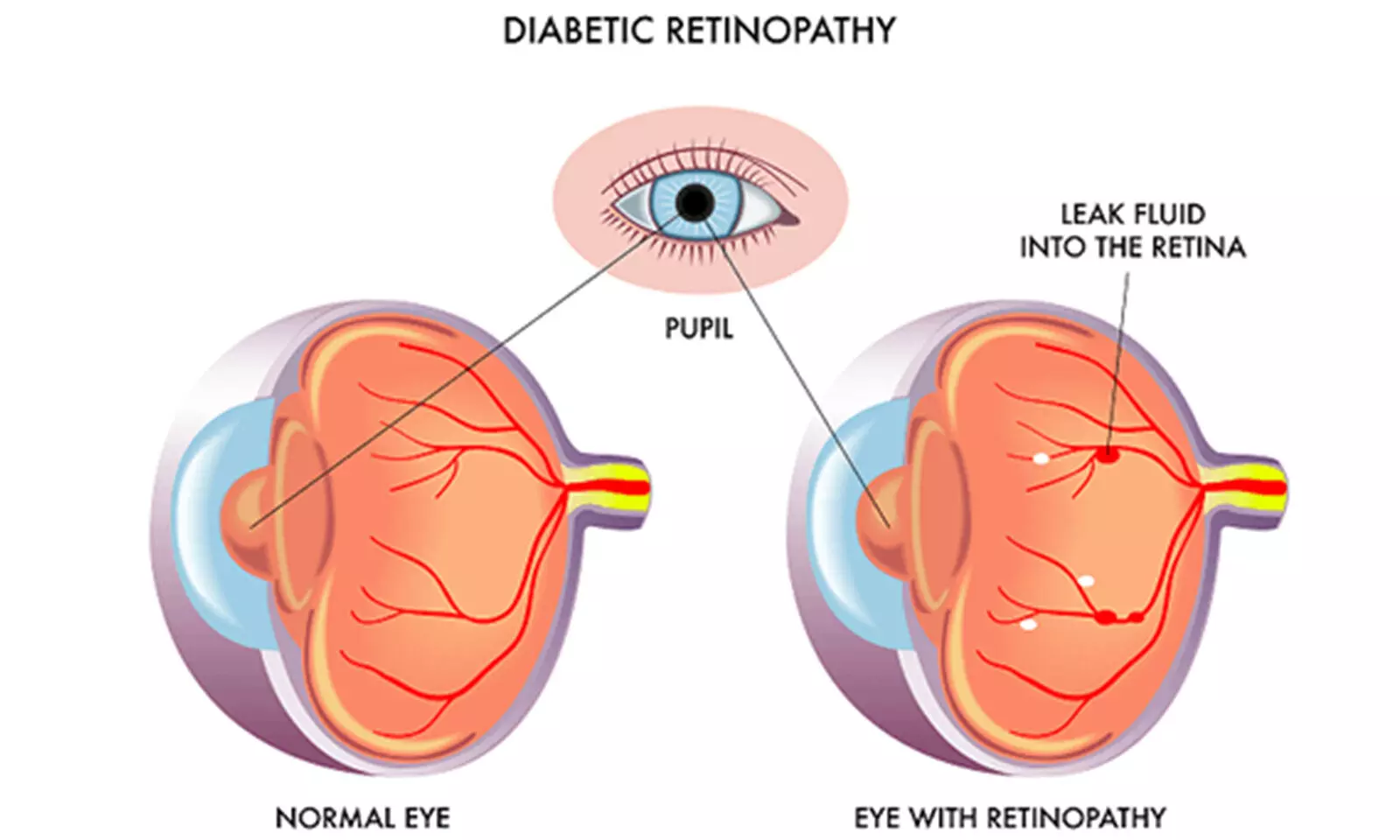
Diabetic retinopathy (DR) is a serious complication of type 1 diabetes (T1D) that can lead to vision impairment or blindness if left untreated. Understanding the impact of diabetes management technologies, such as continuous glucose monitoring (CGM) and insulin pumps, on DR outcomes is crucial for improving patient care and outcomes. The use of CGM and insulin pumps has revolutionized diabetes management by providing real-time glucose data and precise insulin delivery.

A recent retrospective cohort study aimed to evaluate the association between CGM, insulin pump use, or both, and the development of DR and proliferative diabetic retinopathy (PDR) in adults with T1D. This study was published in JAMA Network Open by T. Y. Alvin and colleagues.

The study included 550 adults with T1D from a tertiary diabetes center and ophthalmology center. Data were analyzed retrospectively from 2013 to 2021. Participants were categorized based on their use of diabetes technologies, including CGM, insulin pump, or both.

Key Findings:

  • 62.7% of patients used CGM, 58.2% used an insulin pump, and 47.5% used both.

  • 44% of participants had DR at any point during the study.

  • CGM use was associated with lower odds of developing DR and PDR.

  • In the longitudinal analysis, 21.8% of patients experienced progression of DR during the study period.

The study suggests that CGM use is associated with a reduced risk of developing DR and PDR in adults with T1D, even after adjusting for other factors like hemoglobin A1c levels. These findings underscore the potential benefits of CGM in diabetes management for mitigating the risk of DR and PDR. Early adoption and utilization of CGM technology may play a crucial role in preventing diabetic eye complications and improving long-term visual outcomes in individuals with T1D.

Reference:

Liu, T. Y. A., Shpigel, J., Khan, F., Smith, K., Prichett, L., Channa, R., Kanbour, S., Jones, M., Abusamaan, M. S., Sidhaye, A., Mathioudakis, N., & Wolf, R. M. Use of diabetes technologies and retinopathy in adults with type 1 diabetes. JAMA Network Open,2024;7(3):e240728. https://doi.org/10.1001/jamanetworkopen.2024.0728

Powered by WPeMatico

Low LDL-C levels tied to increased risk of bleeding in stroke patients receiving DAPT: JAMA

China: A cohort study of 7440 patients revealed an association between low LDL-C levels and an increased bleeding risk within three months in patients with high-risk transient ischemic attack or minor ischemic stroke who are receiving dual antiplatelet therapy, particularly those taking ticagrelor-aspirin.

“At three months, there were 270 (3.63%) bleeding events, and LDL-C below 70 mg/dL was linked with an increased risk of any bleeding and severe or moderate bleeding, with the risk of any bleeding risk increased at lower LDL-cholesterol levels in the ticagrelor-aspirin group.” the researchers wrote in their study published in JAMA Neurology.

No increased risk of any bleeding was seen in the clopidogrel-aspirin group. There was no significant association between LDL-C levels and the risk of severe or moderate bleeding in either the clopidogrel-aspirin or ticagrelor-aspirin group.

Low-density lipoprotein cholesterol (LDL-C) is associated independently with ischemic stroke risk. Despite the efficacy of low LDL-C levels in reducing recurrent stroke risk, some observational studies have revealed that lowering LDL-C might contribute to an increased risk of hemorrhagic events, particularly hemorrhagic stroke. Additionally, the findings of a Mendelian randomization study support a possible causal relationship between LDL-C and intracranial hemorrhage (ICH).

Evidence remains sparse on the bleeding risk associated with LDL-C levels in patients receiving dual antiplatelet therapy (DAPT). Therefore, Aichun Cheng, Capital Medical University, Beijing, China, and colleagues aimed to investigate the association of LDL-C levels with bleeding risk in patients with high-risk transient ischemic attack (HRTIA) or minor ischemic stroke (MIS) receiving DAPT.

For this purpose, the researchers analyzed pooled data from 2 randomized, double-blind, placebo-controlled clinical trials in China comprising patients with MIS or HRTIA who were receiving DAPT: the CHANCE trial enrolled patients at 114 sites from 2009 to 2012, and HANCE-2 enrolled patients at 202 centers from 2019 to 2021. Patients from both the trials were followed for 90 days.

Exposures were baseline LDL-cholesterol levels and receipt of clopidogrel-aspirin and ticagrelor-aspirin DAPT.

The study’s primary outcome was any bleeding, and the secondary outcome included severe or moderate bleeding within three months after randomization. The association of LDL-C levels and all outcomes was assessed.

In the two trials, 8996 patients with acute MIS or HRTIA who were receiving DAPT were included, 1066 without serum specimens, and 490 patients with missing baseline LDL-C value were excluded. Finally, the study included 7440 patients with DAPT (4486 in the clopidogrel-aspirin group and 2954 in the ticagrelor-aspirin group). The median age was 64.32 years, and 33.32% were women.

Based on the study, the researchers reported the following findings:

  • A total of 270 (3.63%) bleeding events were reported at three months, and LDL-C less than 70 mg/dL was associated with an increased risk of both any bleeding (aHR, 1.48) and severe or moderate bleeding (aHR, 2.78).
  • The risk of any bleeding was raised at lower LDL-C levels in the ticagrelor-aspirin group (aHR, 1.71). However, an increased risk of any bleeding was not seen in the clopidogrel-aspirin group (aHR, 1.30).
  • There was no significant association between LDL-C levels and the risk of severe or moderate bleeding in either the ticagrelor-aspirin or clopidogrel-aspirin group.

In conclusion, the findings suggest that low levels of LDL-cholesterol are associated with an increased risk of bleeding within three months among MIS or HRTIA patients receiving DAPT, particularly those taking ticagrelor-aspirin.

“Weighing the benefits and risks is important when simultaneously considering the selection of LDL-C target strategies and DAPT regimens among these patients,” the researchers wrote.

Reference:

Cheng A, Xue J, Wang A, et al. LDL-C Levels and Bleeding Risk in Patients Taking DAPT After Minor Ischemic Stroke or TIA. JAMA Neurol. Published online March 04, 2024. doi:10.1001/jamaneurol.2024.0086

Powered by WPeMatico

Herbal Medicine Schisanhenol: A potential drug for treatment of cytokine storm

Cytokine storm (CS) is an acute systemic inflammatory response with limited effective interventions up to now. The treatment experience of the COVID-19 pandemic suggests great potential in the intervention of CS by herbal medicine. This study aimed to investigate whether Schisanhenol (SSH), an active component of the Chinese herbal medicine Schisandra chinensis, has the potential to interfere with CS.

The effect of SSH on nuclear factor-kappa B (NF-κB) signaling pathway activity was observed with THP-1/NF-κB cells. THP-1 and abdominal macrophages were used as cell models to observe the effect of SSH on inflammatory responses. The lipopolysaccharide-induced acute inflammatory response in mice was used to observe the effect of SSH on systemic inflammatory response and induced acute lung injury. The potential biological mechanism of SSH against inflammatory storm was explored by network pharmacology and molecular docking methods.

SSH significantly inhibited NF-κB pathway activity and suppressed macrophage and systemic inflammatory responses in mice. SSH also effectively alleviated lipopolysaccharide-induced acute lung injury. The network pharmacology results showed that estimated glomerular filtration rate, matrix metalloproteinase 9, proto-oncogene tyrosine-protein kinase Src, and mammalian target of rapamycin are potential key target proteins of SSH.

The findings of this study demonstrate that SSH inhibited the macrophage inflammatory response and cytokine production at both the systemic and local levels in mice. Additionally, SSH effectively mitigated acute lung injury resulting from CS. Furthermore, network pharmacological analysis revealed that SSH has the ability to suppress inflammatory response through multiple mechanisms.

Reference:

Wenjun Qi, Dandan Shi, Ruojia Zhang, Xuehong Lin, Mengxue Lü1, Yuang Zhang, Jihong Pan, Wei Chen, Luna Ge, and Lin Wang, Schisanhenol: A Potential Drug for the Treatment of Cytokine Storm, Exploratory Research and Hypothesis in Medicine, doi: 10.14218/ERHM.2023.00054.

Powered by WPeMatico

Genetic Study Suggests Link Between Liver Disease and Vascular Dementia

Metabolic-dysfunction associated steatotic liver disease (MASLD) has been increasingly recognized as a significant health concern worldwide, with potential implications beyond liver health. Recent studies have explored the relationship between MASLD-related diseases and cognitive function, yet findings have been inconsistent. A recent study aimed to provide clarity on the effects of MASLD-related diseases on cognition and dementia.

This study was published in PloS One journal by Yao-Shuang Li and colleagues. MASLD encompasses a spectrum of liver conditions, including non-alcoholic fatty liver disease (NAFLD), which has been linked to various metabolic disorders.

Researchers utilized mendelian randomization (MR) analysis, a method leveraging genetic variants associated with MASLD-related traits, to investigate causal relationships with cognition and dementia. Three MR methods (inverse-variance weighted, weighted median, and MR-Egger) were employed, along with sensitivity tests to assess the robustness of the results.

Key Findings:

  • MR analysis suggested a positive causal association between MASLD confirmed by liver biopsy and liver fibrosis/cirrhosis with vascular dementia (VD).

  • However, there was no evidence of a causal link between MASLD-related diseases and cognitive performance or other types of dementia (e.g., Alzheimer’s disease, dementia with Lewy bodies).

  • Sensitivity tests supported the robustness of the results, indicating the reliability of the findings.

This study highlights a potential link between MASLD-related diseases, particularly liver fibrosis/cirrhosis, and an increased risk of vascular dementia. While further research is needed to confirm these findings and elucidate underlying mechanisms, this study contributes to our understanding of the broader health implications of MASLD.

Healthcare professionals should consider monitoring cognitive function in individuals with MASLD-related liver conditions, particularly those at risk for vascular dementia. Continued investigation into the relationship between liver health and cognitive outcomes is warranted to inform comprehensive patient care strategies.

Reference:

Li Y-S, Xia Y-G, Liu Y-L, Jiang W-R, Qiu H-N, Wu F, et al. (2024) Metabolic-dysfunction associated steatotic liver disease-related diseases, cognition and dementia: A two-sample mendelian randomization study. PLoS ONE 19(2): e0297883. https://doi.org/10.1371/journal.pone.0297883

Powered by WPeMatico

New cardiovascular imaging approach provides better view of dangerous plaques: Study

Researchers have developed a new catheter-based device that combines two powerful optical techniques to image the dangerous plaques that can build up inside the arteries that supply blood to the heart. By providing new details about plaque, the device could help clinicians and researchers improve treatments for preventing heart attacks and strokes.

Atherosclerosis occurs when fats, cholesterol and other substances accumulate on the artery walls, which can cause these vessels to become thick and stiff. A heart attack or stroke may occur if an atherosclerotic plaque inside the blood vessels ruptures or parts of it break off.

“Atherosclerosis, leading to heart attacks and strokes, is the number one cause of death in Western societies-exceeding all combined cancer types-and, therefore, a major public health issue,” said research team member leader Laura Marcu from University of California, Davis . “Better clinical management made possible by advanced intravascular imaging tools will benefit patients by providing more accurate information to help cardiologists tailor treatment or by supporting the development of new therapies.”

In the Optica Publishing Group journal Biomedical Optics Express, researchers describe their new flexible device, which combines fluorescence lifetime imaging (FLIM) and polarization-sensitive optical coherence tomography (PSOCT) to capture rich information about the composition, morphology and microstructure of atherosclerotic plaques. The work was a collaborative project with Brett Bouma and Martin Villiger, experts in OCT from the Wellman Center for Photomedicine at Massachusetts General Hospital.

“With further testing and development, our device could be used for longitudinal studies where intravascular imaging is obtained from the same patients at different timepoints, providing a picture of plaque evolution or response to therapeutic interventions,” said Julien Bec, first author of the paper. “This will be very valuable to better understand disease evolution, evaluate the efficacy of new drugs and treatments and guide stenting procedures used to restore normal blood flow.”

Gaining an unprecedented view

Most of what scientists know about how atherosclerosis forms and develops over time comes from histopathology studies of postmortem coronary specimens. Although the development of imaging systems such as intravascular ultrasound and intravascular OCT has made it possible to study plaques in living patients, there is still a need for improved methods and tools to investigate and characterize atherosclerosis.

To address this need, the researchers embarked on a multi-year research project to develop and validate multispectral FLIM as an intravascular imaging modality. FLIM can provide insights into features such as the composition of the extracellular matrix (the protein scaffolding between cells), the presence of inflammation and the degree of calcification inside an artery. In earlier work, they combined FLIM with intravascular ultrasound, and in this new work they combined it with PSOCT. PSOCT provides high-resolution morphological information along with birefringence and depolarization measurements. When used together, FLIM and PSOCT provide an unprecedented amount of information on plaque morphology, microstructure and biochemical composition.

“Birefringence provides information about the plaque collagen, a key structural protein that helps with lesion stabilization, and depolarization is related to lipid content that contributes to plaque destabilization,” said Bec. “Holistically, this hybrid approach can provide the most detailed picture of plaque characteristics of all intravascular imaging modalities reported to date.”

Getting two imaging modalities into one device

The development of multimodal intravascular imaging systems compatible with coronary catheterization is technologically challenging. It requires very thin — less than 1 mm-flexible catheters that can operate in vessels with sharp twists and turns. A high imaging speed of around 100 frames/second is also necessary to limit cardiac motion artifacts and ensure proper imaging inside an artery.

To integrate FLIM and PSOCT into a single device without compromising the performance of either imaging modality, the researchers used optical components previously developed by Marcu’s lab and other research groups. Key to achieving high PSOCT performance was a newly designed rotary collimator with high light throughput and a high return loss-the ratio of power reflected back toward the light source compared to the power incident on the device. The catheter system they developed has similar dimensions and flexibility as the intravascular imaging devices that are currently in clinical use.

After testing the new system with artificial tissue to demonstrate basic functionality on well characterized samples, the researchers also showed that it could be used to measure properties of a healthy coronary artery removed from a pig. Finally, in vivo testing in swine hearts demonstrated that the hybrid catheter system’s performance was sufficient to support work toward clinical validation. These tests all showed that the FLIM-PSOCT catheter system could simultaneously acquire co-registered FLIM data over four distinct spectral bands and PSOCT backscattered intensity, birefringence and depolarization information.

Next, the researchers plan to use the intravascular imaging system to image plaques in ex vivo human coronary arteries. By comparing the optical signals acquired using the system with plaque characteristics identified by expert pathologists, they can better understand which features can be identified by FLIM-PSOCT and use this to develop prediction models. They also plan to move forward with testing in support of clinical validation of the system in patients.

Reference:

Julien Bec, Xiangnan Zhou, Martin Villiger, Jeffrey A. Southard, Brett Bouma, and Laura Marcu, Dual modality intravascular catheter system combining pulse-sampling fluorescence lifetime imaging and polarization-sensitive optical coherence tomography, Biomedical Optics Express, https://doi.org/10.1364/BOE.516515

Powered by WPeMatico