Case Study: Acute Kidney Injury Post-Pancreatectomy Linked to Oxalate Nephropathy

A recent case study published in the BMC Nephrology reported a 75-year-old man who developed acute kidney injury (AKI) stage 3, as classified by the Kidney Disease: Improving Global Outcomes (KDIGO) criteria, weeks after undergoing Whipple surgery for distal cholangiocarcinoma. This case highlighted the critical connection between post-surgical malabsorption, inadequate pancreatic enzyme replacement therapy and excessive vitamin C intake that leads to oxalate nephropathy, a condition marked by kidney damage due to oxalate crystal deposition.

Following the Whipple procedure the patient experienced a significant decline in kidney function, a complication which initially was confusing to his healthcare providers. The detection of acute kidney injury came at a critical juncture when the patient was due to start adjuvant chemotherapy, that highlighted an unexpected hurdle in his cancer treatment journey. The laboratory tests revealed markedly increased creatinine levels along with an increase in urinary oxalate levels, the absence of the typical signs like the hematuria or leucocyturia.

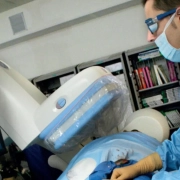
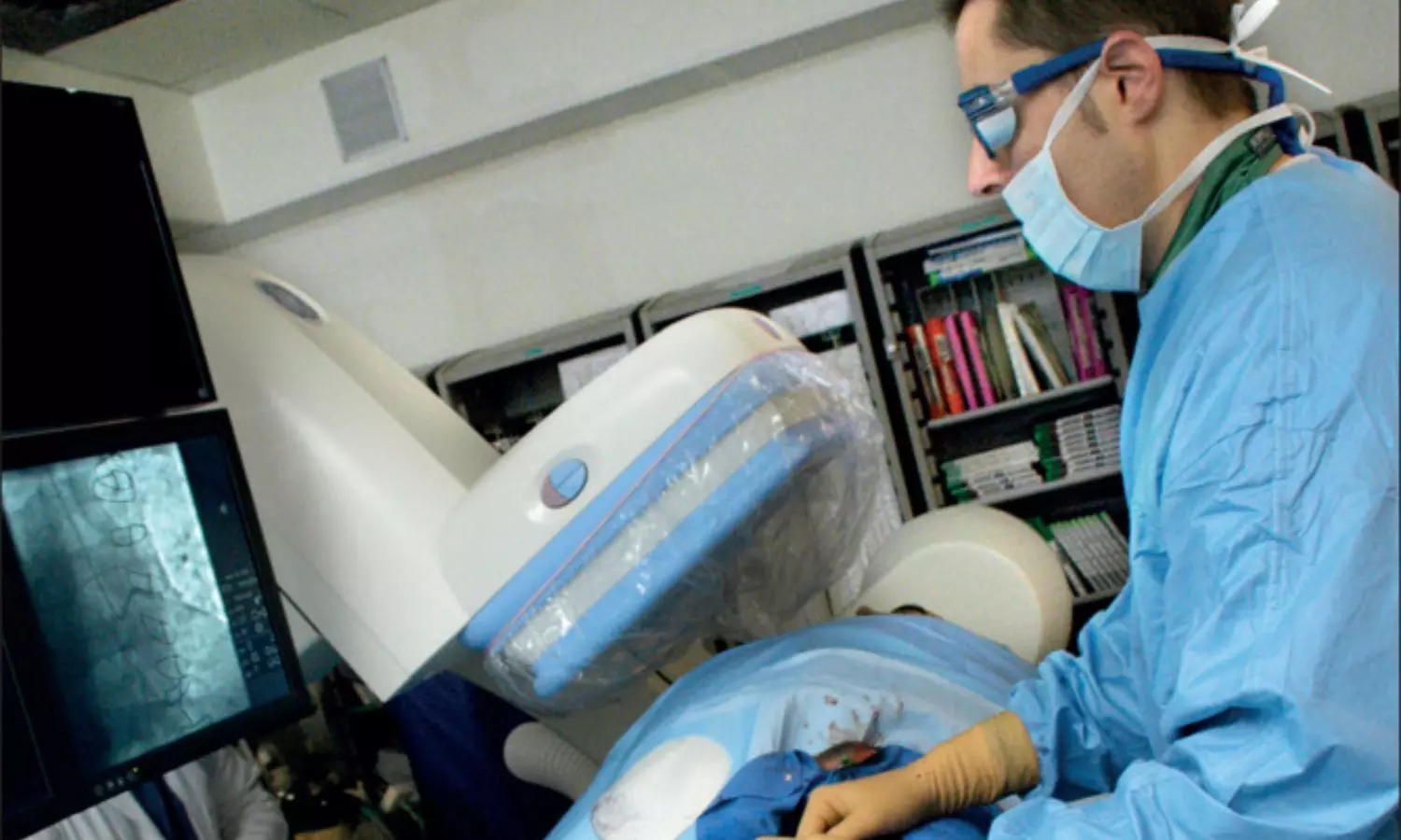
A meticulous kidney biopsy was instrumental in diagnosing oxalate nephropathy which uncovered the presence of numerous intratubular oxalate crystals, despite their absence in urinalysis. The surgical removal of part of the pancreas led to exocrine pancreatic insufficiency and subsequent fat malabsorption, while the non-compliance of the patient with pancreatic enzyme replacement therapy and daily consumption of vitamin C supplements further exacerbated the condition.

The study underlined the pathophysiology of secondary hyperoxaluria and its progression to oxalate nephropathy which emphasized the need for increased awareness of this condition as a potential cause of acute kidney injury post-pancreatectomy. The secondary hyperoxaluria can stem from increased oxalate or precursor intake, fat malabsorption or diminished intestinal oxalate degradation which often culminates in a perfect storm for patients with specific surgical histories and dietary habits.

The prompt intervention including the resumption of pancreatic enzyme replacement therapy and the introduction of calcium carbonate resulted in the improvement of the patient’s kidney function. However, the subsequent advancement of his cancer illuminated the complexities of managing post-surgical complications amidst the ongoing oncological concerns.

This case report contributes significantly to the medical literature by marking as the first reported instance of acute oxalate nephropathy post-pancreatectomy compounded by vitamin C intake. It prompts a reevaluation of post-operative care strategies in ensuring compliance with enzyme replacement therapy and rethinking nutritional supplements in vulnerable patients. Overall, this report emphasizes the importance of considering oxalate nephropathy in differential diagnoses for AKI following the pancreatectomy. 

Reference:

Barani, C., Aydin, S., Demoulin, N., & Jadoul, M. (2024). Oxalate nephropathy after pancreaticoduodenectomy: a case report. In BMC Nephrology (Vol. 25, Issue 1). Springer Science and Business Media LLC. https://doi.org/10.1186/s12882-024-03543-9

Powered by WPeMatico

Probiotic-rich Kombucha Tea may reduce fat, triglyceride and positively impact Host Metabolism: Study

Kombucha Tea (KT) has gained popularity for its potential health benefits, including protection against metabolic diseases. However, scientific evidence supporting these claims has been lacking. A recent study aimed to investigate how the probiotic microbes in Kombucha Tea impact host metabolism using the model organism Caenorhabditis elegans.

This study was published in the journal PLoS Genetics. The study was conducted by Rachel N. and colleagues.

Despite its long-standing use in traditional medicine, the precise mechanisms underlying the purported health benefits of Kombucha Tea remain elusive. This study sought to elucidate how Kombucha Tea-associated microbes (KTMs) influence host physiology, particularly in relation to lipid metabolism, using C. elegans as a model system.

Researchers established a method to exclusively feed C. elegans a diet consisting of Kombucha Tea-associated microbes (KTM). They observed the colonization of KTM in the nematode’s gut and investigated its effects on host development, fecundity, and lipid metabolism. Transcriptomic analysis was performed to understand the molecular mechanisms underlying the observed metabolic changes.

The key findings of the study were:

• Colonization of KTMs: C. elegans fed a diet of KTM displayed robust colonization of the gut by KTMs, with no adverse effects on host development or reproduction.

• Reduction in Lipid Accumulation: Consumption of KTMs led to a significant reduction in total lipid stores and lipid droplet size in C. elegans, indicating an impact on host lipid metabolism.

• Metabolic Rewiring: Transcriptomic analysis revealed widespread transcriptional changes in core lipid metabolism pathways, including upregulation of lysosomal lipase genes involved in lipid degradation, resembling a fasting-like response.

The study sheds light on how Kombucha Tea influences host metabolism through its probiotic microbes. By colonizing the gut of C. elegans, KTMs induce a metabolic shift towards increased lipid utilization, resulting in reduced fat accumulation. These findings align with the reported health benefits of Kombucha Tea in humans, suggesting its potential as a complementary health intervention.

Understanding the mechanisms by which Kombucha Tea affects host metabolism provides insights into its potential therapeutic applications for metabolic disorders like obesity and diabetes. Incorporating Kombucha Tea or its probiotic components into dietary interventions may offer novel approaches for managing metabolic diseases.

In conclusion, the study illuminates how Kombucha Tea-associated probiotic microbes influence host metabolism, leading to reduced fat accumulation in C. elegans. These findings contribute to our understanding of the health benefits associated with Kombucha Tea consumption and pave the way for future research exploring its therapeutic potential in human metabolic disorders. Further studies are warranted to validate these findings and translate them into clinical applications.

Reference:

DuMez-Kornegay, R. N., Baker, L. S., Morris, A. J., DeLoach, W. L. M., & Dowen, R. H. (2024). Kombucha Tea-associated microbes remodel host metabolic pathways to suppress lipid accumulation. PLoS Genetics, 20(3), e1011003. https://doi.org/10.1371/journal.pgen.1011003

Powered by WPeMatico

Probiotic Therapy Shows Promise in Treatment of Autoimmune Diseases: Study

Autoimmune diseases pose significant challenges in healthcare, affecting millions worldwide. Recent research has suggested a potential link between gut microbiota and autoimmune conditions, paving the way for innovative therapeutic approaches. A study published in BMC Medicine aimed to systematically review the efficacy of probiotic therapy in managing various autoimmune diseases. The study was conducted by Zeng L. and colleagues.

Autoimmune diseases, including fibromyalgia, psoriasis, juvenile idiopathic arthritis (JIA), lupus nephritis, systemic lupus erythematosus, ulcerative colitis, and Crohn’s disease, result from dysregulation of the immune system. Genetic, environmental, and microbial factors, particularly gut microbiota, are implicated in their pathogenesis. Probiotics, defined as beneficial microorganisms that colonize the gut and modulate host immunity and metabolism, offer a promising avenue for treatment.

The study conducted a systematic review and meta-analysis of randomized controlled trials (RCTs) evaluating the efficacy of probiotic therapy in autoimmune diseases. Researchers searched multiple databases for eligible trials up to June 2022 and assessed outcomes such as Disease Activity Score at 28 joints (DAS28), Psoriasis Area and Severity Index (PASI), and Systemic Lupus Erythematosus Disease Activity Measure (SLEDAI).

The key findings of the study were

• Out of 5799 studies screened, 80 RCTs encompassing 14 autoimmune diseases were included, originating from 27 countries.

• Probiotic therapy demonstrated efficacy in various autoimmune diseases, including improvements in pain for fibromyalgia patients and reductions in Psoriasis Area and Severity Index (PASI) scores for psoriasis patients.

• Adverse events associated with probiotic therapy were minimal, with only one trial reporting non-significant differences compared to controls.

• While no significant differences were observed in Disease Activity Score (DAS28) for certain diseases, improvements were noted in hemoglobin A1C (HbA1c) levels for type 1 diabetes mellitus (T1Dm) patients.

The meta-analysis highlights the potential of probiotic therapy in managing autoimmune diseases, with notable improvements in disease activity, symptomatology, and biochemical markers. Importantly, the safety profile of probiotics appears favorable, with minimal adverse events reported across trials.

These findings underscore the significance of gut microbiota modulation in autoimmune disease management. Probiotic therapy represents a promising adjunctive treatment strategy, particularly for conditions such as ulcerative colitis, where improvements in endoscopic scores and inflammatory markers were observed.

The study provides compelling evidence for the efficacy and safety of probiotic therapy in autoimmune diseases, suggesting its potential as a complementary therapeutic approach. However, further well-designed RCTs are warranted to validate these findings and elucidate optimal probiotic strains, dosages, and treatment durations for specific autoimmune conditions.

Reference:

Zeng L, Yang K, He Q, et al. Efficacy and safety of gut microbiota-based therapies in autoimmune and rheumatic diseases: a systematic review and meta-analysis of 80 randomized controlled trials. BMC Med. 2024;22(1):110. Published 2024 Mar 13. doi:10.1186/s12916-024-03303-4

Powered by WPeMatico

Culprit lesion PCI before diagnostic coronary angiography effectively reduces reperfusion time in STEMI patients: JAMA

A recent study published in the Journal of American Medical Association suggests a new approach to treat patients with ST-elevation myocardial infarction (STEMI) to improve the patient outcomes. The research focused on the timing and sequence of procedures during primary percutaneous coronary intervention (PCI), a critical treatment for STEMI patients to enhance the reperfusion process which is the restoration of blood flow to the heart.

Traditionally, the complete diagnostic coronary angiography (CAG) is performed before addressing the culprit lesion with PCI. However, this study, carried out from April 1, 2021 to August 31, 2022, proposed a radical shift in this sequence. By randomizing 216 patients into two groups, researchers evaluated the effects of performing culprit lesion PCI immediately, before a complete CAG, versus the conventional method of full CAG followed by PCI.

The findings found that the patients who received immediate PCI had significantly shorter needle-to-balloon times and the period from STEMI diagnosis to reperfusion, with an average time of 11.4 minutes when compared to 17.3 minutes in the traditional approach group. Over half of the patients in the immediate PCI group achieved a needle-to-balloon time of 10 minutes or less when compared to only 19.1% in the conventional treatment group.

These results are not just numbers but represent a potential shift in clinical practice that could save lives and reduce the severity of heart damage in STEMI patients. The rapid reperfusion achieved through this innovative approach suggests that immediate identification and treatment of the culprit lesion before conducting a full coronary angiography can significantly enhance the effectiveness of STEMI treatment.

This study reported that the rates of adverse events were similar between the two groups which indicates the accelerated approach does not compromise patient safety. Additionally, there were no significant differences in in-hospital, 30-day and 1-year all-cause mortality rates between the groups that underscore the efficacy and safety of this procedure. While the outcome of this study call for larger trials to validate these promising results and further evaluate the impact on clinical outcomes, this research presents a potential shift in the treatment of STEMI patients.

Source:

Levi, N., Wolff, R., Jubeh, R., Shuvy, M., Steinmetz, Y., Perel, N., Maller, T., Amsalem, I., Hitter, R., Asher, E., Turyan, A., Karmi, M., Orlev, A., Dratva, D., Khoury, Z., Hasin, T., Wolak, A., Glikson, M., & Dvir, D. (2024). Culprit Lesion Coronary Intervention Before Complete Angiography in ST-Elevation Myocardial Infarction. In JAMA Network Open (Vol. 7, Issue 3, p. e243729). American Medical Association (AMA). https://doi.org/10.1001/jamanetworkopen.2024.3729

Powered by WPeMatico

Fiber-Rich Diet Found Crucial in Preventing lethal colitis in susceptible individuals: Study

Inflammatory bowel diseases (IBDs), including ulcerative colitis and Crohn’s disease, present significant challenges for affected individuals, with symptoms ranging from mild discomfort to severe complications. Despite extensive research, the exact etiology of IBDs remains elusive, but recent findings shed light on the intricate interplay between diet, gut microbiota, and genetic factors.

The study, published in Cell Host & Microbe, investigated the role of dietary fiber in modulating gut microbiota and its impact on colonic health in mice lacking interleukin-10 (Il10−/−), a cytokine associated with IBDs. Prior research has highlighted the importance of diet and gut bacteria in IBD development, but the underlying mechanisms are still being elucidated. This study was conducted by Gabriel and colleagues.

Researchers subjected Il10−/− mice to a fiber-deprived diet to mimic dietary habits prevalent in industrialized populations. They observed the progression of colonic inflammation and analyzed changes in gut microbiota composition, mucus thickness, and immune responses.

• Mucosal Deterioration: The study found that a fiber-deprived diet led to the deterioration of colonic mucus in Il10−/− mice, precipitating lethal colitis.

• Immune Response: Inflammation was initiated by the expansion of natural killer (NK) cells and alterations in bacterial IgA coating, preceding the onset of colitis.

• Microbial Influence: Th1 immune responses were triggered by increased activities of mucin-degrading bacteria, particularly in regions with thinner mucus, driving the progression of colitis.

• Dietary Intervention: Exclusive enteral nutrition with a fiber-free diet exacerbated mucus erosion but mitigated inflammation by increasing the production of an anti-inflammatory bacterial metabolite, isobutyrate.

The findings underscore the critical role of dietary fiber in maintaining colonic health and preventing the onset of lethal colitis, particularly in individuals genetically predisposed to IBDs. By promoting the development of healthy mucus thickness and inhibiting inflammatory responses, fiber-rich diets may offer a preventive strategy against IBDs.

These findings have significant implications for the management and prevention of IBDs in clinical settings. Incorporating fiber-rich foods into the diet may help modulate gut microbiota composition and enhance colonic barrier function, thereby reducing the risk of inflammatory flare-ups and disease progression.

In conclusion, the study highlights the importance of dietary fiber in preserving colonic health and preventing lethal colitis in susceptible individuals. Further research is warranted to elucidate the precise mechanisms underlying the interplay between diet, gut microbiota, and genetic factors in IBD development. These insights pave the way for targeted interventions aimed at optimizing dietary habits and promoting gut health to mitigate the burden of inflammatory bowel diseases.

Reference:

Pereira, G. V., Boudaud, M., Wolter, M., Alexander, C., De Sciscio, A., Grant, E. T., Trindade, B. C., Pudlo, N. A., Singh, S., Campbell, A., Shan, M., Zhang, L., Yang, Q., Willieme, S., Kim, K., Denike-Duval, T., Fuentes, J., Bleich, A., Schmidt, T. M., … Martens, E. C. (2024). Opposing diet, microbiome, and metabolite mechanisms regulate inflammatory bowel disease in a genetically susceptible host. Cell Host & Microbe. https://doi.org/10.1016/j.chom.2024.03.001

Powered by WPeMatico

Deucravacitinib improves outcomes in patients with psoriasis arthritis: Study

Psoriatic arthritis is a chronic and debilitating autoimmune disease which leads to significant physical disability and diminished quality of life. The traditional treatments often come with various side effects and this highlights the need for safer and more effective options. A recent phase 2 trial published in the Arthritis Care & Research journal showed the potential benefits of Deucravacitinib, a TYK-2 inhibitor, from the perspective of the patient with psoriatic arthritis (PsA).

This double-blind trial included a total of 203 participants to assess the efficacy and patient-reported outcomes (PROs) of the two dosage levels of Deucravacitinib when compared to a placebo. The participants in the trial were randomly assigned to receive either 6 mg or 12 mg of Deucravacitinib once daily or a placebo, over a 16-week period.

The study primarily focused on the change from baseline in the Health Assessment Questionnaire Disability Index (HAQ-DI) and the 36-item Short Form Health Survey (SF-36) physical component summary (PCS) score at Week 16. These measures were crucial in assessing physical function and overall health status that provides insight into the impact of treatment from the patient’s view.

The results found that both dosages of Deucravacitinib significantly outperformed the placebo in improving HAQ-DI and SF-36 PCS scores. The adjusted mean difference in HAQ-DI score at Week 16 was −0.26 (P = 0.0020) for the 6 mg dose and −0.28 (P = 0.0008) for the 12 mg dose indicated a notable improvement in physical function. Similarly, the SF-36 PCS scores improved by 3.3 points (P = 0.0062) and 3.5 points (P = 0.0042) for the 6 mg and 12 mg doses, respectively which suggests enhanced quality of life.

Moreover, a significant percentage of patients reported improvements in patient-reported outcomes that included fatigue, pain and mental health after reaching or exceeding the minimum clinically important differences (MCIDs) or achieving normative values. This indicates that Deucravacitinib not only improve the physical health but also positively affects the overall wellbeing.

Overall, these findings underscore the potential of Deucravacitinib as a transformative treatment for psoriatic arthritis. The significant and clinically meaningful improvements in patient-reported outcomes that highlight its capacity to address both the physical and psychological aspects of the disease.

Reference:

Strand, V., Gossec, L., Coates, L. C., Ogdie, A., Choi, J., Becker, B., Zhuo, J., Lehman, T., Nowak, M., Elegbe, A., Mease, P. J., & Deodhar, A. (2024). Improvements in patient‐reported outcomes after treatment with deucravacitinib in patients with psoriatic arthritis: results from a randomized phase 2 trial. In Arthritis Care & Research. Wiley. https://doi.org/10.1002/acr.25333

Powered by WPeMatico

Artificial Intelligence tool successfully predicts ventricular arrhythmia from standard heart tracings: Study

In a Leicester study that looked at whether artificial intelligence (AI) can be used to predict whether a person was at risk of a lethal heart rhythm, an AI tool correctly identified the condition 80 per cent of the time.

The findings of the study, led by Dr Joseph Barker working with Professor Andre Ng, Professor of Cardiac Electrophysiology and Head of Department of Cardiovascular Sciences at the University of Leicester and Consultant Cardiologist at the University Hospitals of Leicester NHS Trust, have been published in the European Heart Journal-Digital Health.

Ventricular arrhythmia (VA) is a heart rhythm disturbance originating from the bottom chambers (ventricles) where the heart beats so fast that blood pressure drops which can rapidly lead to loss of consciousness and sudden death if not treated immediately.

NIHR Academic Clinical Fellow Dr Joseph Barker co-ordinated the multicentre study at the National Institute for Health and Care Research (NIHR) Leicester Biomedical Research Centre,  and co-developed an AI tool with Dr Xin Li, Lecturer in Biomedical Engineering, School of Engineering. The tool examined Holter electrocardiograms (ECGs) of 270 adults taken during their normal daily routine at home.  

These adults had the Holter ECGs taken as part of their NHS care between 2014 and 2022. Outcomes for these patients were known, and 159 had sadly experienced lethal ventricular arrhythmias, on average 1.6 years following the ECG.

The AI tool, VA-ResNet-50, was used to retrospectively examine ‘normal for patient’ heart rhythms to see if their heart was capable of the lethal arrythmias.

Professor Ng said: “Current clinical guidelines that help us to decide which patients are most at risk of going on to experience ventricular arrhythmia, and who would most benefit from the life-saving treatment with an implantable cardioverter defibrillator are insufficiently accurate, leading to a significant number of deaths from the condition.

“Ventricular arrhythmia is rare relative to the population it can affect, and in this study we collated the largest Holter ECG dataset associated with longer term VA outcomes.

“We found the AI tool performed well compared with current medical guidelines, and correctly predicted which patient’s heart was capable of ventricular arrhythmia in 4 out of every 5 cases.

“If the tool said a person was at risk, the risk of lethal event was three times higher than normal adults.

“These findings suggest that using artificial intelligence to look at patients’ electrocardiograms while in normal cardiac rhythm offers a novel lens through which we can determine their risk, and suggest appropriate treatment; ultimately saving lives.”

He added: “This is important work, which wouldn’t have been possible without an exceptional team in Dr Barker and Dr Xin Li, and their belief and dedication to novel methods of analysis of historically disregarded data.”

Reference:

Joseph Barker, Xin Li, Ahmed Kotb, Akash Mavilakandy, Ibrahim Antoun, Chokanan Thaitirarot, Ivelin Koev, Sharon Man, Fernando S Schlindwein, Harshil Dhutia, Shui Hao Chin, Ivan Tyukin, William B Nicolson, G Andre Ng, Artificial intelligence for ventricular arrhythmia capability using ambulatory electrocardiograms, European Heart Journal – Digital Health, 2024;, ztae004, https://doi.org/10.1093/ehjdh/ztae004

.

Powered by WPeMatico

Retinal Ischemic Perivascular lesions Linked to Myocardial Infarction Risk in CAD Patients: Study

Coronary artery disease (CAD) remains a leading cause of morbidity and mortality worldwide, with myocardial infarction (MI) posing a significant risk for affected individuals. While various cardiovascular risk factors are well-documented, emerging evidence suggests a potential link between retinal ischemic perivascular lesions (RIPLs) and MI in CAD patients.

Retinal ischemic perivascular lesions (RIPLs) have garnered attention as potential biomarkers for retinal ischemia, reflecting underlying vascular pathology. However, their association with MI among CAD patients remains poorly understood. A recent retrospective cross-sectional study aimed to investigate this relationship using macular spectral domain optical coherence tomography (SD-OCT) imaging. This study was published in the American Journal Of Ophthalmology. The study was conducted by Elodie B. and colleagues.

The study included 317 consecutive CAD patients who underwent SD-OCT imaging of the macula. Patients with a history of MI were compared to those without MI. RIPLs were assessed by two independent graders, and medical records were reviewed. Multivariate logistic regression analysis was conducted to evaluate the association between RIPLs and MI, adjusting for relevant covariates.

The key findings of the study were:

• Prevalence of RIPLs: Among CAD patients, 17% had a history of MI, with a significantly higher prevalence of RIPLs observed in the MI group compared to the non-MI group (59.3% vs. 35.7%; p<0.001).

• Association with MI: Presence of RIPLs was strongly associated with MI, with an odds ratio of 3 (95% CI: 1.91-4.74; p<0.001), after adjusting for age, gender, smoking status, hypertension, diabetes, dyslipidemia, and body mass index.

The study findings highlight a significant association between RIPLs detected via SD-OCT imaging and MI in CAD patients. RIPLs may serve as valuable biomarkers for identifying individuals at heightened risk of cardiovascular events within this population. The incorporation of RIPL evaluation into routine clinical assessments could enhance risk stratification and inform targeted interventions to mitigate cardiovascular risk in CAD patients.

These findings underscore the importance of comprehensive cardiovascular evaluations in CAD patients, including assessment of retinal vascular changes. Early detection of RIPLs may facilitate proactive management strategies aimed at reducing the incidence of MI and improving patient outcomes in this high-risk population.

In conclusion, the presence of retinal ischemic perivascular lesions (RIPLs), as assessed by SD-OCT, is significantly associated with myocardial infarction in patients with coronary artery disease. These findings underscore the potential clinical utility of RIPL evaluation as part of the medical management of CAD, offering valuable insights into cardiovascular risk assessment and personalized intervention strategies. Further research is warranted to validate these findings and elucidate the underlying mechanisms linking RIPLs to MI in CAD patients.

Reference:

Bousquet, E., Santina, A., Au, A., Somisetty, S., Abraham, N., Voichanski, S., Estawro, R., Fouad, Y. A., Romero-Morales, V., Bakhoum, M. F., & Sarraf, D. (2024). Retinal Ischemic Perivascular Lesions are associated with myocardial infarction in patients with coronary artery disease. American Journal of Ophthalmology. https://doi.org/10.1016/j.ajo.2024.03.017

Powered by WPeMatico

Compared to single-layer closure, double-layer closure may not exhibit superior reproductive outcomes after first cesarean delivery: Study

The increasing prevalence of cesarean deliveries has raised concerns about long-term complications, including niche formation in the uterine scar. Niche development is associated with various gynecologic issues and obstetric complications in subsequent pregnancies. Despite its importance, there’s uncertainty regarding the optimal uterine closure technique.

Niche formation, or defects in the uterine scar, can lead to gynecologic complaints and complications in future pregnancies. The debate over single-layer vs. double-layer uterine closure techniques persists, with conflicting evidence on which approach is superior for preventing long-term complications.

This study was published in the Journal Of Obstretrics & Gynaecology by Carry Verberkt and colleagues. A multicenter, double-blind, randomized controlled trial conducted in the Netherlands aimed to assess the impact of single-layer vs. double-layer closure of the uterine incision on long-term outcomes in women undergoing their first cesarean delivery. The primary outcome was live birth rate at a 3-year follow-up, with secondary outcomes including fertility, gynecologic, and obstetrical outcomes.

Key Findings:

• The study included 2292 women, with 830 in the single-layer closure group and 818 in the double-layer closure group.

• At the 3-year follow-up, there were no significant differences in live birth rates between the two closure techniques.

• Secondary outcomes, including pregnancy rate, need for fertility treatments, mode of delivery, and obstetrical complications, also showed no significant differences between the groups.

• Both groups reported high rates of gynecologic symptoms, including spotting, dysmenorrhea, and sexual dysfunction.

The study did not find superiority of double-layer closure over single-layer closure in terms of reproductive outcomes after a first cesarean delivery. This challenges the current recommendation favoring double-layer closure, suggesting that surgeons can choose their preferred technique. Additionally, the high prevalence of gynecologic symptoms after cesarean delivery highlights the importance of discussing these risks with patients.

Surgeons should consider individual patient factors and preferences when choosing uterine closure techniques during cesarean deliveries. Patient counseling regarding the potential for gynecologic symptoms post-delivery is crucial for informed decision-making. Further research may be needed to elucidate the long-term effects of different closure methods on maternal health and reproductive outcomes.

Reference:

Verberkt, C., Stegwee, S. I., Van der Voet, L. F., Van Baal, W. M., Kapiteijn, K., Geomini, P. M. A. J., Van Eekelen, R., de Groot, C. J. M., de Leeuw, R. A., Huirne, J. A. F., van Baal, M., Klerkx, W., Bekker, M. N., de Boer, K., Boormans, E. M. A., van Eijndhoven, H. W. F., Feitsma, H., Hehenkamp, W. J. K., Hemelaar, M., … de Vleeschouwer, M. Single-layer vs double-layer uterine closure during cesarean delivery: 3-year follow-up of a randomized controlled trial (2Close study). American Journal of Obstetrics and Gynecology,2023. https://doi.org/10.1016/j.ajog.2023.12.032

Powered by WPeMatico

ChatGPT may help dental students retain knowledge and enhance performance, suggests research

 A study aiming to utilize current dental course materials, syllabi, and textbooks to investigate ChatGPT’s potential benefits for dental students. was presented at the 102nd General Session of the IADR, which was held in conjunction with the 53rd Annual Meeting of the American Association for Dental, Oral, and Craniofacial Research and the 48th Annual Meeting of the Canadian Association for Dental Research, on March 13-16, 2024, in New Orleans, LA, USA.

The abstract, “ChatGPT to Help Dental Students Retain Knowledge and Enhance Performance” was presented during the “SCADA: Clinical Science/Public Health Research” Poster Session that took place on Thursday, March 14, 2024 at 11 a.m. Central Standard Time (UTC-6).

The study, by Ihunna Amugo of Meharry Medical College, Nashville, TN, USA, conducted an extensive literature review, analyzing previous studies in different educational contexts to evaluate ChatGPT’s efficacy in dental education. Questions aligned with dental course materials and objectives were formulated to ensure ChatGPT’s relevance to the specific needs of dental students. The research encompassed various dental courses, including operative dentistry, nutrition, periodontics, oral radiology, and biology of disease.

By applying ChatGPT in these courses, its capabilities were harnessed to generate step-by-step instructions, summaries, and questions to supplement traditional learning methods. Data on students’ interactions with ChatGPT were collected and analyzed, assessing usage patterns, engagement levels, and perceived benefits.

The study’s results indicated that ChatGPT has the potential to enhance students’ understanding and knowledge retention. In addition, ChatGPT can effectively provide instructional support and generate relevant content for dental students. 

Powered by WPeMatico